This website uses cookies to ensure you get the best experience on our website.


- Table of Contents
Plan KRT10 chromogenic IHC in paraffin sections using skin as a positive tissue (HPA tissue IHC). Use the catalog antibody at 1:100–1:300 (datasheet A02305-1), then score cytoplasmic staining in corneal-layer cells (HPA tissue IHC).

Expected localisation, validated protocols, controls and antibodies — the at-a-glance facts below, then the full design guide.
| Expected localisation | Cytoplasmic in skin squamous epithelium (HPA tissue IHC) | |
| Staining pattern | Corneal-layer cells show selective cytoplasmic staining (HPA tissue IHC) | |
| Antigen retrieval | Tris-EDTA pH 9.0 HIER, heat-mediated (datasheet A02305-1) | |
| Positive control | Skin | |
| Negative control | Adipose tissue+4 more · see all |
| Fixation | Keep paraffin-section fixation consistent across samples. (standard IHC practice; not target-specific) | |
| Caveat | A secreted variant may complicate RNA–stain concordance (HPA tissue IHC) | |
| Regulation | Expressed during suprabasal differentiation (UniProt) | |
| Isoform / epitope | No isoforms; extracellular vs cytoplasmic epitope may matter (UniProt) |
The catalog antibody has an IHC-P protocol (datasheet A02305-1). Published KRT10 IHC methods provide cervical tissue and human corneal epithelium examples (PMC9530870; PMC12395845).
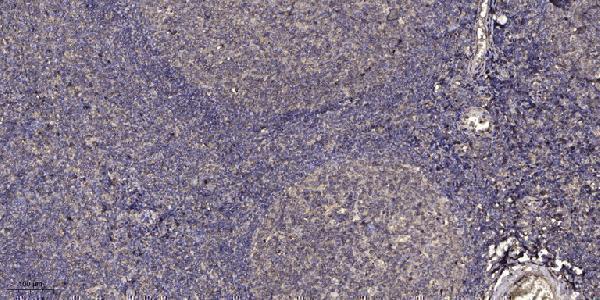
| Sample | Paraffin-embedded human tonsil tissue; fixative not specified (datasheet A02305-1) |
| Fixation | Image fixative and duration unreported (datasheet A02305-1); verify before use. |
| Sectioning | 4–5 µm sections on charged slides (standard) |
| Deparaffinisation | Xylene, graded ethanol series to water (standard) |
| Antigen retrieval | Heat retrieval: Tris-EDTA pH 9.0 (datasheet A02305-1); 20 min, 95–100 °C (standard) |
| Peroxidase block | 3% H2O2, 10 min, room temperature (standard) |
| Blocking | 10% normal serum of the secondary host, 30 min, room temperature (standard) |
| Primary antibody | Rabbit anti-KRT10, 1:100-1:300 (datasheet A02305-1) |
| Primary incubation | Overnight at 4 °C (standard) |
| Detection | HRP-polymer secondary, DAB chromogen 5–10 min (standard) |
| Counterstain | Hematoxylin, blue, dehydrate and mount (standard) |
| Expected result | KRT10-positive staining in cells in corneal layer of skin (HPA tissue IHC: Medium). HPA tissue profile: Selective cytoplasmic expression in squamous epithelium of skin. No signal in the no-primary control. |
KRT10 is an intermediate filament protein of suprabasal keratinocytes (UniProt P13645 function). In paraffin sections, expect selective cytoplasmic staining in skin squamous epithelium (HPA tissue IHC, Enhanced reliability). UniProt also lists cell surface and extracellular locations, but KRT10 has no transmembrane segment (UniProt P13645 subcellular location and topology). Interpret those locations in the context of the observed tissue pattern.
| Cytoplasmic staining follows the suprabasal layers of skin squamous epithelium; cells in the corneal layer stain. | This fits KRT10 expression during keratinocyte differentiation (UniProt P13645 tissue specificity and function). HPA reports selective cytoplasmic skin staining and medium staining in corneal-layer cells (HPA tissue IHC). Assess the layer pattern alongside the counterstain; HPA does not assign one intensity to every suprabasal layer. |
| Predominantly nuclear staining appears in skin epithelium. | A nuclear-dominant result does not match HPA's selective cytoplasmic tissue pattern or UniProt's principal intermediate-filament function (HPA tissue IHC; UniProt P13645 function). Treat it as suspect and inspect the counterstain, chromogen deposition, and omission control before scoring it as KRT10 (general IHC practice). |
| The main signal is in adipocytes, glandular cells, or respiratory epithelial cells. | HPA reports KRT10 as not detected in adipocytes of adipose tissue, glandular cells of adrenal gland and appendix, and bronchial respiratory epithelium (HPA tissue IHC). Consider antibody cross-reactivity or endogenous detection activity; compare with a reagent-omission control rather than assigning specificity from staining alone (general IHC practice). |
| Diffuse chromogen covers epithelium, stroma, and blank slide areas without a discernible cell pattern. | A field-wide deposit cannot establish the selective cytoplasmic pattern reported for skin (HPA tissue IHC). Inspect reagent-omission controls and background across the section; adjust blocking, washes, and detection conditions according to the assay workflow (general IHC practice). |
| Skin epithelium, including the suprabasal region, has no interpretable signal. | That conflicts with UniProt's suprabasal expression and HPA's skin staining (UniProt P13645 tissue specificity; HPA tissue IHC). First confirm that the section contains intact, assessable epithelium, then check antibody and detection controls (general IHC practice). A negative run alone cannot establish absent KRT10 expression. |
| Anatomic layer and tissue | UniProt places KRT10 throughout suprabasal layers, including stratum corneum (UniProt P13645 tissue specificity). HPA's observed paraffin-section profile is selective cytoplasmic expression in skin squamous epithelium, with medium corneal-layer staining (HPA tissue IHC). Use layer and tissue identity together when judging a positive. |
| Compartment and assembly | KRT10 forms keratin intermediate filaments, primarily with KRT1, supporting a cytoplasmic epithelial pattern (UniProt P13645 function and subunit). UniProt also lists surface and extracellular locations and no transmembrane segment (UniProt P13645 subcellular location and topology). Those annotations do not establish that a surface-only paraffin-section pattern is the expected HPA tissue-IHC result. |
| Evidence for antibody interpretation | HPA rates tissue staining Enhanced and records IHC Enhanced status for HPA012014 and CAB000132 (HPA tissue IHC; HPA antibodies). This supports the reported skin pattern as a reference, while staining outside that pattern still needs controls and tissue context (general IHC practice). The record supplies no epitope map or working dilution. |
| Processing and retrieval limits | UniProt lists a single KRT10 chain, no signal peptide or propeptide, no annotated isoforms, and phosphorylation sites (UniProt P13645 processing and modified residues). These facts do not identify the antibody epitope or establish KRT10-specific retrieval or fixation sensitivity. If signal fails, assess retrieval against the antibody's own instructions and run controls (general IHC practice). |
| IF/ICC question: should fluorescence match this IHC pattern? | HPA provides a secreted subcellular summary but no ICC-IF image-bearing cell lines, and neither listed antibody has ICC validation recorded (HPA subcellular; HPA antibodies). An IF/ICC pattern therefore cannot be verified from this payload; use the separate IF/ICC guide for assay design. |
| Situation | Likely cause | Next action |
|---|---|---|
| No staining in an intact skin section | The result conflicts with the expected suprabasal skin expression (UniProt P13645 tissue specificity; HPA tissue IHC); the failed step is undetermined. | Verify tissue orientation and epithelial preservation, then review the antibody, detection, and run controls (general IHC practice). Check retrieval against the antibody's documented instructions; no KRT10-specific retrieval condition is supplied. |
| Only basal epithelial cells stain | Basal-restricted staining does not fit UniProt's suprabasal distribution or HPA's selective skin squamous-epithelium profile (UniProt P13645 tissue specificity; HPA tissue IHC). | Recheck layer assignment with morphology and the counterstain. Compare another skin section and assay controls before interpreting basal signal as target-specific (general IHC practice). |
| Strong nuclear or stromal staining dominates | The dominant compartment differs from HPA's cytoplasmic epithelial profile (HPA tissue IHC). Its source cannot be identified from appearance alone. | Inspect a reagent-omission control and section morphology; review detection background and antibody conditions before scoring (general IHC practice). |
| A nominally negative cell population stains | HPA reports not detected for several sampled non-squamous populations, including bronchial respiratory epithelium (HPA tissue IHC). Cross-reactivity or endogenous detection activity is possible (general IHC practice). | Confirm cell identity, compare the skin reference pattern, and test appropriate omission or endogenous-activity controls for the chosen detection system (general IHC practice). |
| Diffuse background obscures epithelial boundaries | Background prevents assessment of HPA's selective cytoplasmic pattern (HPA tissue IHC); the deposit's mechanism remains uncertain. | Review blocking, washes, detection exposure, and reagent-omission controls, changing one assay condition at a time (general IHC practice). |
| Surface-like or extracellular staining appears without cytoplasmic epithelial staining | UniProt lists surface and extracellular locations, but HPA's observed tissue-IHC profile is cytoplasmic in skin squamous epithelium (UniProt P13645 subcellular location; HPA tissue IHC). | Record the compartment separately, check morphology and omission controls, and avoid calling an isolated surface deposit the expected paraffin-section positive pattern (general IHC practice). |




Comprehensive Human Protein Atlas IHC scoring per tissue (reliability: Enhanced — High consistency between antibody staining and RNA expression data. At least one protein variant secreted, tissue location of RNA and protein might differ and correlation is complex.). Rows are taken directly from the HPA tissue chart — click any row's HPA link to view the source.
| Tissue | Cell type | Level | Evidence | Source |
|---|---|---|---|---|
| Skin | Cells in corneal layer | Medium | Protein (IHC) | HPA → |
| Tissue | Cell type | Level | Evidence | Source |
|---|---|---|---|---|
| Adipose tissue | Adipocytes | Not detected | Protein (IHC) | HPA → |
| Adrenal gland | Glandular cells | Not detected | Protein (IHC) | HPA → |
| Appendix | Glandular cells | Not detected | Protein (IHC) | HPA → |
| Bone marrow | Hematopoietic cells | Not detected | Protein (IHC) | HPA → |
| Breast | Adipocytes | Not detected | Protein (IHC) | HPA → |
Troubleshoot chromogenic KRT10 IHC by checking retrieval, tissue architecture, and compartment-specific staining before comparing scores across sections.
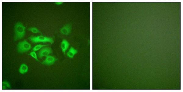
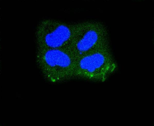
The catalog lists anti-KRT10 antibodies for human, mouse and rat IHC and IF (catalog: A02305-1, M02305-1, A02305-2), with human tissue IHC and HeLa ICC images (catalog: image captions).
A02305-1 shows paraffin-embedded human tonsil IHC at 1:200 after Tris-EDTA pH 9.0 retrieval (A02305-1 image caption); M02305-1 shows paraffin-embedded human bladder IHC (M02305-1 image caption). A02305-2 shows ICC in paraformaldehyde-fixed, permeabilised HeLa cells (A02305-2 image caption), while its catalog also lists IHC (catalog: A02305-2 applications).
Which to pick: For tissue IHC, choose A02305-1 when its documented tonsil staining and retrieval conditions are useful (A02305-1 image caption), or M02305-1 for a monoclonal antibody with a bladder IHC image (catalog: M02305-1 clone EEF-11; M02305-1 image caption); neither IHC caption reports the fixative (A02305-1 and M02305-1 image captions). For IF/ICC, A02305-2 has a HeLa ICC image with stated fixation and permeabilisation (A02305-2 image caption). For human, mouse or rat samples, all three list reactivity with those species (catalog: A02305-1, M02305-1, A02305-2 reactivity); the pictured IHC tissues are human (A02305-1 and M02305-1 image captions).