This website uses cookies to ensure you get the best experience on our website.


- Table of Contents
Plan MTHFR staining in paraffin sections using the reported cytoplasmic tissue pattern (HPA tissue IHC). Compare glandular and kidney tubule cells with documented low or undetected cell populations when assessing signal (HPA tissue IHC).

Expected localisation, validated protocols, controls and antibodies — the at-a-glance facts below, then the full design guide.
| Expected localisation | Cytoplasmic tissue staining (HPA tissue IHC) | |
| Staining pattern | Cytoplasmic staining in glandular and kidney tubule cells (HPA tissue IHC) | |
| Antigen retrieval | Tris-EDTA pH 9.0 HIER, heat-mediated (datasheet A00336) | |
| Positive control | Bronchus+4 more · see all | |
| Negative control | Adipose tissue+4 more · see all |
| Fixation | Keep fixation consistent across sections. (standard IHC practice; not target-specific) | |
| Caveat | Antibody staining has only medium consistency with RNA expression (HPA tissue IHC) | |
| Regulation | Staining-linked regulation is unreported (UniProt) | |
| Isoform / epitope | 2 isoforms; epitope coverage is unspecified (UniProt) |
The catalog antibody protocol uses heat-mediated Tris-EDTA pH 9.0 retrieval (datasheet A00336). The published IHC options below report tissue staining methods from three MTHFR studies (PMC10290629; PMC7353690; PMC9353118).
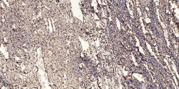
| Sample | Paraffin-embedded human oophoroma tissue; fixative not specified (datasheet A00336) |
| Fixation | Image fixative and duration unreported (datasheet A00336); verify before use. |
| Sectioning | 4–5 µm sections on charged slides (standard) |
| Deparaffinisation | Xylene, graded ethanol series to water (standard) |
| Antigen retrieval | Heat retrieval: Tris-EDTA pH 9.0 (datasheet A00336); 20 min, 95–100 °C (standard) |
| Peroxidase block | 3% H2O2, 10 min, room temperature (standard) |
| Blocking | 10% normal serum of the secondary host, 30 min, room temperature (standard) |
| Primary antibody | Rabbit anti-MTHFR, 1:50 recommended; image 1:200 (datasheet A00336) |
| Primary incubation | Overnight at 4 °C (standard) |
| Detection | HRP-polymer secondary, DAB chromogen 5–10 min (standard) |
| Counterstain | Hematoxylin, blue, dehydrate and mount (standard) |
| Expected result | MTHFR-positive staining in ciliated cells (cilia axoneme) of bronchus (HPA tissue IHC: High). HPA tissue profile: Cytoplasmic expression in several tissues, including epididymis, testis, kidney, anterior pituitary gland, lymphoid and neuronal tissues. No signal in the no-primary control. |
In paraffin-section IHC, expect predominantly cytoplasmic MTHFR staining in selected glandular and tubular cells, including epididymal glandular cells and kidney tubule cells (HPA: tissue IHC). Bronchial ciliated cells can show ciliary axoneme staining (HPA: High). Tissue IHC reliability is Enhanced, with medium consistency between staining and RNA data (HPA). UniProt assigns no subcellular location and reports no transmembrane segment (UniProt P42898 topology).
| Clear cytoplasmic staining in kidney tubule cells or epididymal glandular cells, with recognizable tissue structure. | This matches the reported high staining in those cell populations (HPA: kidney and epididymis, High). Score the named cells and their compartment; adjacent unstained cells need not invalidate the result (standard IHC practice). |
| Predominantly nuclear staining, or another unexpected compartment, replaces the expected cytoplasmic pattern. | Treat this as a possible localization artefact and check controls before assigning it to MTHFR (HPA: cytoplasmic tissue IHC profile). UniProt supplies no subcellular annotation that would independently confirm a new compartment (UniProt P42898). |
| Strong staining appears in lung alveolar cells or adipocytes, while expected positive cells stain weakly. | Those cell populations are reported as not detected (HPA: lung alveolar cells; adipose adipocytes). Consider cross-reactivity or endogenous detection activity; compare cell-resolved controls before interpreting the signal (standard IHC practice). |
| Color is spread across stroma, empty spaces or most cells without a discernible cellular pattern. | Diffuse background cannot establish MTHFR localization. Assess reagent-only and tissue background, washing, blocking and chromogen development (standard IHC practice); HPA's cell-specific observations do not validate uniform staining (HPA: tissue IHC). |
| No signal is visible in kidney tubule cells or epididymal glandular cells on an otherwise interpretable section. | This conflicts with reported high staining in those cells (HPA: kidney and epididymis, High). Check whether the assay worked using a known-positive section; absence alone cannot distinguish technical failure from specimen variation (standard IHC practice). |
| Choice of IHC evidence | HPA076180 and HPA077255 carry Enhanced IHC validation (HPA: antibodies); the overall tissue profile has medium staining-to-RNA consistency (HPA: reliability description). Compare the actual slide with the reported cells. |
| Cell population within a tissue | Interpret cells separately: bronchial cilia axonemes are High, whereas lung alveolar cells are not detected (HPA: bronchus and lung). A tissue-level positive label cannot be applied to every cell. |
| Molecular features | UniProt lists two isoforms, no signal peptide or propeptide, and a single chain spanning residues 1–656 (UniProt P42898). These facts do not establish isoform-specific IHC staining or a processing-related compartment change. |
| Detection background | With an enzyme-based chromogen, endogenous tissue activity can mimic antibody staining (standard IHC practice). Use the appropriate enzyme control and assess staining with the primary antibody omitted before assigning atypical color to MTHFR. |
| Q: Should IF/ICC show the same localization as paraffin-section IHC? | A: HPA IF/ICC reports mainly cell junctions, with centrosome and basal-body staining (HPA: subcellular ICC-IF). This is assay-specific evidence; it does not replace the cytoplasmic tissue IHC reference (HPA: tissue IHC). |
| Situation | Likely cause | Next action |
|---|---|---|
| Expected positive cells are blank. | Assay sensitivity or detection may be inadequate; kidney tubule and epididymal glandular cells are reported High (HPA: tissue IHC). | Run a known-positive section alongside the specimen and review antigen retrieval and detection controls (standard IHC practice). No MTHFR-specific retrieval condition or fixation sensitivity is established by these sources. |
| Only the section edge or damaged areas are strongly colored. | Uneven reagent access or local background can distort interpretation (standard IHC practice). | Judge intact interior cells, inspect an antibody-omission control, and repeat with even reagent coverage if needed (standard IHC practice). Do not score edge color as a cell-specific MTHFR pattern. |
| Unexpected nuclear staining dominates. | The pattern disagrees with the reported cytoplasmic tissue profile (HPA: tissue IHC); the source data do not establish a nuclear IHC pattern. | Compare with a positive tissue and an antibody-omission control, then review antibody concentration and detection background (standard IHC practice). Record the discrepancy rather than reclassifying localization. |
| Lung alveolar cells or adipocytes appear strongly positive. | These cells are reported as not detected (HPA: lung alveolar cells; adipose adipocytes); cross-reactivity or endogenous detection activity is possible. | Check cell identity and compare antibody-omission and enzyme-background controls where relevant (standard IHC practice). Interpret discordant staining cautiously, even when another cell population in the section stains. |
| Most cells show weak, uniform chromogen. | Diffuse background may obscure the cell-resolved cytoplasmic pattern reported by HPA (HPA: tissue IHC; standard IHC practice). | Review blocking, washing, primary-antibody concentration and chromogen development with matched controls (standard IHC practice). Reassess named positive and negative cell populations after background is reduced. |
| A negative-control tissue shows little color, but so does the positive tissue. | A clean negative alone does not show that the assay can detect MTHFR (standard IHC practice). HPA reports high staining in kidney tubules and epididymal glandular cells (HPA: tissue IHC). | Confirm that a known-positive section develops cell-specific signal before interpreting absent specimen staining; document the tested cell population and controls (standard IHC practice). |


Comprehensive Human Protein Atlas IHC scoring per tissue (reliability: Enhanced — Medium consistency between antibody staining and RNA expression data.). Rows are taken directly from the HPA tissue chart — click any row's HPA link to view the source.
| Tissue | Cell type | Level | Evidence | Source |
|---|---|---|---|---|
| Bronchus | Ciliated cells (cilia axoneme) | High | Protein (IHC) | HPA → |
| Duodenum | Glandular cells | High | Protein (IHC) | HPA → |
| Epididymis | Glandular cells | High | Protein (IHC) | HPA → |
| Kidney | Cells in tubules | High | Protein (IHC) | HPA → |
| Pituitary gland | Cells in anterior | High | Protein (IHC) | HPA → |
| Tissue | Cell type | Level | Evidence | Source |
|---|---|---|---|---|
| Adipose tissue | Adipocytes | Not detected | Protein (IHC) | HPA → |
| Breast | Adipocytes | Not detected | Protein (IHC) | HPA → |
| Cervix | Glandular cells | Not detected | Protein (IHC) | HPA → |
| Esophagus | Squamous epithelial cells | Not detected | Protein (IHC) | HPA → |
| Heart muscle | Cardiomyocytes | Not detected | Protein (IHC) | HPA → |
Troubleshoot MTHFR staining in paraffin sections by checking retrieval, cell type, staining pattern, and controls alongside the catalog antibody’s tissue image.
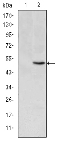
A00336 and M00336 have real IHC images of paraffin-embedded human tissues (catalog IHC captions). Listed species reactivity covers human, monkey, mouse, and rat; IF is listed for M00336 (catalog applications and reactivity).
A00336 has an IHC image of paraffin-embedded human oophoroma and lists human, monkey, and mouse reactivity (A00336 IHC caption; catalog reactivity). M00336 has a DAB IHC image of paraffin-embedded human lung cancer tissue, lists human and rat reactivity, and includes IF among its applications (M00336 IHC caption; catalog applications and reactivity).
Which to pick: For paraffin-section IHC, choose A00336 for its rabbit host and human, monkey, and mouse reactivity, with human oophoroma IHC evidence; choose M00336 for a mouse monoclonal (clone 5D3) with human and rat reactivity and human lung cancer IHC evidence (catalog host, clone, and reactivity; each SKU’s IHC caption). For IF planning, choose M00336 because IF is listed at 1:50; ICC validation is unreported (M00336 catalog applications and dilution). The fixative is unreported for both IHC images (A00336 and M00336 IHC captions).