ORAI3 / Protein orai-3 · IHC design guide

Design Immunohistochemistry for ORAI3

Plan ORAI3 chromogenic IHC on paraffin sections using the reported nuclear and cytoplasmic tissue pattern (HPA tissue IHC). Interpret that pattern cautiously: UniProt places ORAI3 at the cell membrane (UniProt), and HPA rates its tissue staining profile uncertain (HPA tissue IHC).

Evidence assembled Oct 2026 · For research use; verify linked source records and product datasheet before use
Immunohistochemistry protocol sheet for ORAI3 (IHC for ORAI3): expected localisation Nuclear and cytoplasmic tissue staining (HPA tissue IHC); cell membrane expected (UniProt), antibody A09399, validated IHC image, and IHC protocol steps
Printable ORAI3 IHC protocol sheet — expected localisation Nuclear and cytoplasmic tissue staining (HPA tissue IHC); cell membrane expected (UniProt), antibody A09399, controls and protocol steps. Open the full ORAI3 IHC guide →

ORAI3 Immunohistochemistry Experimental Design Guide

Expected localisation, validated protocols, controls and antibodies — the at-a-glance facts below, then the full design guide.

Must know before staining
Expected localisation Nuclear and cytoplasmic tissue staining (HPA tissue IHC); cell membrane expected (UniProt)
Staining pattern General nuclear staining with additional cytoplasmic signal (HPA tissue IHC)
Antigen retrieval Citrate pH 6.0 HIER, 95–98 °C, 20 min (rule: cytoplasmic / membrane antigen)
Positive control ⓘ Adrenal gland+4 more · see all
Negative control ⓘ None in HPA (detected in all 45 tissues); use no-primary + isotype controls
Important caveats
Reasons your staining may differ from the expected pattern.
Fixation Keep fixation conditions consistent across sections. (standard IHC practice; not target-specific)
Caveat Nuclear tissue signal conflicts with membrane location (HPA tissue IHC; UniProt)
Regulation Higher in effector than naive T helper cells (UniProt)
Isoform / epitope 0 isoforms annotated; check epitope side across 4 TM segments (UniProt)
Section 1

Recommended ORAI3 IHC & IF Protocols

The catalog antibody’s IHC-P protocol is accompanied by 3 published ORAI3 IHC protocols (PMC5348414; PMC8150283; PMC3772818).

Recommended immunohistochemistry (IHC-P) protocol parameters
SampleTissue sections; selected-image fixative not specified (standard IHC workflow)
FixationImage fixative and duration unreported (datasheet A09399); verify before use.
Sectioning4–5 µm sections on charged slides (standard)
DeparaffinisationXylene, graded ethanol series to water (standard)
Antigen retrievalHeat-induced epitope retrieval in citrate buffer, pH 6.0, 20 min at 95–98 °C (standard rule: cytoplasmic / membrane antigen)
Peroxidase block3% H2O2, 10 min, room temperature (standard)
Blocking10% normal serum of the secondary host, 30 min, room temperature (standard)
Primary antibodyRabbit anti-ORAI3, 2 μg/mL (datasheet A09399)
Primary incubationOvernight at 4 °C (standard)
DetectionHRP-polymer secondary, DAB chromogen 5–10 min (standard)
CounterstainHematoxylin, blue, dehydrate and mount (standard)
Expected resultORAI3-positive staining in glandular cells of adrenal gland (HPA tissue IHC: High). HPA tissue profile: General nuclear expression with additional cytoplasmic expression. No signal in the no-primary control.
💡Decision noteStart with citrate pH 6.0 retrieval at 95–98 °C for 20 min (page retrieval rule); the first published protocol instead specifies CC1 (PMC5348414).
Section 2

What Is the Expected ORAI3 Staining Pattern?

ORAI3 is a four-pass cell-membrane protein (UniProt Q9BRQ5 topology and subcellular location). In paraffin IHC, examine the HPA High cell populations, including adrenal glandular cells, bone-marrow hematopoietic cells and cervical squamous epithelial cells (HPA tissue IHC). HPA instead reports general nuclear and additional cytoplasmic staining, but rates its tissue-IHC localisation Uncertain because external data contradict it (HPA tissue IHC).

What am I looking at on my slide?
Defined staining in the expected cells, with a cell-edge component and limited surrounding haze.A cell-edge pattern is consistent with ORAI3 at the plasma membrane (UniProt Q9BRQ5 subcellular location). Compare its intensity with adjacent cells: HPA calls adrenal glandular cells and bone-marrow hematopoietic cells High, but its IHC localisation remains Uncertain (HPA tissue IHC).
Strong staining confined to nuclei, without a convincing cell-edge component.HPA describes general nuclear staining and approved nucleoplasm in ICC-IF, yet its tissue-IHC localisation is Uncertain and UniProt places ORAI3 at the cell membrane (HPA tissue IHC; HPA subcellular; UniProt Q9BRQ5). Treat nuclear-only IHC as an unresolved specificity concern, not proof of ORAI3.
Strong staining dominates cells outside the expected HPA High populations.Unexpected intensity can indicate antibody cross-reactivity or endogenous detection activity (standard IHC practice). HPA records Low staining in duodenal glandular cells and liver cholangiocytes; that comparison is a warning signal, not a claim that either tissue must be negative (HPA tissue IHC).
A broad, poorly bounded deposit covers cells and intervening tissue.Diffuse chromogen obscures cell-level localisation and can reflect nonspecific binding, endogenous enzyme activity or excessive detection signal (standard chromogenic IHC practice). It cannot resolve the membrane-versus-nuclear discrepancy between UniProt and HPA (UniProt Q9BRQ5; HPA tissue IHC).
No staining appears in the chosen HPA High population.First suspect an assay or detection failure and check an appropriate positive section and the staining run (standard IHC practice). HPA reports high staining in the named populations, but its IHC reliability is Uncertain; one negative section cannot establish absent ORAI3 protein (HPA tissue IHC).
💡Expected ORAI3 appearanceA credible positive is defined, cell-associated staining with a membrane component in a cell population HPA rates High, while uniform nuclear-only or diffuse staining is a specificity concern rather than a confirmed ORAI3 result (UniProt Q9BRQ5 subcellular location; HPA tissue IHC: High, Uncertain).
How each factor affects the staining
Compartment disagreementUniProt assigns ORAI3 to the cell membrane, whereas HPA tissue IHC describes nuclear and additional cytoplasmic staining and labels localisation Uncertain (UniProt Q9BRQ5; HPA tissue IHC). Score compartment and cell identity separately; the HPA pattern alone does not settle specificity.
Topology and epitope accessORAI3 has four transmembrane segments at residues 63–82, 95–115, 157–177 and 244–264 (UniProt Q9BRQ5 topology). The supplied sources do not locate the antibody epitope, so they cannot establish whether a particular retrieval or permeabilisation condition exposes it.
Choice of comparison tissueHPA lists several High cell populations and Low staining in duodenal glandular cells and liver cholangiocytes (HPA tissue IHC). Use these as relative staining comparisons; HPA gives no negative tissue in this payload and rates tissue-IHC reliability Uncertain (HPA tissue IHC).
Antibody validationThe listed antibody, HPA015022, is rated Uncertain for IHC and Approved for ICC (HPA antibodies). Neither rating resolves the tissue-IHC localisation conflict; an independent specificity control can strengthen an interpretation (HPA tissue IHC; standard IHC practice).
IF/ICC Q: Does its location confirm the IHC pattern?A: HPA ICC-IF reports approved nucleoplasm and cytosol localisation in A-431, U-251MG and U2OS (HPA subcellular). That observation provides context, but cannot validate nuclear staining in paraffin tissue where HPA flags contradictory external data (HPA tissue IHC).
Why is my staining missing, weak or wrong?
SituationLikely causeNext action
Only nuclei stain in the intended positive cells.The observed pattern matches HPA's nuclear report but conflicts with UniProt's membrane assignment (HPA tissue IHC; UniProt Q9BRQ5).Record nuclear and membrane scores separately; examine an independent specificity control before calling nuclear staining ORAI3 (standard IHC practice).
Strong signal appears in nearly every cell.Overdeveloped detection, nonspecific binding or endogenous activity can flatten real cell-to-cell differences (standard chromogenic IHC practice).Review a no-primary control, blocking and detection development; then compare HPA High and Low populations (standard IHC practice; HPA tissue IHC).
Duodenal glandular cells or liver cholangiocytes stain as strongly as selected High cells.HPA lists those two populations as Low, so an equally strong result raises a specificity or scoring concern (HPA tissue IHC).Compare sections processed together and reassess cell identity, background and controls; do not treat the HPA Low calls as absolute negatives (HPA tissue IHC; standard IHC practice).
Diffuse chromogen masks cell borders.Background from nonspecific binding or endogenous detection activity can obscure the predicted membrane component (standard IHC practice; UniProt Q9BRQ5).Inspect no-primary and detection controls, then adjust blocking, antibody concentration or development time as indicated by those controls (standard chromogenic IHC practice).
The selected High tissue shows no signal.A missed detection step or unsuitable assay settings are possible; HPA's High call is an observation with Uncertain IHC reliability (standard IHC practice; HPA tissue IHC).Confirm tissue identity and run performance with controls, then review the catalog antibody's IHC-P instructions before changing retrieval or dilution (standard IHC practice).
Membrane staining is weak while cytoplasmic staining predominates.HPA reports additional cytoplasmic tissue staining, while UniProt places ORAI3 at the cell membrane; the sources do not explain this difference (HPA tissue IHC; UniProt Q9BRQ5).Score each compartment separately and seek independent specificity evidence before interpreting cytoplasmic intensity as ORAI3 abundance (standard IHC practice).

Sample controls for ORAI3 IHC & IF

🧪Run bone marrow first and assess staining in hematopoietic cells (HPA: High in bone marrow hematopoietic cells). HPA detects ORAI3 in all 45 scored tissues, so there is no HPA-supported negative tissue; use no-primary and isotype controls for the negative comparison, and treat any unstained neighboring cells as background references rather than confirmed ORAI3-negative cells (HPA: no negative rows).
Positive control tissue: Adrenal gland (Glandular cells, HPA High)
Negative control tissue: None in HPA: ORAI3 is detected in all 45 scored tissues. Use a no-primary (secondary-only) and an isotype control instead.
ICC-IF cell lines (HPA subcellular resource): HPA ICC-IF images show ORAI3 in A-431, U-251MG, U2OS, with annotated localisation: Nucleoplasm (approved), Cytosol (approved) (HPA subcellular).
Technical controls: Include a no-primary, secondary-only slide, an irrelevant IgG control matched to the primary antibody’s host species and isotype where applicable, and a knockout specimen or validated peptide-blocking control to assess target specificity (standard IHC practice). Block endogenous peroxidase and check for residual chromogenic signal in bone marrow hematopoietic cells (HPA: High in bone marrow hematopoietic cells; standard IHC practice).
⚠️Feasibility: A target-specific fixation window and antigen-retrieval dependency are unreported in the supplied evidence; the selected A09399 mouse-spleen IHC caption also does not state a fixative (selected-SKU tissue-IHC caption). The evidence does not establish whether frozen sections or IF are easier for ORAI3. In bone marrow, endogenous peroxidase can produce chromogenic background, so assess it with the no-primary slide (HPA: High in bone marrow hematopoietic cells; standard IHC practice).

HPA tissue IHC evidence for ORAI3

Comprehensive Human Protein Atlas IHC scoring per tissue (reliability: Uncertain — Subcellular location is contradicted by external data. Pending external verification.). Rows are taken directly from the HPA tissue chart — click any row's HPA link to view the source.

Positive expression · recommended positive controls

TissueCell typeLevelEvidenceSource
Adrenal gland Glandular cells High Protein (IHC) HPA →
Bone marrow Hematopoietic cells High Protein (IHC) HPA →
Caudate Glial cells High Protein (IHC) HPA →
Cerebellum Cells in molecular layer High Protein (IHC) HPA →
Cerebral cortex Glial cells High Protein (IHC) HPA →

Undetected expression · recommended negative controls

TissueCell typeLevelEvidenceSource
None in HPA: ORAI3 is detected in all 45 scored tissues. Use a no-primary (secondary-only) and an isotype control instead.
Section 3

Advanced ORAI3 IHC Tips

These answers focus on ORAI3 chromogenic IHC in paraffin sections, with one IF/ICC follow-up for localisation.

Which retrieval condition should I try first for ORAI3 in paraffin sections?
Use citrate pH 6.0 HIER at 95–98 °C for 20 min (page retrieval setting). Apply that condition across matched sections, then compare cell-associated signal and morphology with a no-primary control (standard IHC practice). The catalog antibody’s mouse spleen IHC caption reports 2 µg/mL, but gives no fixation or retrieval method (catalog antibody A09399 caption). If staining is weak, test EDTA pH 9.0 on a matched section after the citrate run, keeping detection constant (standard IHC optimization). Judge any gain against tissue damage and staining at section edges (standard IHC practice).
How should I assess fixation effects when the ORAI3 caption omits the fixative?
Target-specific fixation sensitivity is unknown because the selected mouse spleen IHC caption gives no fixative (catalog antibody A09399 caption). For paraffin IHC, record the fixative, fixation duration, processing schedule, and block age before comparing specimens (standard IHC practice). When establishing a workflow, compare matched sections from routinely processed material using citrate pH 6.0 HIER at 95–98 °C for 20 min (page retrieval setting; standard IHC practice). Differences in fixation can affect epitope access and morphology, so compare the same cell compartment with matched controls (standard IHC practice). Check preservation and batch effects before interpreting signal differences as biological (standard IHC practice).
Which cellular compartment should I score for ORAI3?
ORAI3 is assigned to the cell membrane and has 4 transmembrane segments at residues 63–82, 95–115, 157–177, and 244–264 (UniProt Q9BRQ5 topology/localisation). Score crisp cell-border staining separately from diffuse cytoplasmic or nuclear staining, checking morphology at high magnification (UniProt Q9BRQ5 localisation; standard IHC practice). HPA reports general nuclear expression with additional cytoplasmic expression, but rates tissue IHC reliability uncertain because external localisation data conflict (HPA tissue IHC). Treat nuclear-only profiles as unresolved and investigate them with no-primary and independent controls (HPA tissue IHC; standard IHC practice). DAB distribution alone cannot establish channel activity (UniProt Q9BRQ5 function; standard IHC interpretation).
How does an unknown antibody epitope affect ORAI3 interpretation?
ORAI3 has 4 transmembrane spans and no annotated isoforms or glycosylation sites (UniProt Q9BRQ5 topology/processing). The supplied caption does not identify the catalog antibody’s epitope, so the stain cannot be assigned to an extracellular or intracellular region (catalog antibody A09399 caption). Phosphoserines at residues 42 and 45 are annotated, but the supplied evidence does not establish whether they affect antibody binding (UniProt Q9BRQ5 modified residues; catalog antibody A09399 caption). Compare citrate-retrieved serial sections with an independently validated antibody against another epitope or an RNA assay before resolving disputed compartments (page retrieval setting; standard IHC validation). Keep counterstaining and image review consistent across sections (standard IHC practice).
How can IF/ICC investigate an equivocal ORAI3 IHC pattern?
Use IF/ICC as a separate localisation check; the selected catalog caption describes tissue IHC and supplies no IF/ICC conditions (catalog antibody A09399 caption). Multiplex ORAI3 with an independently validated hematopoietic-cell marker when examining bone marrow, and include single-stain controls (HPA: high in bone-marrow hematopoietic cells; standard IF practice). Choose a far-red fluorophore if tissue autofluorescence obscures shorter wavelengths, and check spectral bleed-through with single-color controls (standard IF practice). Because the antibody’s epitope side is unreported, map it before choosing permeabilisation: omit detergent for an accessible extracellular epitope or permeabilise for an intracellular one (catalog antibody A09399 caption; standard IF practice).
How can I distinguish background from weak ORAI3 DAB staining?
Block endogenous peroxidase before DAB and include a no-primary slide to identify detection background (standard chromogenic IHC practice). Inspect hematopoietic-rich areas carefully: the catalog IHC caption uses mouse spleen, and HPA reports high staining in bone-marrow hematopoietic cells (catalog antibody A09399 caption; HPA tissue IHC). Apply a protein block and optimize the IHC-validated antibody around the caption’s 2 µg/mL concentration on matched sections (catalog antibody A09399 caption; standard IHC optimization). Score diffuse precipitate, pigment, and edge staining separately from cell-associated signal, since HPA rates tissue localisation uncertain (standard IHC practice; HPA tissue IHC). Confirm suspected background against the no-primary slide (standard IHC practice).
How should I quantify ORAI3 staining across paraffin specimens? ⚠ ANSWER MARKED FOR VERIFICATION
Define the eligible cell population and membrane compartment before scoring percent positive cells and intensity as an H-score from 0–300 (UniProt Q9BRQ5 localisation; standard IHC scoring). Alternatively, count positive cells per mm² of annotated viable tissue, excluding edges and necrosis (standard digital IHC practice). Normalize to total eligible cells or viable tissue area, and keep retrieval, chromogen development, counterstain, and image acquisition settings consistent across batches (standard IHC practice). Report nuclear and cytoplasmic signals separately because HPA describes both while rating its tissue IHC localisation uncertain (HPA tissue IHC). Record compartment-specific scores alongside the staining controls (standard IHC practice).
Which ORAI3 IHC patterns warrant further validation?
Favor reproducible cell-border staining in intact cells, consistent with ORAI3’s membrane assignment and 4 transmembrane segments (UniProt Q9BRQ5 localisation/topology; standard IHC interpretation). The catalog antibody’s mouse spleen image at 2 µg/mL provides a tissue reference, although its fixative is unreported (catalog antibody A09399 caption). Treat isolated nuclear staining cautiously: HPA reports it but rates tissue IHC localisation uncertain because external data conflict (HPA tissue IHC). Flag staining confined to section edges or necrotic areas, and use no-primary controls to identify endogenous-enzyme-associated DAB (standard IHC practice). Resolve disputed compartments with an independent assay before calling cells positive (standard IHC validation).
Boster reagents

Best ORAI3 / Protein orai-3 IHC Antibodies

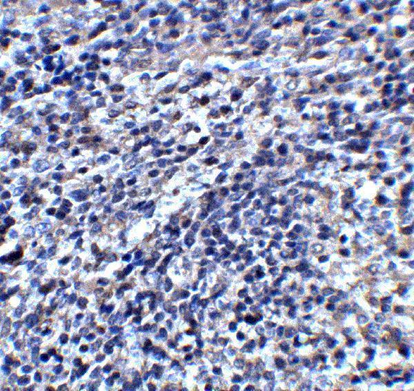
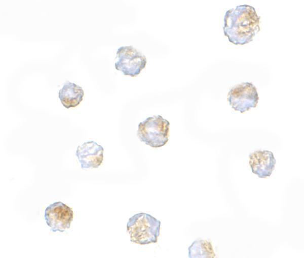
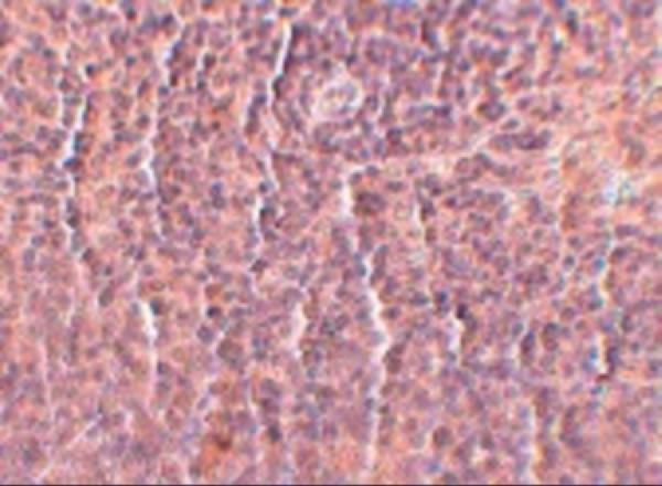
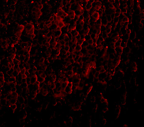
Anti-ORAI3 antibodies have IHC images from mouse and rat spleen and IF/ICC images from rat and human spleen and A20 cells (catalog image captions).

Real IHC data Immunohistochemistry of ORAI3 in mouse spleen tissue with ORAI3 antibody at 2 μg/mL.
Anti-Protein orai-3 ORAI3 Antibody
Cat # A09399
Real IHC data Immunohistochemistry of ORAI3 in rat spleen tissue with ORAI3 antibody at 2.5 μg/mL.
Anti-ORAI3 Monoclonal Antibody [2H2G9]
Cat # M09399

A09399 has a mouse spleen IHC image and an A20 cell ICC image (A09399 image captions). M09399 has a rat spleen IHC image and rat and human spleen IF images (M09399 image captions).

Which to pick: For paraffin-section IHC, choose A09399 for mouse spleen or M09399 for rat spleen; both list IHC-P, and their respective IHC captions show those tissues (catalog applications; A09399 and M09399 IHC image captions). For IF/ICC, M09399 lists IF and shows rat and human spleen, while A09399 lists ICC and shows A20 cells (catalog applications; IF/ICC image captions). For broader listed species reactivity, choose A09399 (Human, Mouse, Rat) over M09399 (Human, Rat); the fixative is unreported in both IHC captions (catalog reactivity; IHC image captions).

Each figure is that product's own IHC / IF validation image from its datasheet.

References

  1. UniProt Consortium. UniProt entry Q9BRQ5 (ORAI3_HUMAN, Protein orai-3).
  2. Human Protein Atlas. ORAI3 tissue IHC expression (reliability: Uncertain).
  3. Human Protein Atlas. ORAI3 subcellular location (ICC-IF): Localized to the nucleoplasm and cytosol..
  4. Human Protein Atlas. ORAI3 antibody validation summary (1 antibodies).
  5. The calcium channel proteins ORAI3 and STIM1 mediate TGF-β induced Snai1 expression. Oncotarget 2018 — PMC6047677.
  6. Orai3 is a predictive marker of metastasis and survival in resectable lung adenocarcinoma. Oncotarget 2016 — PMC5348414.
  7. Orai3-Mediates Cisplatin-Resistance in Non-Small Cell Lung Cancer Cells by Enriching Cancer Stem Cell Population through PI3K/AKT Pathway. Cancers 2021 — PMC8150283.
  8. Orai3 constitutes a native store-operated calcium entry that regulates non small cell lung adenocarcinoma cell proliferation. PloS one 2013 — PMC3772818.
  9. PubMed PMID:15489334 — UniProt-cited evidence.
  10. PubMed PMID:16582901 — UniProt-cited evidence.
  11. PubMed PMID:16807233 — UniProt-cited evidence.