PIK3CB / Phosphatidylinositol 4,5-bisphosphate 3-kinase catalytic subunit beta isoform · IHC design guide

Design Immunohistochemistry for PIK3CB

Plan chromogenic PIK3CB staining in paraffin sections around the general cytoplasmic tissue pattern (HPA tissue IHC). This guide covers consistent fixation, catalog antibody dilution, detection and cell-level interpretation (standard IHC practice; datasheet M01091-1).

Evidence assembled Oct 2026 · For research use; verify linked source records and product datasheet before use
Immunohistochemistry protocol sheet for PIK3CB (IHC for PIK3CB): expected localisation Cytoplasmic tissue staining; nuclear location annotated (HPA tissue IHC; UniProt), antibody M01091-1, validated IHC image, and IHC protocol steps
Printable PIK3CB IHC protocol sheet — expected localisation Cytoplasmic tissue staining; nuclear location annotated (HPA tissue IHC; UniProt), antibody M01091-1, controls and protocol steps. Open the full PIK3CB IHC guide →

PIK3CB Immunohistochemistry Experimental Design Guide

Expected localisation, validated protocols, controls and antibodies — the at-a-glance facts below, then the full design guide.

Must know before staining
Expected localisation Cytoplasmic tissue staining; nuclear location annotated (HPA tissue IHC; UniProt)
Staining pattern General cytoplasmic staining across cell types (HPA tissue IHC)
Antigen retrieval EDTA pH 8.0 HIER, heat-mediated (datasheet M01091-1)
Positive control ⓘ Adrenal gland+4 more · see all
Negative control ⓘ Adipose tissue
Important caveats
Reasons your staining may differ from the expected pattern.
Fixation Keep fixation consistent across sections (standard IHC practice; not target-specific)
Caveat Adipocytes may lack detectable staining (HPA tissue IHC)
Regulation Broad tissue expression (UniProt)
Isoform / epitope No listed isoforms; one unprocessed chain (UniProt)
Section 1

Recommended PIK3CB IHC & IF Protocols

The catalog antibody’s IHC-P protocol is followed by published PIK3CB staining conditions from gastric cancer, head and neck cancer, and in vivo tumor studies (PMC10861831; PMC12102897; PMC6178240).

Recommended immunohistochemistry (IHC-P) protocol parameters
SampleParaffin-embedded human colon tissue; fixative not specified (datasheet M01091-1)
FixationImage fixative and duration unreported (datasheet M01091-1); verify before use.
Sectioning4–5 µm sections on charged slides (standard)
DeparaffinisationXylene, graded ethanol series to water (standard)
Antigen retrievalHeat retrieval: EDTA pH 8.0 (datasheet M01091-1); 20 min, 95–100 °C (standard)
Peroxidase block3% H2O2, 10 min, room temperature (standard)
Blocking10% goat serum (datasheet M01091-1)
Primary antibodyRabbit monoclonal (clone 30P53) anti-PIK3CB, 1:50-1:200 (datasheet M01091-1)
Primary incubationOvernight at 4 °C (datasheet M01091-1)
DetectionHRP-conjugated secondary, DAB chromogen (datasheet M01091-1)
CounterstainHematoxylin, blue, dehydrate and mount (standard)
Expected resultPIK3CB-positive staining in glandular cells of adrenal gland (HPA tissue IHC: Medium). HPA tissue profile: General cytoplasmic expression. No signal in the no-primary control.
💡Decision noteStart with heat-mediated EDTA pH 8.0 retrieval for the catalog antibody (datasheet: M01091-1). The cited articles do not specify a retrieval buffer (PMC10861831; PMC12102897; PMC6178240).
Section 2

What Is the Expected PIK3CB Staining Pattern?

In paraffin-section IHC, expect broad cellular staining with a predominantly cytoplasmic pattern (HPA tissue IHC: general cytoplasmic expression; Approved reliability, with medium consistency between staining and RNA data). Glandular, respiratory epithelial, hematopoietic and selected neural cells are documented at medium intensity (HPA tissue IHC). PIK3CB can also localize to the nucleus and has no transmembrane segment (UniProt P42338: subcellular location and topology).

What am I looking at on my slide?
Moderate cytoplasmic staining in breast or appendix glandular cells, bronchial respiratory epithelial cells, or bone marrow hematopoietic cells.This matches documented medium staining in those cell types and the general cytoplasmic IHC profile (HPA tissue IHC: Medium; general cytoplasmic expression). Compare cells within the section, since ubiquitous expression does not imply equal signal in every cell (UniProt P42338: ubiquitous expression).
Signal appears only as extracellular deposits or sharply confined to tissue spaces, without recognizable cellular staining.That distribution does not match the reported cellular cytoplasmic IHC pattern (HPA tissue IHC: general cytoplasmic expression). Treat it as a possible staining artefact and review tissue morphology and controls (general IHC practice). Nuclear signal alone needs separate evaluation because nuclear localization is reported (UniProt P42338: nucleus).
Adipocytes stain strongly while the expected positive cell population is weak or absent.HPA reports adipocytes as not detected, so this reversal raises concern for cross-reactivity or endogenous detection activity (HPA tissue IHC: adipocytes Not detected; general IHC practice). It is a warning rather than proof: assess a matched negative control and the expected positive cells on the same run.
Brown chromogen spreads uniformly across stroma, empty spaces and cells, obscuring cell boundaries.This is background rather than a readable cellular pattern (general IHC practice). HPA's cytoplasmic profile cannot validate diffuse deposits outside identifiable cells (HPA tissue IHC: general cytoplasmic expression). Judge specificity only after the background is reduced and tissue architecture remains visible.
No cellular signal is visible in an otherwise interpretable breast, appendix or bronchus section.Those tissues contain documented medium-staining cell populations, making an absent result worth checking (HPA tissue IHC: breast and appendix glandular cells; bronchial respiratory epithelial cells, Medium). Verify that the relevant cells are present, then review the antibody and detection workflow (general IHC practice).
💡Expected PIK3CB appearanceCall a result positive when identifiable glandular or respiratory epithelial cells show predominantly cytoplasmic staining around medium intensity (HPA tissue IHC: Medium; general cytoplasmic expression); strong staining limited to adipocytes or acellular deposits is suspect (HPA tissue IHC: adipocytes Not detected; general IHC practice).
How each factor affects the staining
Cell population and reference tissueUse the named cell population when judging a section: breast and appendix glandular cells, bronchial respiratory epithelial cells, and bone marrow hematopoietic cells are Medium (HPA tissue IHC). Adipocytes are Not detected, while ovarian stroma cells, skeletal myocytes and lymph-node germinal-center cells are Low (HPA tissue IHC).
Subcellular interpretationA cytoplasmic IHC pattern is the tissue reference (HPA tissue IHC: general cytoplasmic expression). Nuclear staining cannot be dismissed solely by compartment: UniProt lists the nucleus and says PIK3R2 interaction is required for nuclear localization and export (UniProt P42338: subcellular location; subunit).
Antibody evidenceThe HPA tissue-IHC entry is Approved with medium consistency against RNA data, and CAB031938 is IHC Approved (HPA tissue IHC: reliability; HPA antibodies: CAB031938). Approved supports pattern comparison but does not establish that every antibody or every section will reproduce the same intensity.
Topology and processingPIK3CB has no transmembrane segment, signal peptide or propeptide, and its recorded chain spans residues 1–1070 (UniProt P42338: topology; processing). Interpret a purely extracellular staining pattern cautiously; these annotations do not identify an antibody epitope or establish retrieval sensitivity.
IF/ICC Q&A: should nuclear fluorescence be expected?Yes: HPA ICC-IF reports supported nucleoplasm and nucleoli, with approved additional vesicle and midbody locations (HPA subcellular ICC-IF). This is an IF/ICC observation, so use it as compartment context rather than a paraffin-IHC intensity standard.
Why is my staining missing, weak or wrong?
SituationLikely causeNext action
Expected positive cells show no chromogenic signal.The sampled area may lack the relevant cell population, or an antibody or detection step may have failed (general IHC practice).Confirm the target cells in the section and run a documented positive population, such as breast glandular cells or bronchial respiratory epithelial cells (HPA tissue IHC: Medium); then check the IHC-validated antibody and detection controls (general IHC practice).
Only adipocytes stain strongly.This conflicts with the HPA adipocyte reference and may reflect nonspecific binding or endogenous detection activity (HPA tissue IHC: adipocytes Not detected; general IHC practice).Inspect a matched negative control and an expected positive cell population; address endogenous enzyme activity if the chromogenic detection system requires it (general IHC practice).
Acellular or uniformly diffuse brown staining hides the cells.Excess background or detection deposits can obscure the cellular pattern (general IHC practice).Check blocking, washes, antibody concentration and chromogen development as general IHC workflow variables; reassess whether cytoplasmic signal is confined to recognizable cells (general IHC practice; HPA tissue IHC: general cytoplasmic expression).
The section shows nuclear staining with little cytoplasmic signal.Nuclear localization is biologically plausible, but it differs from the general tissue-IHC profile (UniProt P42338: nucleus; HPA tissue IHC: general cytoplasmic expression).Review morphology and controls, then report the observed compartment separately from the expected IHC pattern; do not label nuclear signal artefactual on location alone (general IHC practice; UniProt P42338: PIK3R2-dependent nuclear localization).
Weak signal is seen in ovarian stroma, myocytes or germinal-center cells.Low staining is recorded for these cell populations, so weak signal alone is inconclusive (HPA tissue IHC: Low).Compare with a documented Medium population in the same run before changing the assay, and score the named cells rather than the whole tissue (HPA tissue IHC: Medium populations; general IHC practice).
Two antibodies give different compartment patterns.Antibody evidence differs by application: CAB031938 is IHC Approved, while HPA042288 and HPA064207 are ICC Supported (HPA antibodies).Compare results within the relevant application and its controls; use the HPA tissue-IHC profile for paraffin IHC and treat ICC-IF locations as separate evidence (HPA tissue IHC; HPA subcellular ICC-IF; general IHC practice).

Sample controls for PIK3CB IHC & IF

🧪Run breast first; glandular cells should stain (HPA: breast glandular cells, Medium). Run adipose tissue as the negative (HPA: adipocytes, Not detected); adipocytes within the breast section, if present, should provide a low-signal internal comparison, while acellular spaces should remain free of chromogen (HPA: adipocytes, Not detected; standard IHC practice).
Positive control tissue: Adrenal gland (Glandular cells, HPA Medium)
Negative control tissue: Adipose tissue (HPA Not detected)
ICC-IF cell lines (HPA subcellular resource): HPA ICC-IF images show PIK3CB in A-431, SiHa, U2OS, SK-MEL-30, RPTEC/TERT1, hTERT-RPE1 (serum starved), with annotated localisation: Nucleoplasm (supported), Nucleoli (supported) (HPA subcellular).
Technical controls: Include a no-primary, secondary-only slide, nonimmune rabbit IgG matched to the primary antibody’s clonality, and PIK3CB-knockout material if available (selected-SKU caption: rabbit primary; standard IHC controls). Quench endogenous peroxidase and check adipose edges for nonspecific DAB signal (selected-SKU caption: HRP/DAB detection; standard chromogenic IHC practice).
⚠️Feasibility: The selected-SKU paraffin-section caption documents heat retrieval in EDTA at pH 8.0, but does not establish whether retrieval is required for PIK3CB (selected-SKU IHC caption). Its fixative is unreported, and the supplied evidence reports no target-specific fixation window or fixation effect (selected-SKU IHC caption). Frozen-section IHC is not evaluated in the supplied evidence; ICC-IF images support cellular IF feasibility, while lipid-rich breast areas warrant attention to processing gaps and edge background (HPA: ICC-IF cell-line images; standard IHC practice).

HPA tissue IHC evidence for PIK3CB

Comprehensive Human Protein Atlas IHC scoring per tissue (reliability: Approved — Medium consistency between antibody staining and RNA expression data.). Rows are taken directly from the HPA tissue chart — click any row's HPA link to view the source.

Positive expression · recommended positive controls

TissueCell typeLevelEvidenceSource
Adrenal gland Glandular cells Medium Protein (IHC) HPA →
Appendix Glandular cells Medium Protein (IHC) HPA →
Bone marrow Hematopoietic cells Medium Protein (IHC) HPA →
Breast Glandular cells Medium Protein (IHC) HPA →
Bronchus Respiratory epithelial cells Medium Protein (IHC) HPA →

Undetected expression · recommended negative controls

TissueCell typeLevelEvidenceSource
Adipose tissue Adipocytes Not detected Protein (IHC) HPA →
Section 3

Advanced PIK3CB IHC Tips

Troubleshoot PIK3CB staining in paraffin sections by checking retrieval, tissue handling, cell type and compartment before comparing signal intensity.

What retrieval should I try first when PIK3CB staining is weak?
Start with heat-mediated retrieval in EDTA pH 8.0 for paraffin-section IHC (datasheet M01091-1). The catalog antibody detected PIK3CB in a human colon paraffin section after this retrieval, followed by 1:50 primary antibody overnight at 4°C (datasheet M01091-1). If staining is weak, first check heating consistency, section adhesion and whether the detection reagents work on a control section (standard IHC practice). If you then test another retrieval condition, compare it side by side with EDTA pH 8.0 and monitor tissue damage and background (standard IHC practice). Keep primary dilution and development time constant during that comparison so retrieval is the variable being assessed (standard IHC practice).
How should I troubleshoot weak staining when fixation may have varied?
The selected PIK3CB caption identifies a paraffin-embedded section but does not state its fixative, so target-specific fixation sensitivity is unknown (datasheet M01091-1). Record the fixative, fixation duration, processing history and section age for each case before comparing staining (standard IHC practice). Use similarly handled sections and a consistent positive control to see whether weak signal follows a processing batch (standard IHC practice). Inspect morphology alongside staining; damaged or poorly preserved regions can make intensity comparisons unreliable (standard IHC practice). If processing varies, optimise retrieval and antibody concentration on representative sections, then keep those conditions fixed for the comparison (standard IHC practice).
Should PIK3CB stain cytoplasm or nuclei in tissue sections?
Expect broad cytoplasmic staining as the main tissue-IHC pattern, while identifying the stained cell type on each section (HPA tissue IHC: general cytoplasmic expression). Nuclear staining is biologically plausible because PIK3CB is annotated in cytoplasm and nucleus, and interaction with PIK3R2 is required for its nuclear localisation and export (UniProt P42338). Cell imaging also supports nucleoplasmic and nucleolar localisation, with vesicles and midbody as additional locations (HPA subcellular). Score cytoplasmic and nuclear staining separately instead of combining them into one intensity value (standard IHC practice). Treat a nuclear-only tissue pattern as a finding to verify against controls and morphology, rather than assuming it represents the usual tissue-IHC pattern (HPA tissue IHC; standard IHC practice).
How can I assess whether staining is specific to PIK3CB?
Confirm which PIK3CB sequence the antibody recognises from its epitope documentation; the supplied caption does not identify an epitope (datasheet M01091-1). The record lists 0 annotated isoforms, which does not establish that the antibody distinguishes PIK3CB from related PI3K proteins (UniProt P42338). PIK3CB has PI3K-ABD, RBD, C2, helical and catalytic regions, so epitope placement matters when interpreting cross-reactivity or altered staining (UniProt P42338). Compare antibody staining with an appropriate loss-of-target control when available, using the same tissue processing and detection conditions (standard IHC practice). Document the tested epitope and control outcome before assigning a compartment-specific signal to PIK3CB (standard IHC practice).
How should I adapt the localisation check for multiplex IF?
For the separate IF/ICC workflow, pair PIK3CB with a marker for the expected cell type and inspect channels individually before interpreting overlap (standard IF practice). Choose fluorophores after checking tissue autofluorescence in unstained controls; place a weaker signal in a channel with lower background where possible (standard IF practice). PIK3CB has no transmembrane segment and is annotated in cytoplasm and nucleus, so intracellular epitope access generally calls for permeabilisation after fixation (UniProt P42338 topology; standard IF practice). Optimise permeabilisation against morphology and background because the antibody epitope is unspecified here (datasheet M01091-1; standard IF practice). Cell imaging reports supported nucleoplasmic and nucleolar localisation, which can guide compartment checks without serving as tissue-IF validation for this antibody (HPA subcellular).
What should I check if DAB staining is diffuse or widespread?
Compare the section with a no-primary control and inspect whether colour follows tissue structure or appears across unrelated regions (standard IHC practice). The selected assay used peroxidase detection and DAB, so check peroxidase blocking and DAB development as general workflow variables (datasheet M01091-1; standard IHC practice). Review blocking and primary concentration against the demonstrated 10% goat serum block and 1:50 overnight incubation (datasheet M01091-1). Shorten development or titrate primary antibody if controls show excess colour, changing one condition at a time (standard IHC practice). Because PIK3CB tissue expression is broad, widespread cytoplasmic staining alone cannot establish background; evaluate cell morphology and control sections together (HPA tissue IHC; standard IHC practice).
How should I quantify PIK3CB IHC across samples? ⚠ ANSWER MARKED FOR VERIFICATION
Define the cell population and scoring compartment before counting, since tissue IHC reports general cytoplasmic expression while nuclear localisation is also annotated (HPA tissue IHC; UniProt P42338). For chromogenic sections, report an H-score from intensity and percentage of positive cells, or report percentage positive within the same defined population (standard IHC practice). For sparse populations, positive-cell density per mm² can be useful if the measured tissue area is recorded (standard IHC practice). Normalise counts to eligible nucleated cells or evaluable tissue area, and exclude folds, edges and necrotic regions using fixed criteria (standard IHC practice). Keep retrieval, imaging and scoring thresholds consistent across groups, and report cytoplasmic and nuclear results separately when both are assessed (standard IHC practice).
How can I distinguish a true PIK3CB signal from staining artefact?
Check that signal lies within intact cells and follows a plausible compartment: tissue IHC is generally cytoplasmic, while nuclear localisation is also documented (HPA tissue IHC; UniProt P42338). Compare positive and weakly stained populations with morphology; HPA reports medium staining in several glandular, hematopoietic and neuronal populations but no detection in adipocytes in its listed tissue examples (HPA tissue IHC). Strong colour confined to section edges or necrotic areas calls for caution, and colour in a no-primary control suggests detection background (standard IHC practice). Endogenous peroxidase can confound a DAB assay, so assess peroxidase blocking with the control section (standard IHC practice). Confirm an unexpected compartment or cell pattern with an independent specificity control before making a biological claim (standard IHC practice).
Boster reagents

Best PIK3CB / Phosphatidylinositol 4,5-bisphosphate 3-kinase catalytic subunit beta isoform IHC Antibodies

Both antibodies have paraffin-section IHC images from human tissue; M01091-1 also has mouse and rat brain images and lists IF/ICC use (catalog: IHC captions; M01091-1 applications).

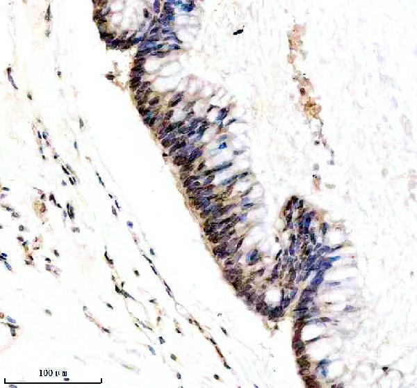
Real IHC data IHC analysis of PI3 Kinase p110 Beta/PIK3CB using anti-PI3 Kinase p110 Beta/PIK3CB antibody (M01091-1). PI3 Kinase p110 Beta/PIK3CB was detected in a paraffin-embedded section of human colon tissue. Heat mediated antigen retrieval was performed in EDTA buffer (pH 8.0, epitope retrieval solution). The tissue section was blocked with 10% goat serum. The tissue section was then incubated with 1: 50 rabbit anti-PI3 Kinase p110 Beta/PIK3CB Antibody (M01091-1) overnight at 4°C. Peroxidase Conjugated Goat Anti-rabbit IgG was used as secondary antibody and incubated for 30 minutes at 37°C. The tissue section was developed using HRP Conjugated Rabbit IgG Super Vision Assay Kit (Catalog # SV0002) with DAB as the chromogen.
Anti-PI3 Kinase p110 beta Rabbit Monoclonal Antibody
Cat # M01091-1
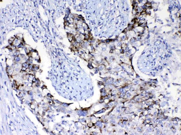
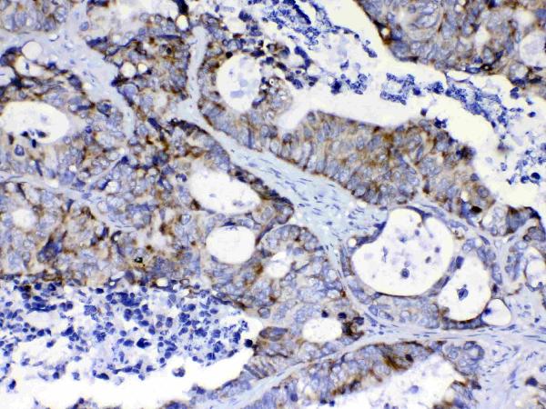
Real IHC data IHC analysis of PIK3CB using anti-PIK3CB antibody (A01091-1). PIK3CB was detected in paraffin-embedded section of human intestinal cancer tissues. Heat mediated antigen retrieval was performed in citrate buffer (pH6, epitope retrieval solution) for 20 mins. The tissue section was blocked with 10% goat serum. The tissue section was then incubated with 1μg/ml rabbit anti-PIK3CB Antibody (A01091-1) overnight at 4°C. Biotinylated goat anti-rabbit IgG was used as secondary antibody and incubated for 30 minutes at 37°C. The tissue section was developed using Strepavidin-Biotin-Complex (SABC)(Catalog # SA1022) with DAB as the chromogen.
Anti-PI3 Kinase p110 beta/PIK3CB Antibody ®
Cat # A01091-1

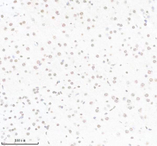
M01091-1 will render with a human colon IHC image; its other captions show human colon cancer and mouse and rat brain, and its listed applications include IF/ICC (catalog: M01091-1 captions and applications). A01091-1 will render with a human intestinal cancer IHC image; another caption shows human lung cancer, and its listed applications include IHC (catalog: A01091-1 captions and applications).

Which to pick: For paraffin-section IHC, choose M01091-1 when a rabbit monoclonal and demonstrated human, mouse and rat tissue staining are useful; its images use EDTA retrieval at pH 8.0 (catalog: M01091-1 clone and IHC captions). Choose A01091-1 for the pictured human intestinal or lung cancer sections; its images use citrate retrieval at pH 6 (catalog: A01091-1 IHC captions). For IF/ICC or cross-species work, M01091-1 lists IF/ICC and human, mouse and rat reactivity, though no IF image is supplied; neither antibody’s IHC captions report the fixative (catalog: M01091-1 applications, reactivity and image lists; both IHC captions).

Each figure is that product's own IHC / IF validation image from its datasheet.

References

  1. UniProt Consortium. UniProt entry P42338 (PK3CB_HUMAN, Phosphatidylinositol 4,5-bisphosphate 3-kinase catalytic subunit beta isoform).
  2. Human Protein Atlas. PIK3CB tissue IHC expression (reliability: Approved).
  3. Human Protein Atlas. PIK3CB subcellular location (ICC-IF): Mainly localized to the nucleoplasm and nucleoli. In addition localized to vesicles and midbody..
  4. Human Protein Atlas. PIK3CB antibody validation summary (3 antibodies).
  5. SP1 Mediated PIK3CB Upregulation Promotes Gastric Carcinogenesis. Journal of Cancer 2024 — PMC10861831.
  6. Targeting PIK3CB/YAP1 improves the sensitivity of paclitaxel by suppressing aging in head and neck squamous tumor cells. Cancer cell international 2025 — PMC12102897.
  7. An Efficient Bivalent Cyclic RGD-PIK3CB siRNA Conjugate for Specific Targeted Therapy against Glioblastoma In Vitro and In Vivo. Molecular therapy. Nucleic acids 2018 — PMC6178240.
  8. USP8-mediated PTK7 promotes PIK3CB-related pathway to accelerate the malignant progression of non-small cell lung cancer. Thoracic cancer 2025 — PMC11729734.
  9. PubMed PMID:8246984 — UniProt-cited evidence.
  10. PubMed PMID:11016459 — UniProt-cited evidence.
  11. PubMed PMID:15489334 — UniProt-cited evidence.