RPSA / Small ribosomal subunit protein uS2 · IHC design guide

Design Immunohistochemistry for RPSA

Plan chromogenic RPSA IHC in paraffin sections with the catalog antibody starting at 0.5–1 μg/mL (datasheet PB10094). Compare staining with the cytoplasmic tissue pattern and consider the described surface 67LR when interpreting results (HPA tissue IHC; UniProt).

Evidence assembled Oct 2026 · For research use; verify linked source records and product datasheet before use
Immunohistochemistry protocol sheet for RPSA (IHC for RPSA): expected localisation Cytoplasmic in tissue; surface 67LR is also described (HPA tissue IHC; UniProt), antibody PB10094, validated IHC image, and IHC protocol steps
Printable RPSA IHC protocol sheet — expected localisation Cytoplasmic in tissue; surface 67LR is also described (HPA tissue IHC; UniProt), antibody PB10094, controls and protocol steps. Open the full RPSA IHC guide →

RPSA Immunohistochemistry Experimental Design Guide

Expected localisation, validated protocols, controls and antibodies — the at-a-glance facts below, then the full design guide.

Must know before staining
Expected localisation Cytoplasmic in tissue; surface 67LR is also described (HPA tissue IHC; UniProt)
Staining pattern Cytoplasmic staining across tissues, including glandular cells (HPA tissue IHC)
Antigen retrieval Citrate pH 6 HIER, heat-mediated (datasheet PB10094)
Positive control ⓘ Adrenal gland+4 more · see all
Negative control ⓘ Soft tissue
Important caveats
Reasons your staining may differ from the expected pattern.
Fixation Keep fixation consistent across sections (standard IHC practice; not target-specific); optimize empirically.
Caveat Broad cytoplasmic staining may obscure surface 67LR (HPA tissue IHC; UniProt)
Regulation No specific expression regulator annotated (UniProt)
Isoform / epitope No isoforms annotated; surface 67LR may alter epitope access (UniProt)
Section 1

Recommended RPSA IHC & IF Protocols

The catalog antibody protocol is accompanied by two published RPSA IHC protocols for paraffin-embedded brain tissue (PMC7898445; PMC7884477).

Recommended immunohistochemistry (IHC-P) protocol parameters
SampleParaffin-embedded human mammary cancer tissue; fixative not specified (datasheet PB10094)
FixationImage fixative and duration unreported (datasheet PB10094); verify before use.
Sectioning4–5 µm sections on charged slides (standard)
DeparaffinisationXylene, graded ethanol series to water (standard)
Antigen retrievalHeat retrieval: Citrate pH 6, 20 min (datasheet PB10094)
Peroxidase block3% H2O2, 10 min, room temperature (standard)
Blocking10% goat serum (datasheet PB10094)
Primary antibodyRabbit anti-RPSA, 0.5-1μg/ml (datasheet PB10094)
Primary incubationOvernight at 4 °C (datasheet PB10094)
DetectionStreptavidin-biotin complex (SABC), DAB chromogen (datasheet PB10094)
CounterstainHematoxylin, blue, dehydrate and mount (standard)
Expected resultRPSA-positive staining in glandular cells of adrenal gland (HPA tissue IHC: High). HPA tissue profile: Cytoplasmic expression in all tissues. No signal in the no-primary control.
💡Decision noteStart with heat-mediated citrate retrieval at pH 6 for the catalog antibody (datasheet PB10094); neither published excerpt specifies retrieval (PMC7898445; PMC7884477).
Section 2

What Is the Expected RPSA Staining Pattern?

RPSA staining should be predominantly cytoplasmic across tissues, with strong staining in selected glandular, hematopoietic, respiratory epithelial and neuronal cells (HPA: tissue IHC). Cell surface staining is biologically plausible, although no transmembrane segment is annotated (UniProt P08865: localization and topology). HPA rates the tissue IHC profile Approved, with medium consistency between staining and RNA expression; interpret unexpected patterns with that limitation in mind (HPA: tissue IHC).

What am I looking at on my slide?
Cytoplasmic signal in colon or breast glandular cells, or in bone marrow hematopoietic cells.This matches cell types rated High by HPA and the broadly cytoplasmic tissue profile (HPA: tissue IHC). Judge the signal within the relevant cells against background; a High designation does not specify that every cell must stain equally (HPA: tissue IHC).
Signal appears mainly nuclear, with little cytoplasmic staining.Review as a compartment mismatch for tissue IHC, which HPA describes as cytoplasmic across tissues (HPA: tissue IHC). UniProt also lists nuclear 37LRP by similarity, so nuclear signal alone does not prove an artefact; check morphology, background and controls before assigning it to RPSA (UniProt P08865: localization).
Prominent staining appears in peripheral nerve within a soft tissue section.HPA reports peripheral nerve as Not detected, making this an unexpected cell type for that comparison (HPA: tissue IHC). Consider cross-reactivity or endogenous detection activity, then compare a primary antibody omission control with correctly identified cells; absence of signal elsewhere on the section cannot validate this staining (general IHC practice).
Color spreads across stroma, lumina or most of the section without clear cell boundaries.This is diffuse background rather than a convincing cytoplasmic cell pattern (HPA: tissue IHC; general IHC practice). Check whether the same distribution appears without primary antibody, and assess blocking, washes and detection chemistry before scoring cells as positive (general IHC practice).
No specific signal appears in a known positive comparison such as colon glandular cells.Colon glandular cells are rated High, so a blank result calls for a technical check before a biological interpretation (HPA: tissue IHC). Confirm that the cells are present and assess antigen retrieval, antibody dilution, detection reagents and counterstain using the chosen IHC workflow (general IHC practice).
💡Expected RPSA appearanceA credible positive result shows discernible cytoplasmic staining in the relevant cells, including cells rated High such as colon glandular cells, against limited background; diffuse color outside identifiable cells or signal confined to an unexpected cell type warrants investigation (HPA: tissue IHC; general IHC practice).
How each factor affects the staining
Tissue and cell contextHPA describes cytoplasmic expression across tissues, but rates skeletal myocytes and adipocytes Low and peripheral nerve in soft tissue Not detected (HPA: tissue IHC). Compare identified cell types rather than treating a whole section as uniformly positive.
Antibody evidenceCAB009561 is Approved for IHC; that status does not establish identical staining with another antibody or resolve every unexpected compartment (HPA: antibody validation). HPA also reports medium consistency between tissue staining and RNA expression (HPA: tissue IHC).
RPSA localization and topologyUniProt places RPSA at the cell membrane, in cytoplasm and in nucleus, while annotating no transmembrane segment (UniProt P08865: localization and topology). Surface staining may be plausible, but the HPA tissue IHC summary supports a cytoplasmic pattern (HPA: tissue IHC).
IF/ICC: where should RPSA appear?Plasma membrane and cytosol are supported locations in HPA ICC-IF images; this is an interpretation answer, not an IF/ICC protocol or a guarantee of the same visual pattern in paraffin IHC (HPA: subcellular ICC-IF).
Why is my staining missing, weak or wrong?
SituationLikely causeNext action
Positive comparison is blank.The result conflicts with a cell type rated High, such as colon glandular cells (HPA: tissue IHC); a failed staining step is possible (general IHC practice).Verify cell identity and section quality, then check retrieval, antibody application and detection with the established IHC workflow (general IHC practice).
Staining is faint in skeletal muscle myocytes or adipocytes.Both cell types are rated Low by HPA; weak signal there alone does not show assay failure (HPA: tissue IHC).Evaluate a High cell type in the same run before changing conditions, and score cell types separately (HPA: tissue IHC; general IHC practice).
Peripheral nerve stains strongly in soft tissue.That cell type is reported Not detected, so cross-reactivity or endogenous detection activity merits investigation (HPA: tissue IHC; general IHC practice).Confirm the cell identity and inspect a primary antibody omission control; review detection blocking if color persists (general IHC practice).
The section has widespread diffuse color.Background can obscure the cellular cytoplasmic pattern reported by HPA (HPA: tissue IHC; general IHC practice).Compare the primary antibody omission control, then review blocking, washes and detection development in the IHC workflow (general IHC practice).
Signal is predominantly nuclear.HPA tissue IHC describes cytoplasmic expression, although UniProt includes nuclear 37LRP localization by similarity (HPA: tissue IHC; UniProt P08865: localization).Check the cytoplasmic compartment and background in a High comparison cell type; avoid calling nuclear staining specific from location alone (HPA: tissue IHC; general IHC practice).
Only a membrane rim is visible.Surface RPSA is plausible from UniProt and supported in HPA ICC-IF, while HPA tissue IHC describes a cytoplasmic profile (UniProt P08865: localization; HPA: subcellular ICC-IF and tissue IHC).Confirm that the rim follows intact cells and assess a cytoplasmic positive comparison before accepting membrane only IHC staining (HPA: tissue IHC; general IHC practice).

Sample controls for RPSA IHC & IF

🧪Run breast first: glandular cells should stain (HPA: High in breast glandular cells). Use peripheral nerve in soft tissue as the negative tissue (HPA: Not detected); record any unstained cells on the breast slide as local background, since the supplied HPA rows do not establish an internal negative cell type.
Positive control tissue: Adrenal gland (Glandular cells, HPA High)
Negative control tissue: Soft tissue (HPA Not detected)
ICC-IF cell lines (HPA subcellular resource): HPA ICC-IF images show RPSA in HEK293, MCF-7, U2OS, with annotated localisation: Plasma membrane (supported), Cytosol (supported) (HPA subcellular).
Technical controls: Run no-primary (secondary-only) and concentration-matched rabbit IgG isotype controls alongside an RPSA knockout sample or, if a suitable immunizing peptide is available, a peptide-block control (selected IHC caption: rabbit primary; standard IHC practice). For the breast slide, check endogenous peroxidase and biotin background when using the caption’s biotin-based DAB detection (selected IHC caption; standard IHC practice).
⚠️Feasibility: No target-specific fixation window or fixation effect is reported, and the selected PB10094 tissue-IHC caption does not state a fixative; its paraffin section therefore does not establish fixation (selected IHC caption). The caption reports citrate pH 6 heat retrieval for 20 minutes, but does not establish that retrieval is required; endogenous peroxidase or biotin could complicate interpretation with its DAB detection workflow (selected IHC caption; standard IHC practice). The supplied evidence does not establish that frozen sections or IF are easier; for IF/ICC, HPA supports plasma-membrane and cytosolic localization but supplies no comparative fixation condition (HPA subcellular). The selected PB10094 tissue-IHC caption documents paraffin sections, but does not specify the fixative (selected-SKU IHC image PB10094).

HPA tissue IHC evidence for RPSA

Comprehensive Human Protein Atlas IHC scoring per tissue (reliability: Approved — Medium consistency between antibody staining and RNA expression data.). Rows are taken directly from the HPA tissue chart — click any row's HPA link to view the source.

Positive expression · recommended positive controls

TissueCell typeLevelEvidenceSource
Adrenal gland Glandular cells High Protein (IHC) HPA →
Appendix Glandular cells High Protein (IHC) HPA →
Bone marrow Hematopoietic cells High Protein (IHC) HPA →
Breast Glandular cells High Protein (IHC) HPA →
Bronchus Respiratory epithelial cells High Protein (IHC) HPA →

Undetected expression · recommended negative controls

TissueCell typeLevelEvidenceSource
Soft tissue Peripheral nerve Not detected Protein (IHC) HPA →
Section 3

Advanced RPSA IHC Tips

Troubleshoot RPSA chromogenic IHC by checking retrieval, compartment-specific staining and controls against the reported tissue and subcellular patterns.

How should I retrieve RPSA in paraffin sections when staining is weak?
Start with heat-mediated citrate retrieval at pH 6 for 20 minutes (datasheet PB10094; PB10094 image caption). The selected paraffin-section image used this condition before overnight incubation at 4°C with 1 µg/mL primary antibody (PB10094 image caption). If staining remains weak, vary heating and cooling conditions on adjacent sections while holding antibody concentration and detection constant (standard IHC practice). Compare signal in glandular cells with expected cytoplasmic staining, and check whether tissue edges stain disproportionately (HPA tissue IHC; standard IHC practice). A stronger DAB signal alone does not establish better retrieval; evaluate cellular detail and background alongside it (standard IHC practice).
Could fixation explain weak or uneven RPSA staining?
Target-specific fixation sensitivity is unknown: the selected paraffin-section caption does not state a fixative (PB10094 image caption). Record the fixative, fixation duration and section age for each specimen before comparing staining across cases (standard IHC practice). On matched sections, keep citrate retrieval at pH 6 for 20 minutes constant while investigating fixation-related variation (datasheet PB10094; PB10094 image caption; standard IHC practice). Inspect morphology and compare staining within intact tissue, since poorly preserved or damaged regions can mislead interpretation (standard IHC practice). The reported cytoplasmic tissue pattern supplies an expected location, but no evidence about this antibody’s fixation tolerance (HPA tissue IHC; PB10094 image caption).
How should I assess cytoplasmic, membrane and nuclear RPSA staining?
Expect widespread cytoplasmic staining in tissue sections (HPA tissue IHC), while plasma-membrane and cytosolic localisation have support from ICC/IF images (HPA subcellular). UniProt also lists nuclear localisation and describes surface-accessible laminin binding by the mature 67LR form (UniProt P08865 localisation). Score cytoplasmic, membrane-associated and nuclear signal separately, using cell outlines and a counterstain to judge the compartment (standard IHC practice). Do not classify every dark cell border as surface RPSA; compare it with adjacent cells and a no-primary control (standard IHC practice). State which compartment drives the result, because these sources support different aspects of RPSA localisation (HPA tissue IHC; HPA subcellular; UniProt P08865 localisation).
Can epitope position explain differing membrane and cytoplasmic results?
RPSA has 0 annotated isoforms, while 37LRP and 67LR are described as monomeric and homodimeric forms (UniProt P08865 isoforms and subunit). Its mature chain spans residues 2–295, and UniProt annotates no transmembrane segment or glycosylation sites (UniProt P08865 processing and topology). Check the antibody’s documented immunogen or epitope before attributing compartment differences to epitope access; none is supplied here (PB10094 image caption). UniProt lists acetylation and phosphorylation sites, but these annotations alone do not establish an effect on this antibody’s IHC signal (UniProt P08865 modified residues). Compare compartment-specific staining under the same retrieval and detection conditions (standard IHC practice).
How can IF help assess an ambiguous RPSA IHC pattern?
Use IF/ICC as a complementary localisation check: supported plasma-membrane and cytosolic signals are reported in images from HEK293, MCF-7 and U2OS (HPA subcellular). For tissue multiplexing, pair RPSA with a marker identifying the expected cell population, such as glandular cells where the tissue-IHC profile reports strong signal (HPA tissue IHC; standard IF practice). Choose a fluorophore away from prominent tissue autofluorescence and include single-stain controls to assess bleed-through (standard IF practice). For an intracellular epitope, compare appropriately permeabilised samples; for a surface-accessible epitope, preserve membrane integrity during staining (UniProt P08865 localisation; standard IF practice). The epitope and IF fixation conditions for the catalog antibody are unspecified, so optimise them experimentally (PB10094 image caption).
What should I check when DAB staining obscures cellular RPSA signal?
Separate diffuse cytoplasmic signal from haze by inspecting cell boundaries, blank areas and tissue edges at the same exposure and microscope settings (HPA tissue IHC; standard IHC practice). The selected image used 10% goat-serum blocking, 1 µg/mL primary antibody and an overnight 4°C incubation (PB10094 image caption). Check a no-primary control, adequate washing and serum compatibility before changing those conditions (standard IHC practice). Include a peroxidase block and inspect a detection-only control when endogenous enzyme activity could produce DAB staining (standard chromogenic IHC practice). If background persists, titrate antibody and detection intensity on adjacent sections while checking that expected cellular staining remains visible (HPA tissue IHC; standard IHC practice).
How should I quantify RPSA IHC across specimens? ⚠ ANSWER MARKED FOR VERIFICATION
Define the compartment and cell population before scoring, because RPSA tissue IHC is broadly cytoplasmic while membrane localisation is also supported (HPA tissue IHC; HPA subcellular). For a cellular endpoint, report the percentage of positive cells and an H-score based on staining intensity; retain the compartment in the score label (standard IHC practice). For spatial comparisons, report positive-cell density per mm² of viable tissue and record the sampled area (standard IHC practice). Normalise comparisons to the same cell population, tissue area, staining batch and analysis threshold (standard IHC practice). Avoid treating whole-section DAB area as a cell-specific measurement when stromal content or cellularity differs (standard IHC practice).
When is an apparent RPSA-positive region likely to be artefactual?
A credible pattern should have identifiable stained cells and a stated compartment; cytoplasmic tissue staining and supported membrane localisation provide comparison points (HPA tissue IHC; HPA subcellular). Question signal confined to cut edges, folds or necrotic areas, where tissue damage can distort chromogenic staining (standard IHC practice). Check a no-primary control and peroxidase blocking if a region shows DAB colour without convincing cellular structure (standard chromogenic IHC practice). Interpret cell identity carefully: HPA reports high glandular-cell staining in breast and low staining in skeletal myocytes, but its tissue-IHC reliability is Approved with medium RNA concordance (HPA tissue IHC). Resolve unexpected nuclear-only or cell-restricted results with morphology, controls and an independent localisation method (UniProt P08865 localisation; standard IHC practice).
Boster reagents

Best RPSA / Small ribosomal subunit protein uS2 IHC Antibodies

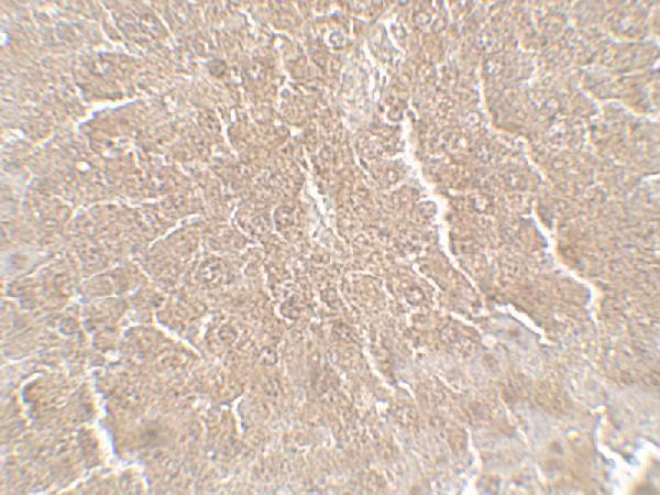
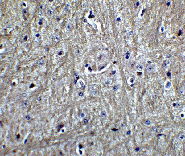
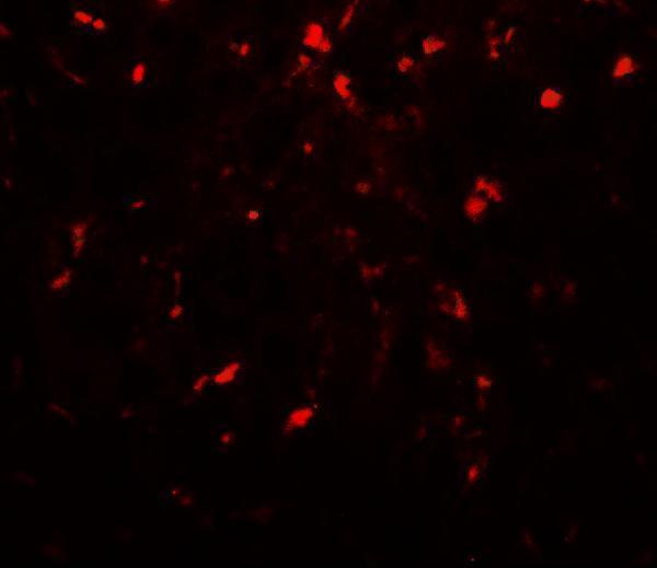
Anti-RPSA IHC images cover human mammary cancer paraffin sections and mouse liver and brain; IF images cover U20S cells and mouse liver and brain (catalog image captions).

Real IHC data IHC analysis of RPSA using anti-RPSA antibody (PB10094). RPSA was detected in paraffin-embedded section of human mammary cancer tissue. Heat mediated antigen retrieval was performed in citrate buffer (pH6, epitope retrieval solution) for 20 mins. The tissue section was blocked with 10% goat serum. The tissue section was then incubated with 1μg/ml rabbit anti-RPSA Antibody (PB10094) overnight at 4°C. Biotinylated goat anti-rabbit IgG was used as secondary antibody and incubated for 30 minutes at 37°C. The tissue section was developed using Strepavidin-Biotin-Complex (SABC)(Catalog # SA1022) with DAB as the chromogen.
Anti-67kDa Laminin Receptor/RPSA Antibody ®
Cat # PB10094
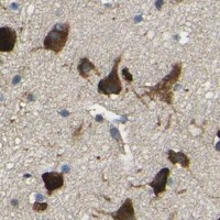
Real IHC data Immunohistochemistry of RPSA in mouse liver tissue with RPSA antibody at 5 μg/mL.
Anti-40S ribosomal protein SA RPSA Antibody
Cat # A01691

PB10094 has IHC data from human mammary cancer paraffin sections and IF data from U20S cells (PB10094 image captions). A01691 has IHC and IF images from mouse liver and brain, and lists human and mouse reactivity (A01691 catalog).

Which to pick: For human paraffin-section IHC, choose PB10094: its caption documents that preparation; the fixative is unreported (PB10094 IHC caption). For IF/ICC, PB10094 lists both applications and shows cellular IF, while A01691 shows tissue IF in mouse liver and brain (catalog applications; PB10094 and A01691 IF captions). For work spanning human and mouse, choose A01691 for its listed reactivity, while recognizing that its pictured IHC data are from mouse tissue; neither rendered antibody has a clone specified (A01691 catalog and IHC captions; catalog clone fields).

Each figure is that product's own IHC / IF validation image from its datasheet.

References

  1. UniProt Consortium. UniProt entry P08865 (RSSA_HUMAN, Small ribosomal subunit protein uS2).
  2. Human Protein Atlas. RPSA tissue IHC expression (reliability: Approved).
  3. Human Protein Atlas. RPSA subcellular location (ICC-IF): Localized to the plasma membrane and cytosol..
  4. Human Protein Atlas. RPSA antibody validation summary (1 antibodies).
  5. Streptococcus suis serotype 2 enolase interaction with host brain microvascular endothelial cells and RPSA-induced apoptosis lead to loss of BBB integrity. Veterinary research 2021 — PMC7898445.
  6. Reconsideration of the laminin receptor 67LR in colorectal cancer cells. Biomolecules & biomedicine 2024 — PMC11378999.
  7. Ribosomal Protein SA-Positive Neutrophil Elicits Stronger Phagocytosis and Neutrophil Extracellular Trap Formation and Subdues Pro-Inflammatory Cytokine Secretion Against Streptococcus suis Serotype 2 Infection. Frontiers in immunology 2020 — PMC7884477.
  8. Expression of the RPSA-Containing and 67EBP Laminin Receptors in Relation to the Debatable Nature of the 67 kDa Laminin Receptor 67LR in Colorectal Cancer. International journal of molecular sciences 2025 — PMC11942345.
  9. PubMed PMID:2970639 — UniProt-cited evidence.
  10. PubMed PMID:2543954 — UniProt-cited evidence.
  11. PubMed PMID:1534510 — UniProt-cited evidence.