This website uses cookies to ensure you get the best experience on our website.


- Table of Contents
SRSF3 shows general nuclear expression in tissue IHC (HPA tissue IHC). This guide covers paraffin-section staining, interpretation of nuclear signal, and the 2 annotated isoforms (UniProt).

Expected localisation, validated protocols, controls and antibodies — the at-a-glance facts below, then the full design guide.
| Expected localisation | Nuclear staining in tissue (HPA tissue IHC) | |
| Staining pattern | General nuclear staining across cell types (HPA tissue IHC) | |
| Antigen retrieval | Tris-EDTA pH 9.0 HIER, heat-mediated (datasheet A30468) | |
| Positive control | Adipose tissue+4 more · see all | |
| Negative control | None in HPA (detected in all 45 tissues); use no-primary + isotype controls |
| Fixation | Keep fixation consistent across paraffin sections. (standard IHC practice; not target-specific) | |
| Caveat | Broad expression may limit cell-type discrimination (HPA tissue IHC) | |
| Regulation | No expression regulator annotated (UniProt) | |
| Isoform / epitope | 2 isoforms; epitope coverage unspecified (UniProt) |
Use the catalog antibody’s IHC-P protocol (datasheet A30468) as the starting point; the published SRSF3 tissue protocols below provide alternative retrieval and detection conditions (PMC4946124; PMC7149931; PMC9221257).
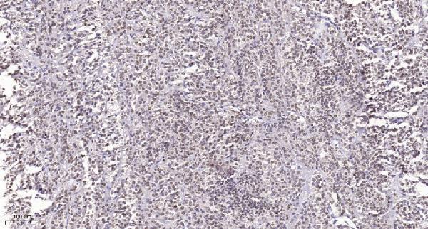
| Sample | Paraffin-embedded human Small intestinal stromal tumor tissue; fixative not specified (datasheet A30468) |
| Fixation | Image fixative and duration unreported (datasheet A30468); verify before use. |
| Sectioning | 4–5 µm sections on charged slides (standard) |
| Deparaffinisation | Xylene, graded ethanol series to water (standard) |
| Antigen retrieval | Heat retrieval: Tris-EDTA pH 9.0 (datasheet A30468); 20 min, 95–100 °C (standard) |
| Peroxidase block | 3% H2O2, 10 min, room temperature (standard) |
| Blocking | 10% normal serum of the secondary host, 30 min, room temperature (standard) |
| Primary antibody | Rabbit anti-SRSF3, 1:100 - 1:300 (datasheet A30468) |
| Primary incubation | Overnight at 4 °C (standard) |
| Detection | HRP-polymer secondary, DAB chromogen 5–10 min (standard) |
| Counterstain | Hematoxylin, blue, dehydrate and mount (standard) |
| Expected result | SRSF3-positive staining in adipocytes of adipose tissue (HPA tissue IHC: High). HPA tissue profile: General nuclear expression. No signal in the no-primary control. |
SRSF3 should give a predominantly nuclear pattern in paraffin-section IHC (HPA: general nuclear expression; reliability Supported). High staining is reported in adipocytes, glandular cells, hematopoietic cells, respiratory epithelial cells, glial cells and cerebellar granular-layer cells in the listed tissues (HPA: tissue IHC). Nuclear speckles and some cytoplasmic localisation are biologically possible (UniProt P84103: subcellular location). A membrane-rim pattern is unexpected (UniProt P84103: no transmembrane segment).
| Distinct nuclear chromogen in cells from an HPA high-staining tissue, with tissue structure still readable. | This fits the reported general nuclear pattern (HPA: tissue IHC, Supported). Judge localisation in individual cells as well as overall intensity; high is an HPA observation in specified cell populations, not a required score for every cell in a section (HPA: tissue IHC). |
| Signal forms a membrane rim, extracellular deposit or tissue-edge accent while nuclei remain unstained. | That distribution conflicts with the expected nuclear IHC pattern and lacks support from SRSF3 topology (HPA: general nuclear expression; UniProt P84103: no transmembrane segment). Check staining controls and section quality before calling it SRSF3. Cytoplasmic staining alone needs more care because cytoplasm is listed as a location (UniProt P84103). |
| An unexpected cell population stains more strongly than nearby expected cells. | Possible causes include nonspecific antibody binding or endogenous detection activity (general IHC practice). HPA reports low tissue RNA specificity and supplies no negative cell list, so an unlisted population is not automatically SRSF3-negative (HPA: tissue IHC). Compare nuclear localisation, morphology and a matched detection control. |
| Chromogen is diffuse across stroma, empty spaces or many compartments, obscuring nuclear boundaries. | Treat this as background until controls support a cellular pattern (general IHC practice). The supported tissue pattern is general nuclear expression, not uniform staining of extracellular space (HPA: tissue IHC). Review blocking, washing, primary concentration and detection chemistry using the antibody's IHC-P instructions. |
| No nuclear signal appears in an HPA high-staining cell population. | A failed positive control is possible; absence in one section does not establish that SRSF3 is absent from the tissue (general IHC practice; HPA: high staining in the specified population). Check specimen integrity and each IHC-P step against the catalog antibody's instructions. No target-specific fixation sensitivity is supplied by UniProt or HPA. |
| Which compartment should guide IHC interpretation? | Use nuclear staining as the primary readout (HPA: general nuclear expression). UniProt also lists nuclear speckles and cytoplasm, including speckle recruitment after YTHDC1 interaction (UniProt P84103). Do not require visible speckles in chromogenic paraffin sections or reject every cytoplasmic component solely on localisation. |
| How broadly can tissue staining occur? | HPA calls tissue RNA specificity low and reports high IHC staining across several listed cell populations (HPA: tissue IHC). These observations support a positive control chosen from those populations. The payload has no negative or low-staining tissue entries; it cannot justify naming a negative-control tissue. |
| What do the protein annotations establish? | The protein has two annotated isoforms, an RRM at residues 10–83, no signal peptide or propeptide, and a chain spanning residues 1–164 (UniProt P84103). These facts do not reveal which isoform an antibody detects or its epitope. Do not infer staining differences from epitope location without antibody-specific evidence. |
| How strong is the supplied antibody evidence? | Both listed antibodies are IHC Supported; CAB012986 is also ICC Supported (HPA: antibody validation). The supplied status is not IHC Enhanced, and no catalog-antibody dilution or retrieval condition is provided. Select detection and working conditions from the actual IHC-P instructions, then assess localisation and controls. |
| What should IF/ICC show? | HPA reports supported nucleoplasmic localisation and ICC/IF images in A-431, U-251MG and U2OS (HPA: subcellular). UniProt additionally lists nuclear speckles and cytoplasm (UniProt P84103). This is an interpretation cue for the separate IF/ICC guide, not an IF protocol or evidence that every image must show each compartment. |
| Situation | Likely cause | Next action |
|---|---|---|
| Nuclei are blank in a chosen positive-control section. | The IHC run, detection reagents or specimen may have failed (general IHC practice); HPA reports high staining only for specified tissue cell populations (HPA: tissue IHC). | Confirm that the expected cell population is present, verify the run controls, and repeat using the catalog antibody's IHC-P instructions. Do not assign a target-specific fixation cause from this payload. |
| Signal is predominantly cytoplasmic, with little nuclear staining. | This departs from HPA's general nuclear IHC pattern, although cytoplasm is an annotated location (HPA: tissue IHC; UniProt P84103). | Compare a known-positive section and detection control, then review cell boundaries and exposure or chromogen development (general IHC practice). Treat the pattern as unresolved until the controls support specificity. |
| A sharp membrane rim or extracellular deposits dominate. | The pattern is inconsistent with general nuclear IHC expression and the absence of a transmembrane segment (HPA: tissue IHC; UniProt P84103). | Inspect a matched control without primary antibody and review washing and detection steps (general IHC practice). Score SRSF3 only where a credible cellular, chiefly nuclear signal remains. |
| Brown haze hides nuclei throughout the section. | Excess background may arise from blocking, antibody concentration, washing or detection conditions (general IHC practice); haze is not the reported HPA nuclear pattern (HPA: tissue IHC). | Check the matched detection control and adjust one IHC-P condition at a time according to the antibody's instructions (general IHC practice). Reassess whether nuclear boundaries become distinguishable. |
| Unexpected cells stain while an expected population also stains. | Cross-reactivity or endogenous detection activity is possible (general IHC practice). An unexpected population is not a defined negative: HPA reports low tissue RNA specificity and no negative cell entries (HPA: tissue IHC). | Check the matched detection control and compare compartment, morphology and intensity across cell populations (general IHC practice). Avoid declaring an unlisted cell type SRSF3-negative from this dataset. |
| Fine nuclear speckles are not resolved in the chromogenic section. | UniProt lists nuclear speckles, while HPA's tissue IHC summary reports general nuclear expression without a speckle requirement (UniProt P84103; HPA: tissue IHC). | Evaluate the overall nuclear pattern and controls for IHC (HPA: tissue IHC; general IHC practice). Use the separate IF/ICC guide when subnuclear distribution is the experimental question (HPA: supported nucleoplasmic ICC/IF location). |


Comprehensive Human Protein Atlas IHC scoring per tissue (reliability: Supported — High consistency between antibody staining and RNA expression data.). Rows are taken directly from the HPA tissue chart — click any row's HPA link to view the source.
| Tissue | Cell type | Level | Evidence | Source |
|---|---|---|---|---|
| Adipose tissue | Adipocytes | High | Protein (IHC) | HPA → |
| Adrenal gland | Glandular cells | High | Protein (IHC) | HPA → |
| Appendix | Glandular cells | High | Protein (IHC) | HPA → |
| Bone marrow | Hematopoietic cells | High | Protein (IHC) | HPA → |
| Breast | Adipocytes | High | Protein (IHC) | HPA → |
| Tissue | Cell type | Level | Evidence | Source |
|---|---|---|---|---|
| None in HPA: SRSF3 is detected in all 45 scored tissues. Use a no-primary (secondary-only) and an isotype control instead. | ||||
Troubleshoot SRSF3 chromogenic IHC in paraffin sections using the catalog antibody’s tissue image and the stated cellular localization evidence.
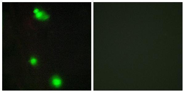
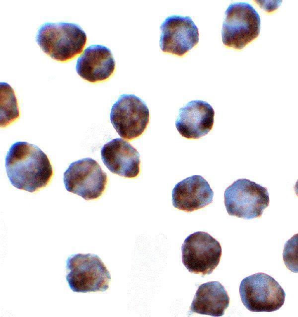
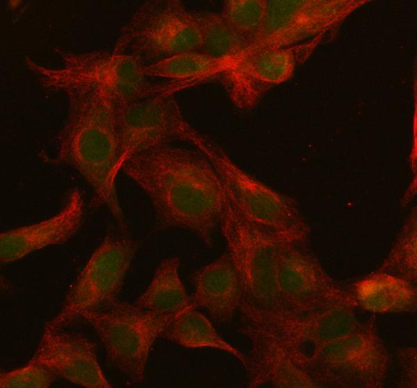
Anti-SRSF3 antibodies have IHC data in human paraffin sections (A30468 image caption) and IF/ICC data in K562 and A549 cells (A03127 and PB9509 image captions); listed reactivity spans human, mouse, and rat (catalog).
A30468 shows IHC in paraffin-embedded human small intestinal stromal tumor, while A03127 shows IF/ICC in K562 cells (respective image captions). PB9509 shows IF in A549 cells (PB9509 image caption).
Which to pick: For tissue IHC, choose rabbit polyclonal A30468, listed for IHC in human and mouse at 1:100–1:300 (A30468 catalog); its own paraffin-section image used Tris-EDTA pH 9 retrieval and 1:200 primary antibody overnight at 4 °C, with fixative unreported (A30468 image caption). For IF/ICC, A03127 has K562 images at 2 μg/mL, and rabbit polyclonal PB9509 has an A549 IF image at 5 μg/mL (A03127 and PB9509 image captions; PB9509 catalog). For the broadest listed species coverage among these three, choose A03127: human, mouse, and rat reactivity is listed, while its pictured IF/ICC data are from K562 cells (A03127 catalog and image captions).