This website uses cookies to ensure you get the best experience on our website.


- Table of Contents
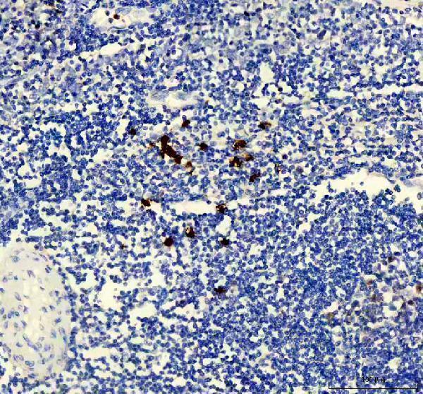
Plan chromogenic TYMS IHC in paraffin sections using tonsil germinal center cells as a positive reference (HPA tissue IHC). Compare nuclear and cytoplasmic staining across matched sections (HPA tissue IHC), and score intensity by cell population.

Expected localisation, validated protocols, controls and antibodies — the at-a-glance facts below, then the full design guide.
| Expected localisation | Nuclear and cytoplasmic staining (HPA tissue IHC) | |
| Staining pattern | Strongest in lymphoid germinal center cells (HPA tissue IHC) | |
| Antigen retrieval | EDTA pH 8.0 HIER, heat-mediated (datasheet A04320-4) | |
| Positive control | Bone marrow+4 more · see all | |
| Negative control | Adipose tissue+4 more · see all |
| Fixation | Keep fixation consistent across sections. (standard IHC practice; not target-specific) | |
| Caveat | Nuclear staining may vary with cell-cycle phase (UniProt) | |
| Regulation | Higher in marrow and lymphoid tissue (HPA tissue IHC) | |
| Isoform / epitope | 3 isoforms; check antibody epitope coverage (UniProt) |
The catalog antibody uses EDTA pH 8.0 heat retrieval (datasheet A04320-4). Published TYMS IHC protocols provide prostate and breast tissue microarray conditions (PMC4480759; PMC7820961).
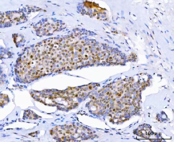
| Sample | Paraffin-embedded human breast cancer tissue; fixative not specified (datasheet A04320-4) |
| Fixation | Image fixative and duration unreported (datasheet A04320-4); verify before use. |
| Sectioning | 4–5 µm sections on charged slides (standard) |
| Deparaffinisation | Xylene, graded ethanol series to water (standard) |
| Antigen retrieval | Heat retrieval: EDTA pH 8.0 (datasheet A04320-4); 20 min, 95–100 °C (standard) |
| Peroxidase block | 3% H2O2, 10 min, room temperature (standard) |
| Blocking | 10% goat serum (datasheet A04320-4) |
| Primary antibody | Rabbit anti-TYMS, 2-5 μg/ml (datasheet A04320-4) |
| Primary incubation | Overnight at 4 °C (datasheet A04320-4) |
| Detection | HRP-conjugated secondary, DAB chromogen (datasheet A04320-4) |
| Counterstain | Hematoxylin, blue, dehydrate and mount (standard) |
| Expected result | TYMS-positive staining in hematopoietic cells of bone marrow (HPA tissue IHC: High). HPA tissue profile: Nuclear and cytoplasmic expression in most tissues, highest expression in lymphoid tissues. No signal in the no-primary control. |
TYMS is reported in the nucleus, cytoplasm and mitochondria, with nuclear localization during S and G2/M and at replication forks during DNA synthesis (UniProt P04818). In paraffin-section IHC, expect nuclear and cytoplasmic staining in many tissues, especially germinal center and bone marrow hematopoietic cells (HPA tissue IHC). HPA rates its tissue pattern Enhanced, while reporting medium consistency between antibody staining and RNA expression (HPA tissue IHC).
| Strong nuclear and cytoplasmic signal in lymph node or tonsil germinal center cells. | This fits the reported high staining in those cells (HPA tissue IHC). Nuclear signal is compatible with TYMS localization during S and G2/M; a single section cannot establish a cell's cycle phase (UniProt P04818; general IHC interpretation). |
| High signal in bone marrow hematopoietic cells or testis preleptotene spermatocytes. | Both are documented high-staining populations (HPA tissue IHC). Compare identifiable cells rather than treating the entire tissue as uniformly positive; neighboring populations need separate assessment (general IHC interpretation). |
| A continuous outline of cell surfaces dominates, with little intracellular signal. | A dominant plasma-membrane pattern does not match the reported nuclear and cytoplasmic tissue pattern (HPA tissue IHC). TYMS has no transmembrane segment, though UniProt also lists mitochondrial locations; check morphology and controls before calling this specific staining (UniProt P04818; general IHC practice). |
| Strong staining appears in adipocytes or prostate glandular cells. | Those specific cell populations are listed as not detected (HPA tissue IHC). Recheck cell identity and controls; cross-reactivity or endogenous detection activity can produce an apparent positive result (general IHC practice). |
| Broad, uniform color covers cells and surrounding section, obscuring nuclei. | Diffuse background prevents a reliable compartment call (general IHC interpretation). It does not reproduce the cell-resolved nuclear and cytoplasmic pattern described for TYMS (HPA tissue IHC). |
| Cell population and tissue | High staining is documented in bone marrow hematopoietic cells, lymphoid germinal center cells and preleptotene spermatocytes (HPA tissue IHC). Adipocytes and prostate glandular cells are listed as not detected; tissue labels alone cannot substitute for identifying the scored cells (HPA tissue IHC; general IHC interpretation). |
| Compartment and cell cycle | UniProt places TYMS in the nucleus, cytoplasm and mitochondria and reports nuclear localization during S and G2/M (UniProt P04818). Mixed nuclear and cytoplasmic staining is therefore plausible; morphology and controls remain necessary to interpret a chromogenic signal (HPA tissue IHC; general IHC practice). |
| Evidence and antibody choice | The tissue profile has Enhanced reliability but only medium antibody-staining/RNA consistency (HPA tissue IHC). HPA lists two antibodies with Enhanced IHC validation, HPA074922 and CAB002784; neither has an ICC validation status in the supplied antibody record (HPA antibodies). |
| Protein forms | UniProt lists three isoforms, a processed chain spanning residues 2–313 and no transmembrane segment (UniProt P04818). The supplied record does not map either antibody's epitope, so these annotations cannot predict which forms it detects or justify a staining difference (UniProt P04818; HPA antibodies). |
| IF/ICC Q: Can this pattern be transferred directly to an IF/ICC result? | A: Use nuclear and cytoplasmic localization as a biological expectation (UniProt P04818; HPA tissue IHC). HPA supplies no main ICC-IF location, image-bearing cell lines or ICC validation status here, so the IHC pattern alone does not validate an IF/ICC readout (HPA subcellular; HPA antibodies). |
| Situation | Likely cause | Next action |
|---|---|---|
| No signal in lymph node or tonsil germinal centers. | These are high-staining reference populations, so an absent result may reflect section sampling or assay performance (HPA tissue IHC; general IHC practice). | Confirm germinal centers are present, then review the IHC-validated antibody's dilution, retrieval, detection reagents and positive control according to its IHC-P instructions (general IHC practice). TYMS-specific retrieval sensitivity is unreported in the supplied sources. |
| Signal appears in adipocytes or prostate glandular cells. | HPA lists those cells as not detected; cell misidentification, nonspecific binding or endogenous detection activity may explain the result (HPA tissue IHC; general IHC practice). | Verify the cell type on the counterstained section, inspect a reagent-only control and evaluate the detection system's endogenous-activity block where applicable (general IHC practice). |
| The section has diffuse chromogenic haze. | Background may arise from nonspecific reagent binding or detection chemistry and makes cell-level interpretation unreliable (general IHC practice). | Check the reagent-only control, blocking and washes; adjust antibody concentration using the catalog antibody's IHC-P instructions (general IHC practice). Do not score haze as TYMS-positive cells. |
| Staining chiefly outlines cell surfaces. | A surface-outline pattern is discordant with HPA's nuclear and cytoplasmic tissue profile; TYMS lacks a transmembrane segment (HPA tissue IHC; UniProt P04818). | Compare the positive-cell morphology and negative control, then reassess antibody concentration and detection background under general IHC practice. Do not infer a new TYMS compartment from one discordant section. |
| Only cytoplasmic staining is visible in an otherwise plausible positive population. | TYMS can be cytoplasmic, and its nuclear localization varies with cell-cycle phase; nuclear signal in every cell is not required (UniProt P04818). | Score identifiable positive cells and both compartments separately, compare with a documented high-staining population, and avoid calling technical failure from nuclear absence alone (HPA tissue IHC; general IHC interpretation). |
| Two antibody stains disagree in the same tissue. | HPA reports medium consistency between staining and RNA despite Enhanced tissue-pattern reliability; antibody-dependent differences remain possible (HPA tissue IHC). | Compare the same identified cell population, appropriate controls and each antibody's IHC-P conditions before assigning a biological difference (general IHC practice). The supplied record gives no epitope map to resolve isoform-specific detection (UniProt P04818; HPA antibodies). |


Comprehensive Human Protein Atlas IHC scoring per tissue (reliability: Enhanced — Medium consistency between antibody staining and RNA expression data.). Rows are taken directly from the HPA tissue chart — click any row's HPA link to view the source.
| Tissue | Cell type | Level | Evidence | Source |
|---|---|---|---|---|
| Bone marrow | Hematopoietic cells | High | Protein (IHC) | HPA → |
| Lymph node | Germinal center cells | High | Protein (IHC) | HPA → |
| Testis | Preleptotene spermatocytes | High | Protein (IHC) | HPA → |
| Tonsil | Germinal center cells | High | Protein (IHC) | HPA → |
| Appendix | Lymphoid tissue | Medium | Protein (IHC) | HPA → |
| Tissue | Cell type | Level | Evidence | Source |
|---|---|---|---|---|
| Adipose tissue | Adipocytes | Not detected | Protein (IHC) | HPA → |
| Bronchus | Respiratory epithelial cells | Not detected | Protein (IHC) | HPA → |
| Endometrium | Cells in endometrial stroma | Not detected | Protein (IHC) | HPA → |
| Fallopian tube | Glandular cells | Not detected | Protein (IHC) | HPA → |
| Heart muscle | Cardiomyocytes | Not detected | Protein (IHC) | HPA → |
Use the IHC-validated TYMS antibody in paraffin sections with compartment-aware controls and cell-level scoring; the selected tissue caption does not report a fixative.
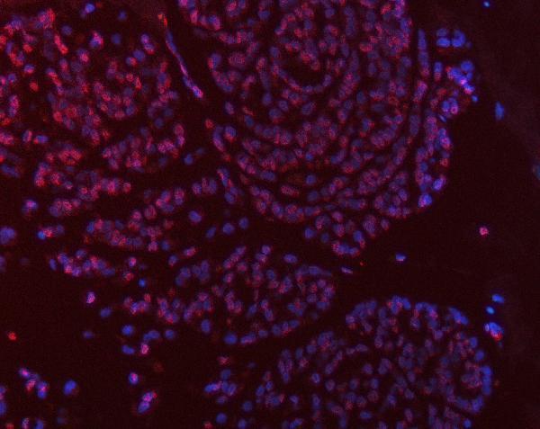
Catalog anti-TYMS antibodies have human paraffin-section IHC images; A04320-4 and A04320-3 also have IF images (catalog image captions). Listed reactivity spans human, monkey, mouse and rat (catalog reactivity).
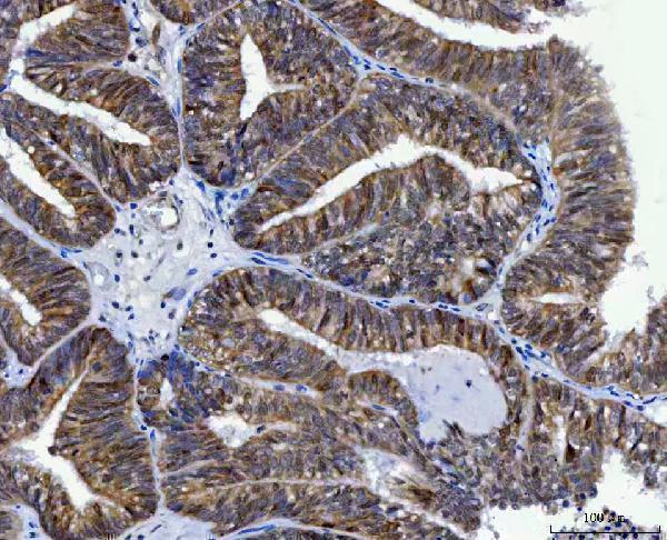
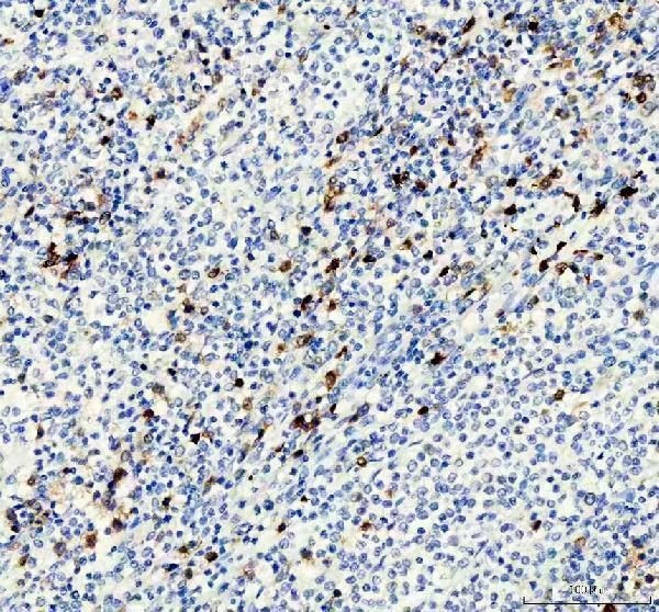
A04320-4 has human breast cancer, endometrial adenocarcinoma, lung cancer and spleen IHC images plus a breast cancer IF image; M04320-1 has human B lymphoblastoma, spleen and tonsil IHC images (catalog image captions). A04320-3 has IHC images from formalin-fixed, paraffin-embedded human colon and breast carcinoma, plus IF images from HeLa and WiDr cells (catalog image captions).
Which to pick: For paraffin-section, paraffin-embedded tissue IHC, choose A04320-3; A04320-4 and M04320-1 have paraffin-section IHC examples with EDTA retrieval, but their captions do not report the fixative (catalog IHC captions). For IF/ICC, A04320-4 has a tissue IF image, A04320-3 has cell IF images, and M04320-1 lists IF/ICC without a supplied IF image (catalog IF captions and applications). For cross-species work, monoclonal M04320-1 lists human, monkey, mouse and rat reactivity, though its supplied IHC images show human samples only (catalog clone, reactivity and IHC captions). The selected A04320-4 tissue-IHC caption documents paraffin sections, but does not specify the fixative (selected-SKU IHC image A04320-4).