XRCC6 / DNA repair protein Ku70 · IHC design guide

Design Immunohistochemistry for XRCC6

Plan XRCC6 IHC in paraffin sections around the widespread nuclear tissue pattern (HPA tissue IHC). High staining in colon glandular cells offers a reference (HPA tissue IHC); assess cytoplasmic signal cautiously because trimethylated XRCC6 can localize there (UniProt).

Evidence assembled Oct 2026 · For research use; verify linked source records and product datasheet before use
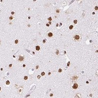
Immunohistochemistry protocol sheet for XRCC6 (IHC for XRCC6): expected localisation Predominantly nuclear in tissue sections (HPA tissue IHC), antibody PB9520, validated IHC image, and IHC protocol steps
Printable XRCC6 IHC protocol sheet — expected localisation Predominantly nuclear in tissue sections (HPA tissue IHC), antibody PB9520, controls and protocol steps. Open the full XRCC6 IHC guide →

XRCC6 Immunohistochemistry Experimental Design Guide

Expected localisation, validated protocols, controls and antibodies — the at-a-glance facts below, then the full design guide.

Must know before staining
Expected localisation Predominantly nuclear in tissue sections (HPA tissue IHC)
Staining pattern Widespread nuclear staining (HPA tissue IHC)
Antigen retrieval EDTA pH 8.0 HIER, heat-mediated (datasheet PB9520)
Positive control ⓘ Adrenal gland+4 more · see all
Negative control ⓘ None in HPA (detected in all 45 tissues); use no-primary + isotype controls
Important caveats
Reasons your staining may differ from the expected pattern.
Fixation Keep fixation conditions consistent across sections. (standard IHC practice; not target-specific)
Caveat Trimethylation may shift XRCC6 to the cytoplasm (UniProt)
Regulation Low tissue specificity (HPA tissue RNA)
Isoform / epitope 2 isoforms; epitope differences are unspecified (UniProt)
Section 1

Recommended XRCC6 IHC & IF Protocols

The catalog antibody’s IHC-P protocol (datasheet PB9520) is accompanied by published XRCC6 IHC protocols from osteosarcoma and colorectal cancer studies (PMC4964557; PMC12623751).

Recommended immunohistochemistry (IHC-P) protocol parameters
SampleParaffin-embedded human bladder cancer tissue; fixative not specified (datasheet PB9520)
FixationImage fixative and duration unreported (datasheet PB9520); verify before use.
Sectioning4–5 µm sections on charged slides (standard)
DeparaffinisationXylene, graded ethanol series to water (standard)
Antigen retrievalHeat retrieval: EDTA pH 8.0 (datasheet PB9520); 20 min, 95–100 °C (standard)
Peroxidase block3% H2O2, 10 min, room temperature (standard)
Blocking10% goat serum (datasheet PB9520)
Primary antibodyRabbit anti-XRCC6, 2-5μg/ml (datasheet PB9520)
Primary incubationOvernight at 4 °C (datasheet PB9520)
DetectionHRP-conjugated secondary, DAB chromogen (datasheet PB9520)
CounterstainHematoxylin, blue, dehydrate and mount (standard)
Expected resultXRCC6-positive staining in glandular cells of adrenal gland (HPA tissue IHC: High). HPA tissue profile: Ubiquitous nuclear expression. No signal in the no-primary control.
💡Decision noteStart with EDTA pH 8.0 heat retrieval for the catalog antibody (datasheet PB9520). Neither published excerpt specifies a retrieval buffer (PMC4964557; PMC12623751).
Section 2

What Is the Expected XRCC6 Staining Pattern?

XRCC6 should stain predominantly in nuclei across many cell types: HPA describes ubiquitous nuclear expression with Enhanced tissue IHC reliability, including High staining in colon glandular and bronchial respiratory epithelial cells (HPA: tissue IHC). UniProt also places XRCC6 on chromosomes and, when trimethylated, in the cytoplasm; it has no transmembrane segment (UniProt P12956: subcellular location and topology).

What am I looking at on my slide?
Clear nuclear staining in colon glandular cells or bronchial respiratory epithelial cells.This matches reported High staining in those cells and the broader ubiquitous nuclear profile (HPA: tissue IHC). Assess nuclear signal against the counterstain and neighboring tissue structures; intensity alone cannot establish antibody specificity (general IHC practice).
Predominantly cytoplasmic staining, with little or no nuclear signal.This conflicts with the usual nuclear tissue pattern (HPA: ubiquitous nuclear expression). UniProt reports cytoplasmic localization when XRCC6 is trimethylated, so cytoplasmic signal is not automatically false (UniProt P12956: subcellular location). Check whether the nuclear positive control stains before interpreting it as biology (general IHC practice).
Strong staining in an unexpected cell population while expected glandular nuclei are blank.For colon or appendix, blank glandular nuclei conflict with reported High glandular staining (HPA: tissue IHC). Cross-reactivity or endogenous chromogenic activity may explain the unexpected signal (general IHC practice); HPA provides no negative cell-type list, so do not declare that population XRCC6-negative.
Diffuse color across nuclei, cytoplasm and tissue spaces.This obscures the expected nuclear distribution (HPA: tissue IHC). Excess detection background, inadequate blocking or incomplete washes are possible general IHC causes; the appearance alone cannot identify which step failed (general IHC practice). Compare a no-primary control with the stained section.
No detectable signal in a well-preserved known-positive tissue.Adrenal gland, breast, colon and duodenum glandular cells are reported High (HPA: tissue IHC). A blank run in one of these tissues raises a technical concern, but is not proof that XRCC6 is absent. Review control performance, detection chemistry and tissue quality (general IHC practice).
💡Expected XRCC6 appearanceA credible positive is predominantly nuclear chromogenic staining in expected cells, including High-staining colon glandular cells (HPA: tissue IHC); diffuse tissue-wide color without nuclear definition suggests background rather than a convincing positive (general IHC practice).
How each factor affects the staining
Compartment and cell contextHPA reports ubiquitous nuclear tissue staining and Enhanced reliability, reflecting high consistency between antibody staining and RNA expression data (HPA: tissue IHC). Score interpretable nuclei in the relevant cell population rather than treating all colored areas as equivalent.
Conditional cytoplasmic localizationUniProt lists nucleus, chromosome and cytoplasm, specifying cytoplasmic localization when XRCC6 is trimethylated (UniProt P12956: subcellular location). This permits a biological explanation for some cytoplasmic signal, but neither source supplies a tissue-specific cytoplasmic IHC pattern.
Antibody validationHPA047549 and HPA062226 have Enhanced IHC and ICC ratings; CAB004254 has Supported IHC status (HPA: antibodies). Those ratings support the reported patterns for those antibodies; they do not validate an unlisted catalog antibody or establish its epitope.
Protein forms and epitope limitsUniProt lists two isoforms, a 2–609 chain and no signal peptide, propeptide or transmembrane segment (UniProt P12956: processing, isoforms and topology). The supplied sources give no antibody epitope, so isoform recognition and effects of processing cannot be predicted from this record.
Retrieval and chromogenic detectionAntigen retrieval, blocking and detection conditions can alter visibility or background in paraffin IHC (general IHC practice). The supplied HPA and UniProt records do not establish XRCC6-specific retrieval needs or fixation sensitivity; optimize against controls without attributing a failed stain to XRCC6-specific masking.
Why is my staining missing, weak or wrong?
SituationLikely causeNext action
Known-positive glandular nuclei are blank.Possible missed detection, ineffective retrieval or unsuitable antibody conditions (general IHC practice); HPA reports High glandular staining in colon (HPA: tissue IHC).Confirm section integrity and positive-control staining, then check retrieval, antibody dilution and detection steps using the IHC-validated antibody's instructions (general IHC practice).
Color is widespread but nuclear boundaries are indistinct.Nonspecific binding or excess chromogenic background can obscure localization (general IHC practice); the expected tissue profile is nuclear (HPA: tissue IHC).Inspect a no-primary control; review blocking, washes, antibody concentration and development time, then score only resolved cellular staining (general IHC practice).
Unexpected cells stain while expected nuclei remain blank.Cross-reactivity or endogenous enzyme activity is possible (general IHC practice); HPA does not designate negative cell types (HPA: tissue IHC).Compare a no-primary control and an established nuclear-positive tissue; evaluate the unexpected cells cautiously rather than assigning specificity from color alone (general IHC practice).
Cytoplasm stains more strongly than nuclei.The usual IHC profile is nuclear (HPA: tissue IHC), although trimethylated XRCC6 can localize to cytoplasm (UniProt P12956: subcellular location).Check nuclear staining in a positive control and background in a no-primary control before interpreting cytoplasmic signal; these controls cannot establish trimethylation (general IHC practice).
Two antibodies give different tissue patterns.HPA assigns antibody-specific IHC status: Enhanced for HPA047549 and HPA062226, Supported for CAB004254 (HPA: antibodies). These ratings do not prove identical epitope recognition.Compare nuclear localization and known-positive cell populations for each antibody; retain the antibody-specific result and investigate discordance with controls (HPA: tissue IHC; general IHC practice).
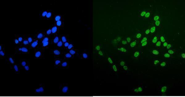
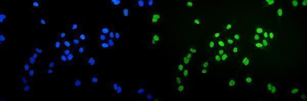
IF/ICC question: Where should XRCC6 signal appear?HPA reports enhanced nucleoplasmic localization in ICC-IF (HPA: subcellular); UniProt also lists conditional cytoplasmic localization (UniProt P12956: subcellular location).Expect predominantly nucleoplasmic signal when interpreting IF/ICC images (HPA: subcellular). Assess cytoplasmic signal cautiously; the tissue IHC pattern does not by itself validate an IF/ICC result.

Sample controls for XRCC6 IHC & IF

🧪Run breast first and score its glandular cells for nuclear staining (HPA: High in breast glandular cells; UniProt P12956: nucleus). HPA detects XRCC6 in all 45 scored tissues, so there is no supported negative tissue; use no-primary and isotype controls, and treat cells showing only counterstain as internal background references rather than confirmed XRCC6-negative cells (HPA: no negative tissue rows).
Positive control tissue: Adrenal gland (Glandular cells, HPA High)
Negative control tissue: None in HPA: XRCC6 is detected in all 45 scored tissues. Use a no-primary (secondary-only) and an isotype control instead.
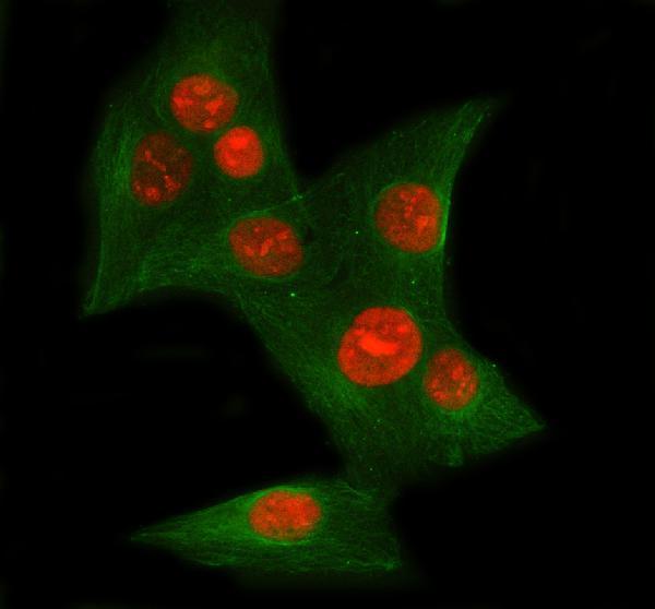
ICC-IF cell lines (HPA subcellular resource): HPA ICC-IF images show XRCC6 in HEK293, PC-3, U2OS, Rh30, U2OS, siRNA 2 (10x), U2OS, siRNA 2 (40x), U2OS, scrambled (10x), U2OS, scrambled (40x), U2OS, siRNA 1 (10x), U2OS, siRNA 1 (40x), HeLa BAC 3840, HeLa , NIH 3T3, with annotated localisation: Nucleoplasm (enhanced) (HPA subcellular).
Technical controls: Run a no-primary, secondary-only slide and a matched rabbit isotype control alongside a validated XRCC6-knockout specimen, if available (selected PB9520 caption: rabbit primary antibody). For chromogenic detection, quench endogenous peroxidase and check for background DAB staining in the breast section (selected PB9520 caption: HRP/DAB detection).
⚠️Feasibility: A target-specific fixation window and fixation effect are unreported, and the selected PB9520 paraffin-section caption does not state a fixative. That caption uses heat retrieval in EDTA at pH 8.0, providing an IHC starting point without establishing that retrieval is required (selected PB9520 tissue-IHC caption). Whether frozen sections or IF are easier is unreported; ICC-IF images show enhanced nucleoplasmic localization, and background chromogen in breast sections should be assessed with the controls (HPA: nucleoplasm enhanced; selected PB9520 caption: HRP/DAB detection).

HPA tissue IHC evidence for XRCC6

Comprehensive Human Protein Atlas IHC scoring per tissue (reliability: Enhanced — High consistency between antibody staining and RNA expression data.). Rows are taken directly from the HPA tissue chart — click any row's HPA link to view the source.

Positive expression · recommended positive controls

TissueCell typeLevelEvidenceSource
Adrenal gland Glandular cells High Protein (IHC) HPA →
Appendix Glandular cells High Protein (IHC) HPA →
Breast Glandular cells High Protein (IHC) HPA →
Bronchus Respiratory epithelial cells High Protein (IHC) HPA →
Cerebellum Cells in molecular layer High Protein (IHC) HPA →

Undetected expression · recommended negative controls

TissueCell typeLevelEvidenceSource
None in HPA: XRCC6 is detected in all 45 scored tissues. Use a no-primary (secondary-only) and an isotype control instead.
Section 3

Advanced XRCC6 IHC Tips

Troubleshoot XRCC6/Ku70 staining by checking retrieval, nuclear localisation and controls before interpreting changes in chromogenic IHC signal.

Which retrieval condition should I start with for Ku70 in paraffin sections?
Use heat-mediated antigen retrieval in EDTA at pH 8.0 for paraffin sections (datasheet PB9520). The selected tissue-IHC image used that condition before incubation with 2 μg/ml primary antibody overnight at 4°C, so it is the most directly supported starting point for this antibody (datasheet PB9520). If nuclear staining is weak, check that sections remained covered during heating and cooling, then compare retrieval times on adjacent sections while holding detection conditions constant (standard IHC practice). Assess recovery in intact cells across the section, because uneven heating or tissue loss can resemble low antigen expression (standard IHC practice; HPA: ubiquitous nuclear expression).
How should I troubleshoot fixation when nuclear Ku70 staining is weak?
Target-specific sensitivity to fixation is unknown: the selected paraffin-section caption does not state a fixative (datasheet PB9520). Record the specimen’s actual fixation history and compare similarly processed sections before attributing weak nuclear signal to XRCC6 biology (standard IHC practice; HPA: ubiquitous nuclear expression). Confirm that EDTA at pH 8.0 was used for heat-mediated retrieval, then inspect morphology and staining across both section centers and edges (datasheet PB9520; standard IHC practice). If processing histories differ, use matched controls and interpret any intensity difference cautiously, since the available tissue image cannot establish an optimal fixation time or a Ku70-specific fixation effect (datasheet PB9520; standard IHC practice).
What staining compartment should I expect, and when is cytoplasmic signal plausible?
Expect predominantly nuclear staining in chromogenic sections: tissue IHC shows ubiquitous nuclear expression, and subcellular data place XRCC6 in the nucleoplasm (HPA: tissue IHC; HPA: subcellular). Assess signal against a nuclear counterstain and compare intact cells in the same section before assigning faint cytoplasmic colour to Ku70 (standard IHC practice). Cytoplasmic localisation is biologically possible when Ku70 is trimethylated, but the supplied tissue-IHC evidence does not establish that state in a particular specimen (UniProt P12956: subcellular localisation; HPA: tissue IHC). Predominantly diffuse cytoplasmic DAB without convincing nuclei therefore warrants checks of background and detection controls before biological interpretation (standard IHC practice; HPA: ubiquitous nuclear expression).
Could isoforms or epitope masking explain discordant Ku70 IHC results?
XRCC6 has 2 listed isoforms, but the supplied antibody caption does not specify its epitope or establish isoform-specific recognition (UniProt P12956: isoforms; datasheet PB9520). Its Ku domain spans residues 261–468 and its SAP domain spans 573–607; these positions describe the protein, not where this antibody binds (UniProt P12956: domains; datasheet PB9520). Ku70 also has annotated modified residues, so epitope accessibility cannot be inferred from sequence position alone without epitope information (UniProt P12956: modified residues; standard IHC practice). When results disagree, repeat the documented EDTA pH 8.0 retrieval with matched sections and controls before assigning the difference to an isoform or modification (datasheet PB9520; standard IHC practice).
How can IF help assess an ambiguous nuclear IHC pattern?
On a separate IF/ICC preparation, compare Ku70 fluorescence with a nuclear stain and a marker identifying the expected cell population; glandular cells are one tissue-IHC population with high expression (HPA: subcellular; HPA: high in glandular cells; standard IF practice). Choose spectrally separated fluorophores, and favor a channel with low measured specimen autofluorescence when evaluating weak signal (standard IF practice). Ku70 has no transmembrane segment and is predominantly nucleoplasmic, so permeabilisation must provide antibody access to the nucleus rather than target a membrane-facing epitope (UniProt P12956: topology; HPA: subcellular; standard IF practice). Include single-label and no-primary controls to distinguish bleed-through or autofluorescence from a reproducible Ku70 pattern; assess IF findings alongside, rather than as a substitute for, the paraffin-section IHC result (standard IF/IHC practice).
How do I reduce diffuse or patchy DAB background?
First inspect a no-primary section and a matched tissue section to separate detection-system colour from primary-antibody staining (standard IHC practice). The selected PB9520 example used 10% goat serum blocking, 2 μg/ml primary antibody overnight at 4°C, and a peroxidase-linked secondary for 30 minutes at 37°C before DAB development (datasheet PB9520). Confirm an appropriate peroxidase block, adequate washes and consistent DAB development across sections; these are general chromogenic IHC controls (standard IHC practice). Judge adjustments by whether intact nuclei remain clear while diffuse colour, edge deposits or signal in the no-primary control decreases (HPA: ubiquitous nuclear expression; standard IHC practice).
How should I score Ku70 staining across samples? ⚠ ANSWER MARKED FOR VERIFICATION
Define the cell population and analysis area before scoring, then exclude folds, necrosis and poorly preserved regions using the same rules for every section (standard IHC practice). For nuclear Ku70, report the percentage of positive nuclei and staining intensity, or calculate an H-score from intensity categories 0–3 for a possible range of 0–300 (HPA: ubiquitous nuclear expression; standard IHC practice). Normalize counts to the number of evaluable nuclei in each specified cell population; if reporting positive-cell density, state the counted tissue area in mm² (standard IHC practice). Keep retrieval, exposure to DAB and scoring thresholds consistent across compared sections, and report cytoplasmic staining separately if it is measured (standard IHC practice; UniProt P12956: subcellular localisation).
When is a strong Ku70 stain likely to be misleading?
A convincing result shows staining in intact nuclei with a reproducible pattern across comparable tissue regions, consistent with ubiquitous nuclear XRCC6 expression (HPA: tissue IHC; standard IHC practice). High staining in glandular or respiratory epithelial cells is compatible with the supplied tissue observations, but intensity alone does not demonstrate active DNA repair (HPA: high in glandular and respiratory epithelial cells; UniProt P12956: function). Reconsider signal confined to cut edges, necrotic areas or the wrong compartment, and compare those regions with the no-primary and peroxidase controls for nonspecific colour (standard IHC practice; HPA: subcellular). Cytoplasmic Ku70 is possible in a trimethylated state, but this IHC pattern alone cannot establish that modification or distinguish it from background (UniProt P12956: subcellular localisation; standard IHC practice).
Boster reagents

Best XRCC6 / DNA repair protein Ku70 IHC Antibodies

Five anti-XRCC6 antibodies have human paraffin-section IHC images (catalog IHC captions); four also have IF images from human tissue or cultured cells (catalog IF captions).

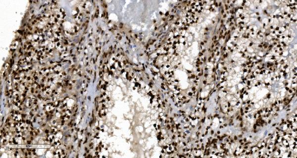
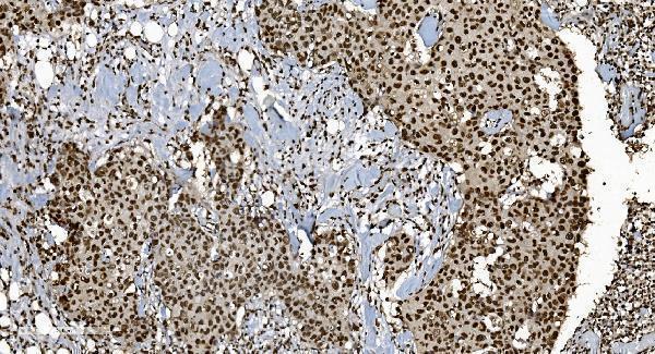
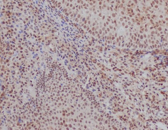
Real IHC data IHC analysis of Ku70/XRCC6 using anti-Ku70/XRCC6 antibody (PB9520). Ku70/XRCC6 was detected in a paraffin-embedded section of human bladder cancer tissue. Heat mediated antigen retrieval was performed in EDTA buffer (pH 8.0, epitope retrieval solution). The tissue section was blocked with 10% goat serum. The tissue section was then incubated with 2 μg/ml rabbit anti-Ku70/XRCC6 Antibody (PB9520) overnight at 4°C. Peroxidase Conjugated Goat Anti-rabbit IgG was used as secondary antibody and incubated for 30 minutes at 37°C. The tissue section was developed using HRP Conjugated Rabbit IgG Super Vision Assay Kit (Catalog # SV0002) with DAB as the chromogen.
Anti-Ku70/XRCC6 Antibody ®
Cat # PB9520
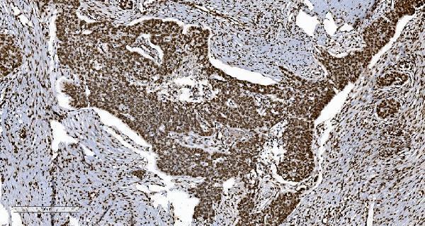
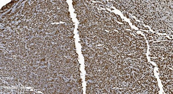
Real IHC data IHC analysis of Ku70 using anti-Ku70 antibody (M01732-4). Ku70 was detected in paraffin-embedded section of human renal clear cell carcinoma tissue. Heat mediated antigen retrieval was performed in EDTA buffer (pH8.0, epitope retrieval solution). The tissue section was blocked with 10% goat serum. The tissue section was then incubated with 2μg/ml mouse anti-Ku70 Antibody (M01732-4) overnight at 4°C. Biotinylated goat anti-mouse IgG was used as secondary antibody and incubated for 30 minutes at 37°C. The tissue section was developed using Strepavidin-Biotin-Complex (SABC) (Catalog # SA1021) with DAB as the chromogen.
Anti-Ku70 Antibody ® (monoclonal, 3D7)
Cat # M01732-4
Real IHC data IHC analysis of Ku70 using anti-Ku70 antibody (M01732-3). Ku70 was detected in paraffin-embedded section of human breast cancer tissue. Heat mediated antigen retrieval was performed in EDTA buffer (pH8.0, epitope retrieval solution). The tissue section was blocked with 10% goat serum. The tissue section was then incubated with 2μg/ml mouse anti-Ku70 Antibody (M01732-3) overnight at 4°C. Biotinylated goat anti-mouse IgG was used as secondary antibody and incubated for 30 minutes at 37°C. The tissue section was developed using Strepavidin-Biotin-Complex (SABC) (Catalog # SA1021) with DAB as the chromogen.
Anti-Ku70 Antibody ® (monoclonal, 9B6)
Cat # M01732-3
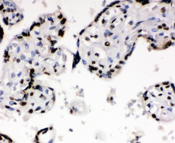
Real IHC data IHC analysis of XRCC6 using anti-XRCC6 antibody (PA1642). XRCC6 was detected in a paraffin-embedded section of Human Placenta tissue. Heat mediated antigen retrieval was performed in EDTA buffer (pH 8.0, epitope retrieval solution). The tissue section was blocked with 10% goat serum. The tissue section was then incubated with 1 μg/ml rabbit anti-XRCC6 Antibody (PA1642) overnight at 4°C. Peroxidase Conjugated Goat Anti-rabbit IgG was used as secondary antibody and incubated for 30 minutes at 37°C. The tissue section was developed using HRP Conjugated Rabbit IgG Super Vision Assay Kit (Catalog # SV0002) with DAB as the chromogen.
Anti-Ku70/XRCC6 Antibody ®
Cat # PA1642
Real IHC data Immunohistochemical analysis of paraffin-embedded human tonsil, using Ku70 Antibody.
Anti-Ku70 XRCC6 Rabbit Monoclonal Antibody
Cat # M01732-1

PB9520 is shown on human bladder cancer, M01732-4 on renal clear cell carcinoma, and M01732-3 on breast cancer paraffin sections (respective catalog IHC captions). PA1642 is shown on human placenta and M01732-1 on human tonsil paraffin sections (respective catalog IHC captions).

Which to pick: For tissue IHC, PB9520 has a human bladder cancer paraffin-section example with EDTA pH 8 retrieval and 2 μg/ml primary antibody (PB9520 IHC caption); its fixative is unreported (PB9520 IHC caption). For IF/ICC, M01732-4 is a mouse monoclonal with IF images from cultured cells and a human colon cancer paraffin section (M01732-4 catalog entry and IF captions). None of these SKUs has documented cross-species reactivity: all list human only (catalog reactivity fields).

Each figure is that product's own IHC / IF validation image from its datasheet.

References

  1. UniProt Consortium. UniProt entry P12956 (XRCC6_HUMAN, DNA repair protein Ku70).
  2. Human Protein Atlas. XRCC6 tissue IHC expression (reliability: Enhanced).
  3. Human Protein Atlas. XRCC6 subcellular location (ICC-IF): Localized to the nucleoplasm..
  4. Human Protein Atlas. XRCC6 antibody validation summary (3 antibodies).
  5. High Expression of XRCC6 Promotes Human Osteosarcoma Cell Proliferation through the β-Catenin/Wnt Signaling Pathway and Is Associated with Poor Prognosis. International journal of molecular sciences 2016 — PMC4964557.
  6. CNOT7 facilitates radiation resistance in colorectal cancer through TRIM21/XRCC6-mediated non-homologous end joining repair. Cell death & disease 2025 — PMC12623751.
  7. A novel NHEJ gene signature based model for risk stratification and prognosis prediction in hepatocellular carcinoma. Cancer cell international 2023 — PMC10071660.
  8. MTHFD2 is required for DNA repair and implicated in LUAD radiotherapy resistance. Journal of translational medicine 2026 — PMC12882504.
  9. PubMed PMID:2917966 — UniProt-cited evidence.
  10. PubMed PMID:2466842 — UniProt-cited evidence.
  11. PubMed PMID:1608402 — UniProt-cited evidence.