Click image to see more details
Product Info Summary
| SKU: | M01617-1 |
|---|---|
| Size: | 100 μg/vial |
| Reactive Species: | Human, Mouse, Rat |
| Host: | Mouse |
| Application: | Flow Cytometry, IHC, WB |
Customers Who Bought This Also Bought

Product info
Product Name
Anti-alpha 1 Catenin/CTNNA1 Antibody Picoband® (monoclonal, 10I2)
SKU/Catalog Number
M01617-1
Size
100 μg/vial
Form
Lyophilized
Description
Boster Bio Anti-alpha 1 Catenin/CTNNA1 Antibody Picoband® (monoclonal, 10I2) catalog # M01617-1. Tested in Flow Cytometry, IHC, WB applications. This antibody reacts with Human, Mouse, Rat. The brand Picoband indicates this is a premium antibody that guarantees superior quality, high affinity, and strong signals with minimal background in Western blot applications. Only our best-performing antibodies are designated as Picoband, ensuring unmatched performance.
Storage & Handling
Store at -20˚C for one year from date of receipt. After reconstitution, at 4˚C for one month. It can also be aliquotted and stored frozen at -20˚C for six months. Avoid repeated freeze-thaw cycles.
Cite This Product
Anti-alpha 1 Catenin/CTNNA1 Antibody Picoband® (monoclonal, 10I2) (Boster Biological Technology, Pleasanton CA, USA, Catalog # M01617-1)
Host
Mouse
Contents
Each vial contains 4mg Trehalose, 0.9mg NaCl, 0.2mg Na2HPO4, 0.05mg NaN3.
Clonality
Monoclonal
Clone Number
10I2
Isotype
Mouse IgG1
Immunogen
E.coli-derived human CTNNA1 recombinant protein (Position: D143-D292). Human CTNNA1 shares 98% amino acid (aa) sequence identity with mouse CTNNA1.
Cross-reactivity
No cross-reactivity with other proteins.
Reactive Species
M01617-1 is reactive to CTNNA1 in Human, Mouse, Rat
Observed Molecular Weight
100 kDa
Calculated molecular weight
100.1 kDa
Background of CTNNA1
CTNNA1, also known as Catenin alpha-1 or Catenin (cadherin-associated protein), alpha 1, is a protein that in humans is encoded by the CTNNA1 gene. It is mapped to 5q31.2. When surface epithelium CTNNA1 was ablated, hair follicle development was blocked and epidermal morphogenesis was dramatically affected, with defects in adherens junction formation, intercellular adhesion, and epithelial polarity. In vitro, CTNNA1 null keratinocytes were poorly contact inhibited and grew rapidly. These differences were not dependent upon intercellular adhesion and were in marked contrast to keratinocytes conditionally null for another essential intercellular adhesion protein, desmoplakin Knockout keratinocytes exhibited sustained activation of the Ras-MAPK cascade due to aberrations in growth factor responses. It is concluded that features of precancerous lesions often attributed to defects in cell cycle regulatory genes can be generated by compromising the function of CTNNA1.
Antibody Validation
Boster validates all antibodies on WB, IHC, ICC, Immunofluorescence, and ELISA with known positive control and negative samples to ensure specificity and high affinity, including thorough antibody incubations.
Application & Images
Applications
M01617-1 is guaranteed for Flow Cytometry, IHC, WB Boster Guarantee
Recommend Dilution
| Application | Dilution | Species |
|---|---|---|
| Western blot | 0.1-0.5μg/ml | Human, Mouse, Rat |
| Immunohistochemistry (Paraffin-embedded Section) | 0.5-1μg/ml | Human |
| Flow Cytometry (Fixed) | 1-3μg/1x106 cells | Human |
Tested application
Suggested blocking solution with 5% non-fat milk or BSA; (*)Recommended protein loading: 20-40 µg per lane
Use TE buffer pH 9.0 for antigen retrieval; (*) citrate buffer pH 6.0 is an alternative.
Validation Images & Assay Conditions
Click image to see more details
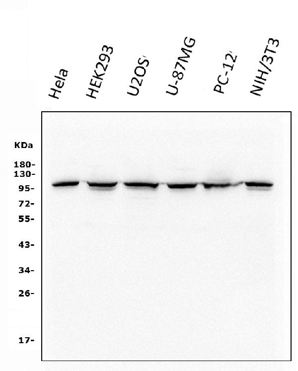
Western blot analysis of CTNNA1 using anti-CTNNA1 antibody (M01617-1).
Electrophoresis was performed on a 5-20% SDS-PAGE gel at 70V (Stacking gel) / 90V (Resolving gel) for 2-3 hours. The sample well of each lane was loaded with 50ug of sample under reducing conditions.
Lane 1: human Hela whole cell lysates;
Lane 2: human HEK293 whole cell lysates;
Lane 3: human U20S whole cell lysates;
Lane 4: human U-87MG whole cell lysates;
Lane 5: rat PC-12 whole cell lysates;
Lane 6: mouse NIH/3T3 whole cell lysates
After Electrophoresis, proteins were transferred to a Nitrocellulose membrane at 150mA for 50-90 minutes. Blocked the membrane with 5% Non-fat Milk/ TBS for 1.5 hour at RT. The membrane was incubated with mouse anti-CTNNA1 antigen affinity purified monoclonal antibody (Catalog # M01617-1) at 0.5 μg/mL overnight at 4°C, then washed with TBS-0.1%Tween 3 times with 5 minutes each and probed with a goat anti-mouse IgG-HRP secondary antibody at a dilution of 1:10000 for 1.5 hour at RT. The signal is developed using an Enhanced Chemiluminescent detection (ECL) kit (Catalog # EK1001) with Tanon 5200 system. A specific band was detected for CTNNA1 at approximately 100KD. The expected band size for CTNNA1 is at 100KD.

Click image to see more details
IHC analysis of CTNNA1 using anti-CTNNA1 antibody (M01617-1).
CTNNA1 was detected in paraffin-embedded section of human mammary cancer tissue. Heat mediated antigen retrieval was performed in EDTA buffer (pH8.0, epitope retrieval solution). The tissue section was blocked with 10% goat serum. The tissue section was then incubated with 1μg/ml mouse anti-CTNNA1 Antibody (M01617-1) overnight at 4°C. Biotinylated goat anti-mouse IgG was used as secondary antibody and incubated for 30 minutes at 37°C. The tissue section was developed using Strepavidin-Biotin-Complex (SABC) (Catalog # SA1021) with DAB as the chromogen.

Click image to see more details
Flow Cytometry analysis of Jurkat cells using anti-CTNNA1 antibody (M01617-1).Overlay histogram showing Jurkat cells stained with M01617-1 (Blue line). The cells were fixed with 4% paraformaldehyde and blocked with 10% normal goat serum. And then incubated with mouse anti-CTNNA1 Antibody (M01617-1,1μg/1x106 cells) for 30 min at 20°C. DyLight®488 conjugated goat anti-mouse IgG (BA1126, 5-10μg/1x106 cells) was used as secondary antibody for 30 minutes at 20°C. Isotype control antibody (Green line) was mouse IgG (1μg/1x106) used under the same conditions. Unlabelled sample without incubation with primary antibody and secondary antibody (Red line) was used as a blank control.
Specific Publications For Anti-alpha 1 Catenin/CTNNA1 Antibody Picoband® (monoclonal, 10I2) (M01617-1)
Loading publications
Recommended Resources
Here are featured tools and databases that you might find useful.
- Boster's Pathways Library
- Protein Databases
- Bioscience Research Protocol Resources
- Data Processing & Analysis Software
- Photo Editing Software
- Scientific Literature Resources
- Research Paper Management Tools
- Molecular Biology Software
- Primer Design Tools
- Bioinformatics Tools
- Phylogenetic Tree Analysis
Customer Reviews
Have you used Anti-alpha 1 Catenin/CTNNA1 Antibody Picoband® (monoclonal, 10I2)?
Share your experimental results or join a short interview to earn up to $1,000 in product credits or other rewards.
0 Reviews For Anti-alpha 1 Catenin/CTNNA1 Antibody Picoband® (monoclonal, 10I2)
Customer Q&As
Have a question?
Find answers in Q&As, reviews.
Can't find your answer?
Submit your question
