Click image to see more details
-

-
-
-
-
+11
Product Info Summary
| SKU: | M00929 |
|---|---|
| Size: | 100 μl |
| Reactive Species: | Human, Mouse |
| Host: | Rabbit |
| Application: | Flow Cytometry, IF, IHC, ICC, WB |
Customers Who Bought This Also Bought


Product info
Product Name
Anti-ASK1 MAP3K5 Rabbit Monoclonal Antibody
SKU/Catalog Number
M00929
Size
100 μl
Form
Liquid
Description
Boster Bio Anti-ASK1 MAP3K5 Rabbit Monoclonal Antibody catalog # M00929. Tested in WB, IHC, ICC/IF, Flow Cytometry applications. This antibody reacts with Human, Mouse.
Storage & Handling
Store at -20°C for one year. For short term storage and frequent use, store at 4°C for up to one month. Avoid repeated freeze-thaw cycles.
Cite This Product
Anti-ASK1 MAP3K5 Rabbit Monoclonal Antibody (Boster Biological Technology, Pleasanton CA, USA, Catalog # M00929)
Host
Rabbit
Contents
Rabbit IgG in phosphate buffered saline, pH 7.4, 150mM NaCl, 0.02% sodium azide and 50% glycerol, 0.4-0.5mg/ml BSA.
Clonality
Monoclonal
Clone Number
CFB-13
Isotype
Rabbit IgG
Immunogen
A synthesized peptide derived from human ASK1
*Blocking peptide can be purchased. Costs vary based on immunogen length. Contact us for pricing.
Reactive Species
M00929 is reactive to MAP3K5 in Human, Mouse
Applications
M00929 is guaranteed for Flow Cytometry, IF, IHC, ICC, WB Boster Guarantee
Observed Molecular Weight
95 kDa
Calculated molecular weight
154.537kDa
Background of ASK1
Catalyzes the first step in leukotriene biosynthesis, and thereby plays a role in inflammatory processes.
Antibody Validation
Boster validates all antibodies on WB, IHC, ICC, Immunofluorescence, and ELISA with known positive control and negative samples to ensure specificity and high affinity, including thorough antibody incubations.
Innovating Scientists Reward
If you are the first to review this product, or if you have results for a special sample, species or application this product is not validated in, share your results with us and receive product credits you can use towards any Boster products! Applicable to all scientists worldwide.
Submit A Review
Assay dilution & Images
Reconsitution
Restore with deionized water (or equivalent) for reconstitution volume of 1.0 mL
Assay Dilutions Recommendation
The recommendations below provide a starting point for assay optimization. The actual working concentration varies and should be decided by the user.
WB 1:500-1:2000
IHC 1:50-1:200
ICC/IF 1:50-1:200
FC 1:50
Validation Images & Assay Conditions
Click image to see more details
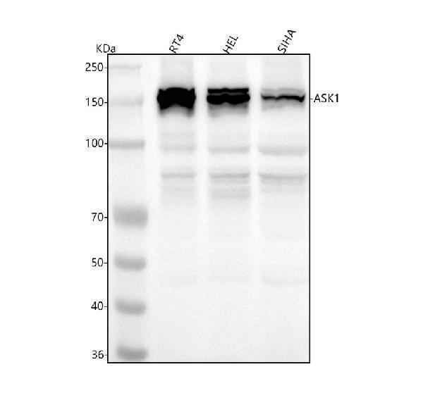
Figure 1. Western blot analysis of ASK1 using anti-ASK1 antibody (M00929).
Electrophoresis was performed on a 5-20% SDS-PAGE gel at 70V (Stacking gel) / 90V (Resolving gel) for 2-3 hours. The sample well of each lane was loaded with 30 ug of sample under reducing conditions.
Lane 1: human RT4 whole cell lysates,
Lane 2: human HEL whole cell lysates,
Lane 3: human SIHA whole cell lysates.
After electrophoresis, proteins were transferred to a nitrocellulose membrane at 150 mA for 50-90 minutes. Blocked the membrane with 5% non-fat milk/TBS for 1.5 hour at RT. The membrane was incubated with rabbit anti-ASK1 antigen affinity purified polyclonal antibody (Catalog # M00929) at 1:500 overnight at 4°C, then washed with TBS-0.1%Tween 3 times with 5 minutes each and probed with a goat anti-rabbit IgG-HRP secondary antibody at a dilution of 1:5000 for 1.5 hour at RT. The signal is developed using an Enhanced Chemiluminescent detection (ECL) kit (Catalog # EK1002) with Tanon 5200 system. A specific band was detected for ASK1 at approximately 95 kDa. The expected band size for ASK1 is at 95 kDa.

Click image to see more details
All lanes use the Antibody at 1:3K dilution for 1 hour at room temperature.

Click image to see more details
All lanes use the Antibody at 1:1K dilution for 1 hour at room temperature.

Click image to see more details
All lanes use the Antibody at 1:1K dilution for 1 hour at room temperature.

Click image to see more details
Immunohistochemical analysis of paraffin-embedded Rat stomach, using the Antibody at 1:50 dilution.

Click image to see more details
Immunohistochemical analysis of paraffin-embedded Mouse skin, using the Antibody at 1:50 dilution.

Click image to see more details
Figure 2. IHC analysis of ASK1 using anti-ASK1 antibody (M00929).
ASK1 was detected in a paraffin-embedded section of human liver cancer tissue. Heat mediated antigen retrieval was performed in EDTA buffer (pH 8.0, epitope retrieval solution). The tissue section was blocked with 10% goat serum. The tissue section was then incubated with 1:50 rabbit anti-ASK1 Antibody (M00929) overnight at 4°C. Peroxidase Conjugated Goat Anti-rabbit IgG was used as secondary antibody and incubated for 30 minutes at 37°C. The tissue section was developed using HRP Conjugated Rabbit IgG Super Vision Assay Kit (Catalog # SV0002) with DAB as the chromogen.

Click image to see more details
Figure 3. IHC analysis of ASK1 using anti-ASK1 antibody (M00929).
ASK1 was detected in a paraffin-embedded section of human liver cancer tissue. Heat mediated antigen retrieval was performed in EDTA buffer (pH 8.0, epitope retrieval solution). The tissue section was blocked with 10% goat serum. The tissue section was then incubated with 1:50 rabbit anti-ASK1 Antibody (M00929) overnight at 4°C. Peroxidase Conjugated Goat Anti-rabbit IgG was used as secondary antibody and incubated for 30 minutes at 37°C. The tissue section was developed using HRP Conjugated Rabbit IgG Super Vision Assay Kit (Catalog # SV0002) with DAB as the chromogen.

Click image to see more details
Figure 4. IHC analysis of ASK1 using anti-ASK1 antibody (M00929).
ASK1 was detected in a paraffin-embedded section of human placenta tissue. Heat mediated antigen retrieval was performed in EDTA buffer (pH 8.0, epitope retrieval solution). The tissue section was blocked with 10% goat serum. The tissue section was then incubated with 1:50 rabbit anti-ASK1 Antibody (M00929) overnight at 4°C. Peroxidase Conjugated Goat Anti-rabbit IgG was used as secondary antibody and incubated for 30 minutes at 37°C. The tissue section was developed using HRP Conjugated Rabbit IgG Super Vision Assay Kit (Catalog # SV0002) with DAB as the chromogen.

Click image to see more details
Figure 5. IHC analysis of ASK1 using anti-ASK1 antibody (M00929).
ASK1 was detected in a paraffin-embedded section of human placenta tissue. Heat mediated antigen retrieval was performed in EDTA buffer (pH 8.0, epitope retrieval solution). The tissue section was blocked with 10% goat serum. The tissue section was then incubated with 1:50 rabbit anti-ASK1 Antibody (M00929) overnight at 4°C. Peroxidase Conjugated Goat Anti-rabbit IgG was used as secondary antibody and incubated for 30 minutes at 37°C. The tissue section was developed using HRP Conjugated Rabbit IgG Super Vision Assay Kit (Catalog # SV0002) with DAB as the chromogen.

Click image to see more details
Figure 6. IHC analysis of ASK1 using anti-ASK1 antibody (M00929).
ASK1 was detected in a paraffin-embedded section of human thyroid papillary carcinoma tissue. Heat mediated antigen retrieval was performed in EDTA buffer (pH 8.0, epitope retrieval solution). The tissue section was blocked with 10% goat serum. The tissue section was then incubated with 1:50 rabbit anti-ASK1 Antibody (M00929) overnight at 4°C. Peroxidase Conjugated Goat Anti-rabbit IgG was used as secondary antibody and incubated for 30 minutes at 37°C. The tissue section was developed using HRP Conjugated Rabbit IgG Super Vision Assay Kit (Catalog # SV0002) with DAB as the chromogen.

Click image to see more details
Figure 7. IHC analysis of ASK1 using anti-ASK1 antibody (M00929).
ASK1 was detected in a paraffin-embedded section of human thyroid papillary carcinoma tissue. Heat mediated antigen retrieval was performed in EDTA buffer (pH 8.0, epitope retrieval solution). The tissue section was blocked with 10% goat serum. The tissue section was then incubated with 1:50 rabbit anti-ASK1 Antibody (M00929) overnight at 4°C. Peroxidase Conjugated Goat Anti-rabbit IgG was used as secondary antibody and incubated for 30 minutes at 37°C. The tissue section was developed using HRP Conjugated Rabbit IgG Super Vision Assay Kit (Catalog # SV0002) with DAB as the chromogen.

Click image to see more details
Figure 8. IHC analysis of ASK1 using anti-ASK1 antibody (M00929).
ASK1 was detected in a paraffin-embedded section of human thyroid papillary carcinoma tissue. Heat mediated antigen retrieval was performed in EDTA buffer (pH 8.0, epitope retrieval solution). The tissue section was blocked with 10% goat serum. The tissue section was then incubated with 1:50 rabbit anti-ASK1 Antibody (M00929) overnight at 4°C. Peroxidase Conjugated Goat Anti-rabbit IgG was used as secondary antibody and incubated for 30 minutes at 37°C. The tissue section was developed using HRP Conjugated Rabbit IgG Super Vision Assay Kit (Catalog # SV0002) with DAB as the chromogen.

Click image to see more details
Figure 9. IHC analysis of ASK1 using anti-ASK1 antibody (M00929).
ASK1 was detected in a paraffin-embedded section of human thyroid papillary carcinoma tissue. Heat mediated antigen retrieval was performed in EDTA buffer (pH 8.0, epitope retrieval solution). The tissue section was blocked with 10% goat serum. The tissue section was then incubated with 1:50 rabbit anti-ASK1 Antibody (M00929) overnight at 4°C. Peroxidase Conjugated Goat Anti-rabbit IgG was used as secondary antibody and incubated for 30 minutes at 37°C. The tissue section was developed using HRP Conjugated Rabbit IgG Super Vision Assay Kit (Catalog # SV0002) with DAB as the chromogen.

Click image to see more details
Immunofluorescent analysis using the Antibody at 1:50 dilution.
Protein Target Info & Infographic
Gene/Protein Information For MAP3K5 (Source: Uniprot.org, NCBI)
Gene Name
MAP3K5
Full Name
Mitogen-activated protein kinase kinase kinase 5
Weight
154.537kDa
Superfamily
protein kinase superfamily
Alternative Names
Apoptosis signal-regulating kinase 1; ASK1; ASK-1; ASK1MEKK 5; EC 2.7.11; MAP/ERK kinase kinase 5; MAP3K5; MAPK/ERK kinase kinase 5; MAPKKK5EC 2.7.11.25; MEK kinase 5; MEKK5; MEKK5apoptosis signal regulating kinase 1; mitogen-activated protein kinase kinase kinase 5 MAP3K5 ASK1, MAPKKK5, MEKK5 mitogen-activated protein kinase kinase kinase 5 mitogen-activated protein kinase kinase kinase 5|ASK-1|MAP/ERK kinase kinase 5|MAPK/ERK kinase kinase 5|MEK kinase 5|MEKK 5|apoptosis signal-regulating kinase 1
*If product is indicated to react with multiple species, protein info is based on the gene entry specified above in "Species".For more info on MAP3K5, check out the MAP3K5 Infographic

We have 30,000+ of these available, one for each gene! Check them out.
In this infographic, you will see the following information for MAP3K5: database IDs, superfamily, protein function, synonyms, molecular weight, chromosomal locations, tissues of expression, subcellular locations, post-translational modifications, and related diseases, research areas & pathways. If you want to see more information included, or would like to contribute to it and be acknowledged, please contact [email protected].
Specific Publications For Anti-ASK1 MAP3K5 Rabbit Monoclonal Antibody (M00929)
Hello CJ!
No publications found for M00929
*Do you have publications using this product? Share with us and receive a reward. Ask us for more details.
Recommended Resources
Here are featured tools and databases that you might find useful.
- Boster's Pathways Library
- Protein Databases
- Bioscience Research Protocol Resources
- Data Processing & Analysis Software
- Photo Editing Software
- Scientific Literature Resources
- Research Paper Management Tools
- Molecular Biology Software
- Primer Design Tools
- Bioinformatics Tools
- Phylogenetic Tree Analysis
Customer Reviews
Have you used Anti-ASK1 MAP3K5 Rabbit Monoclonal Antibody?
Submit a review and receive an Amazon gift card.
- $30 for a review with an image
Be the first to review Anti-ASK1 MAP3K5 Rabbit Monoclonal Antibody
*The first user to submit a review for a product is eligible for Boster's Innovating Scientists Reward, which gives product credits. This is in addition to the gift card reward.
Customer Q&As
Have a question?
Find answers in Q&As, reviews.
Can't find your answer?
Submit your question
7 Customer Q&As for Anti-ASK1 MAP3K5 Rabbit Monoclonal Antibody
Question
See below the WB image, lot number and protocol we used for platelet using anti-ASK1 Rabbit Monoclonal antibody M00929. Please let me know if you require anything else.
Verified Customer
Verified customer
Asked: 2020-04-09
Answer
Thank you very much for the data. Our lab team are working to resolve this as quickly as possible, and we appreciate your patience and understanding! You have provided everything we needed. Please let me know if there is anything you need in the meantime.
Boster Scientific Support
Answered: 2020-04-09
Question
Would anti-ASK1 Rabbit Monoclonal antibody M00929 work for IHC with platelet?
Verified Customer
Verified customer
Asked: 2019-11-20
Answer
According to the expression profile of platelet, MAP3K5 is highly expressed in platelet. So, it is likely that anti-ASK1 Rabbit Monoclonal antibody M00929 will work for IHC with platelet.
Boster Scientific Support
Answered: 2019-11-20
Question
We appreciate helping with my inquiry over the phone. Here are the WB image, lot number and protocol we used for platelet using anti-ASK1 Rabbit Monoclonal antibody M00929. Let me know if you need anything else.
Verified Customer
Verified customer
Asked: 2019-09-19
Answer
We appreciate the data. You have provided everything we needed. Our lab team are working to resolve your inquiry as quickly as possible, and we appreciate your patience and understanding! Please let me know if there is anything you need in the meantime.
Boster Scientific Support
Answered: 2019-09-19
Question
We are currently using anti-ASK1 Rabbit Monoclonal antibody M00929 for human tissue, and we are well pleased with the ICC results. The species of reactivity given in the datasheet says human, mouse. Is it likely that the antibody can work on bovine tissues as well?
Verified Customer
Verified customer
Asked: 2019-07-08
Answer
The anti-ASK1 Rabbit Monoclonal antibody (M00929) has not been tested for cross reactivity specifically with bovine tissues, though there is a good chance of cross reactivity. We have an innovator award program that if you test this antibody and show it works in bovine you can get your next antibody for free. Please contact me if I can help you with anything.
Boster Scientific Support
Answered: 2019-07-08
Question
I was wanting to use your anti-ASK1 Rabbit Monoclonal antibody for IHC for mouse platelet on frozen tissues, but I want to know if it has been tested for this particular application. Has this antibody been tested and is this antibody a good choice for mouse platelet identification?
Verified Customer
Verified customer
Asked: 2017-11-08
Answer
As indicated on the product datasheet, M00929 anti-ASK1 Rabbit Monoclonal antibody has been validated for Flow Cytometry, IF, IHC, ICC, WB on human, mouse tissues. We have an innovator award program that if you test this antibody and show it works in mouse platelet in IHC-frozen, you can get your next antibody for free.
Boster Scientific Support
Answered: 2017-11-08
Question
I am interested in to test anti-ASK1 Rabbit Monoclonal antibody M00929 on mouse platelet for research purposes, then I may be interested in using anti-ASK1 Rabbit Monoclonal antibody M00929 for diagnostic purposes as well. Is the antibody suitable for diagnostic purposes?
Verified Customer
Verified customer
Asked: 2017-08-23
Answer
The products we sell, including anti-ASK1 Rabbit Monoclonal antibody M00929, are only intended for research use. They would not be suitable for use in diagnostic work. If you have the means to develop a product into diagnostic use, and are interested in collaborating with us and develop our product into an IVD product, please contact us for more discussions.
Boster Scientific Support
Answered: 2017-08-23
Question
I see that the anti-ASK1 Rabbit Monoclonal antibody M00929 works with IHC, what is the protocol used to produce the result images on the product page?
N. Krishna
Verified customer
Asked: 2014-05-23
Answer
You can find protocols for IHC on the "support/technical resources" section of our navigation menu. If you have any further questions, please send an email to [email protected]
Boster Scientific Support
Answered: 2014-05-23
