Click image to see more details
-

-
-
-
-
+1
Product Info Summary
| SKU: | A01772-2 |
|---|---|
| Size: | 100 μg/vial |
| Reactive Species: | Human |
| Host: | Rabbit |
| Application: | ELISA, Flow Cytometry, IHC, WB |
Customers Who Bought This Also Bought

Product info
Product Name
Anti-Axin 2/AXIN2 Antibody Picoband™
SKU/Catalog Number
A01772-2
Size
100 μg/vial
Form
Lyophilized
Description
Boster Bio Anti-Axin 2/AXIN2 Antibody Picoband™ catalog # A01772-2. Tested in ELISA, Flow Cytometry, IHC, WB applications. This antibody reacts with Human.
Storage & Handling
At -20°C for one year from date of receipt. After reconstitution, at 4°C for one month. It can also be aliquotted and stored frozen at -20°C for six months. Avoid repeated freezing and thawing.
Cite This Product
Anti-Axin 2/AXIN2 Antibody Picoband™ (Boster Biological Technology, Pleasanton CA, USA, Catalog # A01772-2)
Host
Rabbit
Contents
Each vial contains 4 mg Trehalose, 0.9 mg NaCl, 0.2 mg Na2HPO4.
Clonality
Polyclonal
Isotype
Rabbit IgG
Immunogen
E.coli-derived human Axin 2/AXIN2 recombinant protein (Position: D614-D843).
*Blocking peptide can be purchased. Costs vary based on immunogen length. Contact us for pricing.
Cross-reactivity
No cross-reactivity with other proteins.
Reactive Species
A01772-2 is reactive to AXIN2 in Human
Applications
A01772-2 is guaranteed for ELISA, Flow Cytometry, IHC, WB Boster Guarantee
Observed Molecular Weight
100 kDa
Calculated molecular weight
93.558kDa
Background of Axin-2
Axin-2 also known as axin-like protein (Axil) or axis inhibition protein 2 (AXIN2) or conductin is a protein that in humans is encoded by the AXIN2 gene. The Axin-related protein, Axin2, presumably plays an important role in the regulation of the stability of beta-catenin in the Wnt signaling pathway, like its rodent homologs, mouse conductin/rat axil. In mouse, conductin organizes a multiprotein complex of APC (adenomatous polyposis of the colon), beta-catenin, glycogen synthase kinase 3-beta, and conductin, which leads to the degradation of beta-catenin. Apparently, the deregulation of beta-catenin is an important event in the genesis of a number of malignancies. The AXIN2 gene has been mapped to 17q23-q24, a region that shows frequent loss of heterozygosity in breast cancer, neuroblastoma, and other tumors. Mutations in this gene have been associated with colorectal cancer with defective mismatch repair.
Antibody Validation
Boster validates all antibodies on WB, IHC, ICC, Immunofluorescence, and ELISA with known positive control and negative samples to ensure specificity and high affinity, including thorough antibody incubations.
Innovating Scientists Reward
If you are the first to review this product, or if you have results for a special sample, species or application this product is not validated in, share your results with us and receive product credits you can use towards any Boster products! Applicable to all scientists worldwide.
Submit A Review
Assay dilution & Images
Reconsitution
Adding 0.2 ml of distilled water will yield a concentration of 500 μg/ml.
Assay Dilutions Recommendation
The recommendations below provide a starting point for assay optimization. The actual working concentration varies and should be decided by the user.
Western blot, 0.25-0.5 μg/ml, Human
Immunohistochemistry(Paraffin-embedded Section), 2-5 μg/ml, Human
Flow Cytometry, 1-3 μg/1x106 cells, Human
Direct ELISA, 0.1-0.5 μg/ml, Human
Validation Images & Assay Conditions
Click image to see more details
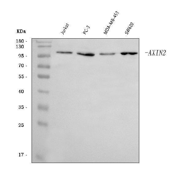
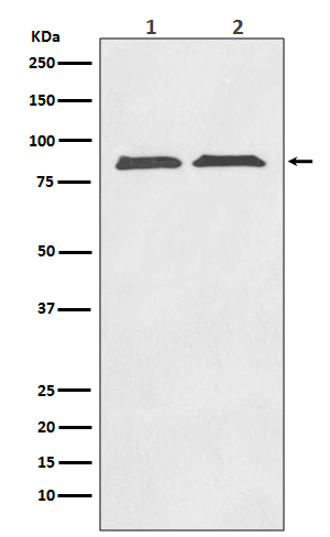
Figure 1. Western blot analysis of Axin 2/AXIN2 using anti-Axin 2/AXIN2 antibody (A01772-2).
Electrophoresis was performed on a 5-20% SDS-PAGE gel at 70V (Stacking gel) / 90V (Resolving gel) for 2-3 hours. The sample well of each lane was loaded with 30 ug of sample under reducing conditions.
Lane 1: human Jurkat whole cell lysates,
Lane 2: human PC-3 whole cell lysates,
Lane 3: human MDA-MB-453 whole cell lysates,
Lane 4: human SW620 whole cell lysates.
After electrophoresis, proteins were transferred to a nitrocellulose membrane at 150 mA for 50-90 minutes. Blocked the membrane with 5% non-fat milk/TBS for 1.5 hour at RT. The membrane was incubated with rabbit anti-Axin 2/AXIN2 antigen affinity purified polyclonal antibody (Catalog # A01772-2) at 0.5 μg/mL overnight at 4°C, then washed with TBS-0.1%Tween 3 times with 5 minutes each and probed with a goat anti-rabbit IgG-HRP secondary antibody at a dilution of 1:5000 for 1.5 hour at RT. The signal is developed using an Enhanced Chemiluminescent detection (ECL) kit (Catalog # EK1002) with Tanon 5200 system. A specific band was detected for Axin 2/AXIN2 at approximately 100 kDa. The expected band size for Axin 2/AXIN2 is at 93 kDa.

Click image to see more details
Figure 2. IHC analysis of Axin 2/AXIN2 using anti-Axin 2/AXIN2 antibody (A01772-2).
Axin 2/AXIN2 was detected in a paraffin-embedded section of human renal carcinoma tissue. Heat mediated antigen retrieval was performed in EDTA buffer (pH 8.0, epitope retrieval solution). The tissue section was blocked with 10% goat serum. The tissue section was then incubated with 2 μg/ml rabbit anti-Axin 2/AXIN2 Antibody (A01772-2) overnight at 4°C. Peroxidase Conjugated Goat Anti-rabbit IgG was used as secondary antibody and incubated for 30 minutes at 37°C. The tissue section was developed using HRP Conjugated Rabbit IgG Super Vision Assay Kit (Catalog # SV0002) with DAB as the chromogen.

Click image to see more details
Figure 3. IHC analysis of Axin 2/AXIN2 using anti-Axin 2/AXIN2 antibody (A01772-2).
Axin 2/AXIN2 was detected in a paraffin-embedded section of human spleen tissue. Heat mediated antigen retrieval was performed in EDTA buffer (pH 8.0, epitope retrieval solution). The tissue section was blocked with 10% goat serum. The tissue section was then incubated with 2 μg/ml rabbit anti-Axin 2/AXIN2 Antibody (A01772-2) overnight at 4°C. Peroxidase Conjugated Goat Anti-rabbit IgG was used as secondary antibody and incubated for 30 minutes at 37°C. The tissue section was developed using HRP Conjugated Rabbit IgG Super Vision Assay Kit (Catalog # SV0002) with DAB as the chromogen.

Click image to see more details
Figure 4. IHC analysis of Axin 2/AXIN2 using anti-Axin 2/AXIN2 antibody (A01772-2).
Axin 2/AXIN2 was detected in a paraffin-embedded section of human thyroid cancer tissue. Heat mediated antigen retrieval was performed in EDTA buffer (pH 8.0, epitope retrieval solution). The tissue section was blocked with 10% goat serum. The tissue section was then incubated with 2 μg/ml rabbit anti-Axin 2/AXIN2 Antibody (A01772-2) overnight at 4°C. Peroxidase Conjugated Goat Anti-rabbit IgG was used as secondary antibody and incubated for 30 minutes at 37°C. The tissue section was developed using HRP Conjugated Rabbit IgG Super Vision Assay Kit (Catalog # SV0002) with DAB as the chromogen.

Click image to see more details
Figure 5. Flow Cytometry analysis of HepG2 cells using anti-Axin 2/AXIN2 antibody (A01772-2).
Overlay histogram showing HepG2 cells stained with A01772-2 (Blue line). The cells were blocked with 10% normal goat serum. And then incubated with rabbit anti-Axin 2/AXIN2 Antibody (A01772-2, 1 μg/1x106 cells) for 30 min at 20°C. DyLight®488 conjugated goat anti-rabbit IgG (BA1127, 5-10 μg/1x106 cells) was used as secondary antibody for 30 minutes at 20°C. Isotype control antibody (Green line) was rabbit IgG (1 μg/1x106) used under the same conditions. Unlabelled sample (Red line) was also used as a control.
Protein Target Info & Infographic
Gene/Protein Information For AXIN2 (Source: Uniprot.org, NCBI)
Gene Name
AXIN2
Full Name
Axin-2
Weight
93.558kDa
Alternative Names
AXIL; axin 2; Axin2; Axin-2; Axin-like protein; Axis inhibition protein 2; Conductin; DKFZp781B0869; MGC10366; MGC126582 AXIN2 AXIL, ODCRCS axin 2 axin-2|axin-like protein|axis inhibition protein 2|conductin
*If product is indicated to react with multiple species, protein info is based on the gene entry specified above in "Species".For more info on AXIN2, check out the AXIN2 Infographic

We have 30,000+ of these available, one for each gene! Check them out.
In this infographic, you will see the following information for AXIN2: database IDs, superfamily, protein function, synonyms, molecular weight, chromosomal locations, tissues of expression, subcellular locations, post-translational modifications, and related diseases, research areas & pathways. If you want to see more information included, or would like to contribute to it and be acknowledged, please contact [email protected].
Specific Publications For Anti-Axin 2/AXIN2 Antibody Picoband™ (A01772-2)
Hello CJ!
No publications found for A01772-2
*Do you have publications using this product? Share with us and receive a reward. Ask us for more details.
Recommended Resources
Here are featured tools and databases that you might find useful.
- Boster's Pathways Library
- Protein Databases
- Bioscience Research Protocol Resources
- Data Processing & Analysis Software
- Photo Editing Software
- Scientific Literature Resources
- Research Paper Management Tools
- Molecular Biology Software
- Primer Design Tools
- Bioinformatics Tools
- Phylogenetic Tree Analysis
Customer Reviews
Have you used Anti-Axin 2/AXIN2 Antibody Picoband™?
Submit a review and receive an Amazon gift card.
- $30 for a review with an image
Be the first to review Anti-Axin 2/AXIN2 Antibody Picoband™
*The first user to submit a review for a product is eligible for Boster's Innovating Scientists Reward, which gives product credits. This is in addition to the gift card reward.
Customer Q&As
Have a question?
Find answers in Q&As, reviews.
Can't find your answer?
Submit your question
