Click image to see more details
Product Info Summary
| SKU: | PB9092 |
|---|---|
| Size: | 100 μg/vial |
| Reactive Species: | Human, Mouse, Rat |
| Host: | Rabbit |
| Application: | IF, ICC, WB |
Customers Who Bought This Also Bought


Product info
Product Name
Anti-c-Myc Antibody Picoband™
SKU/Catalog Number
PB9092
Size
100 μg/vial
Form
Lyophilized
Description
Boster Bio Anti-c-Myc Antibody Picoband™ catalog # PB9092. Tested in IF, ICC, WB applications. This antibody reacts with Human, Mouse, Rat.
Storage & Handling
Store at -20˚C for one year from date of receipt. After reconstitution, at 4˚C for one month. It can also be aliquotted and stored frozen at -20˚C for six months. Avoid repeated freeze-thaw cycles.
Cite This Product
Anti-c-Myc Antibody Picoband™ (Boster Biological Technology, Pleasanton CA, USA, Catalog # PB9092)
Host
Rabbit
Contents
Each vial contains 4 mg Trehalose, 0.9 mg NaCl and 0.2 mg Na2HPO4.
Clonality
Polyclonal
Isotype
Rabbit IgG
Immunogen
E.coli-derived human c-Myc recombinant protein (Position: E257-A439). Human c-Myc shares 91% amino acid (aa) sequences identity with both mouse and rat c-Myc.
*Blocking peptide can be purchased. Costs vary based on immunogen length. Contact us for pricing.
Cross-reactivity
No cross-reactivity with other proteins
Reactive Species
PB9092 is reactive to MYC in Human, Mouse, Rat
Applications
PB9092 is guaranteed for IF, ICC, WB Boster Guarantee
Observed Molecular Weight
60-65 kDa
Calculated molecular weight
48.804kDa
Background of c-Myc
C-Myc is an oncogene that functions both in the stimulation of cell proliferation and in apoptosis. C-Myc elicits its oncogenic activity by causing immortalization, and to a lesser extent the transformation of cells, in addition to several other mechanisms. The c-MYC proto-oncogene encodes a transcription factor that is critical for cell growth and proliferation. It is one of the genes frequently altered in cancer cells in which it exhibits constitutive activity. Downregulation of c-Myc is critical for 2-Methoxyestradiol (2ME2)-induced oxidative stress and apoptosis in AML cells. And its up-regulation is important for promoting lymphocyte cell division, and demonstrating that GFP-c-Myc expression is a marker of proliferating lymphocytes in vivo.
Antibody Validation
Boster validates all antibodies on WB, IHC, ICC, Immunofluorescence, and ELISA with known positive control and negative samples to ensure specificity and high affinity, including thorough antibody incubations.
Innovating Scientists Reward
If you are the first to review this product, or if you have results for a special sample, species or application this product is not validated in, share your results with us and receive product credits you can use towards any Boster products! Applicable to all scientists worldwide.
Submit A Review
Assay dilution & Images
Reconsitution
Add 0.2ml of distilled water will yield a concentration of 500ug/ml.
Assay Dilutions Recommendation
The recommendations below provide a starting point for assay optimization. The actual working concentration varies and should be decided by the user.
Western blot, 0.1-0.5μg/ml, Human, Mouse, Rat
Immunocytochemistry/Immunofluorescence, 5 μg/ml, Human
Validation Images & Assay Conditions
Click image to see more details
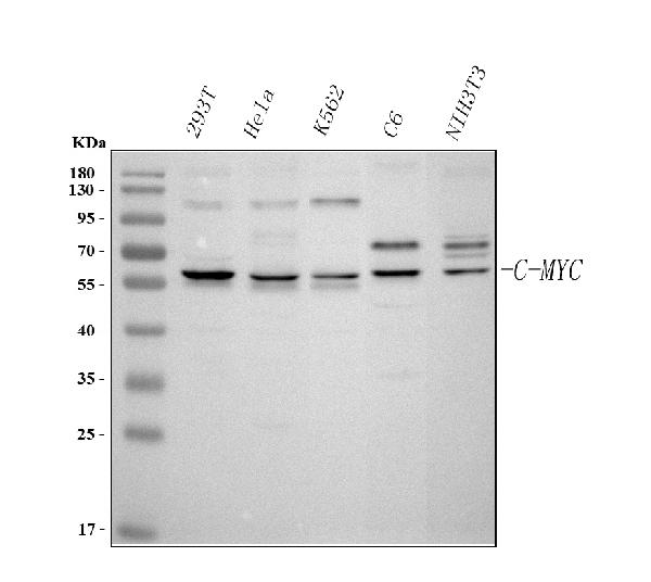
Figure 1. Western blot analysis of c-Myc using anti-c-Myc antibody (PB9092).
Electrophoresis was performed on a 5-20% SDS-PAGE gel at 70V (Stacking gel) / 90V (Resolving gel) for 2-3 hours. The sample well of each lane was loaded with 30 ug of sample under reducing conditions.
Lane 1: human 293T whole cell lysates,
Lane 2: human Hela whole cell lysates,
Lane 3: human K562 whole cell lysates,
Lane 4: rat C6 whole cell lysates,
Lane 5: mouse NIH/3T3 whole cell lysates.
After electrophoresis, proteins were transferred to a nitrocellulose membrane at 150 mA for 50-90 minutes. Blocked the membrane with 5% non-fat milk/TBS for 1.5 hour at RT. The membrane was incubated with rabbit anti-c-Myc antigen affinity purified polyclonal antibody (Catalog # PB9092) at 0.5 μg/mL overnight at 4°C, then washed with TBS-0.1%Tween 3 times with 5 minutes each and probed with a goat anti-rabbit IgG-HRP secondary antibody at a dilution of 1:5000 for 1.5 hour at RT. The signal is developed using an Enhanced Chemiluminescent detection (ECL) kit (Catalog # EK1002) with Tanon 5200 system. A specific band was detected for c-Myc at approximately 60-65 kDa. The expected band size for c-Myc is at 49 kDa.

Click image to see more details
Figure 2. IF analysis of c-Myc using anti-c-Myc antibody (PB9092) and anti-Beta Tubulin antibody (M01857-3).
c-Myc was detected in immunocytochemical section of Hela cell. Enzyme antigen retrieval was performed using IHC enzyme antigen retrieval reagent (AR0022) for 15 mins. The cells were blocked with 10% goat serum. And then incubated with 5 μg/mL rabbit anti-c-Myc Antibody (PB9092) and mouse anti-Beta Tubulin antibody (M01857-3) overnight at 4°C. Cy3 Conjugated Goat Anti-Rabbit IgG (BA1032) and DyLight®488 Conjugated Goat Anti-Mouse IgG (BA1126) were used as secondary antibody at 1:500 dilution and incubated for 30 minutes at 37°C. Visualize using a fluorescence microscope and filter sets appropriate for the label used.
Protein Target Info & Infographic
Gene/Protein Information For MYC (Source: Uniprot.org, NCBI)
Gene Name
MYC
Full Name
Myc proto-oncogene protein
Weight
48.804kDa
Alternative Names
9E10 Myc; 9E10 Myc; avian myelocytomatosis viral oncogene homolog; BHLHE39; bHLHe39MRTL; Class E basic helix-loop-helix protein 39; cMyc; c-Myc; myc proto-oncogene protein; Myc; Myc2; MYCC; myc-related translation/localization regulatory factor; Niard; Nird; Proto-oncogene c-Myc; Transcription factor p64; v-myc avian myelocytomatosis viral oncogene homolog; v-myc myelocytomatosis viral oncogene homolog (avian) MYC MRTLC, bHLHe39, c-Myc, MYC MYC proto-oncogene, bHLH transcription factor myc proto-oncogene protein|avian myelocytomatosis viral oncogene homolog|class E basic helix-loop-helix protein 39|myc-related translation/localization regulatory factor|proto-oncogene c-Myc|transcription factor p64|v-myc avian myelocytomatosis viral oncogene homolog|v-myc myelocytomatosis viral oncogene homolog
*If product is indicated to react with multiple species, protein info is based on the gene entry specified above in "Species".For more info on MYC, check out the MYC Infographic

We have 30,000+ of these available, one for each gene! Check them out.
In this infographic, you will see the following information for MYC: database IDs, superfamily, protein function, synonyms, molecular weight, chromosomal locations, tissues of expression, subcellular locations, post-translational modifications, and related diseases, research areas & pathways. If you want to see more information included, or would like to contribute to it and be acknowledged, please contact [email protected].
Specific Publications For Anti-c-Myc Antibody Picoband™ (PB9092)
Hello CJ!
PB9092 has been cited in 19 publications:
*The publications in this section are manually curated by our staff scientists. They may differ from Bioz's machine gathered results. Both are accurate. If you find a publication citing this product but is missing from this list, please let us know we will issue you a thank-you coupon.
Let-7a microRNA functions as a potential tumor suppressor in human laryngeal cancer
Positional and expressive alteration of prohibitin during the induced differentiation of human hepatocarcinoma SMMC-7721 cells
Long non-coding RNA LINC01503 predicts worse prognosis in glioma and promotes tumorigenesis and progression through activation of Wnt/β-catenin signaling H Wang 1, Z-G Sheng, L-Z Dai Eur Rev Med Pharmacol Sci. 2019 Feb;23(4):1600-1609. doi: 10.26355/e
Inhibition of N-myc downstream-regulated gene 2 in prostatic carcinoma
Tumor suppressor NDRG2 inhibits glycolysis and glutaminolysis in colorectal cancer cells by repressing c-Myc expression
Blockage of Glyoxalase I Inhibits Colorectal Tumorigenesis and Tumor Growth via Upregulation of STAT1, p53, and Bax and Downregulation of c-Myc and Bcl-2
Zhang C, Chang C, Zhao W, Gao H, Wang Q, Li D, Zhang F, Zhang S, Xu C,. PLoS One. 2017 Jul 27;12(7):e0180607. doi: 10.1371/journal.pone.0180607. eCollection 2017. The novel protein C9orf116 promotes rat liver cell line BRL-3A proliferation
Zhang C, Chang C, Li D, Zhang F, Xu C. Cell Mol Biol Lett. 2017 Sep 16;22:21. doi: 10.1186/s11658-017-0051-3. eCollection 2017. The novel protein C3orf43 accelerates hepatocyte proliferation
Wang Ph, Gu Zh, Wan Dh, Zhu Wb, Qiu W, Weng Sp, Yu Xq, He Jg. Plos One. 2013;8(2):E52088. Doi: 10.1371/Journal.Pone.0052088. Epub 2013 Feb 6. Litopenaeus Vannamei Sterile-Alpha And Armadillo Motif Containing Protein (Lvsarm) Is Involved In Regulat...
Long Xb, Sun Gb, Hu S, Liang Gt, Wang N, Zhang Xh, Cao Pp, Zhen Ht, Cui Yh, Liu Z. Oncol Rep. 2009 Nov;22(5):1189-95. Let-7A Microrna Functions As A Potential Tumor Suppressor In Human Laryngeal Cancer.
Recommended Resources
Here are featured tools and databases that you might find useful.
- Boster's Pathways Library
- Protein Databases
- Bioscience Research Protocol Resources
- Data Processing & Analysis Software
- Photo Editing Software
- Scientific Literature Resources
- Research Paper Management Tools
- Molecular Biology Software
- Primer Design Tools
- Bioinformatics Tools
- Phylogenetic Tree Analysis
Customer Reviews
Have you used Anti-c-Myc Antibody Picoband™?
Submit a review and receive an Amazon gift card.
- $30 for a review with an image
Be the first to review Anti-c-Myc Antibody Picoband™
*The first user to submit a review for a product is eligible for Boster's Innovating Scientists Reward, which gives product credits. This is in addition to the gift card reward.
Customer Q&As
Have a question?
Find answers in Q&As, reviews.
Can't find your answer?
Submit your question
5 Customer Q&As for Anti-c-Myc Antibody Picoband™
Question
We bought anti-c-Myc antibody for Flow Cytometry on cervix last year. I am using human, and We intend to use the antibody for IHC-P next. I would like examining cervix as well as uterus in our next experiment. Could you please give me some suggestion on which antibody would work the best for IHC-P?
Verified Customer
Verified customer
Asked: 2019-11-05
Answer
I have checked the website and datasheets of our anti-c-Myc antibody and it appears that PB9092 has been validated on human in both Flow Cytometry and IHC-P. Thus PB9092 should work for your application. Our Boster satisfaction guarantee will cover this product for IHC-P in human even if the specific tissue type has not been validated. We do have a comprehensive range of products for IHC-P detection and you can check out our website bosterbio.com to find out more information about them.
Boster Scientific Support
Answered: 2019-11-05
Question
My colleagues were happy with the WB result of your anti-c-Myc antibody. However we have been able to see positive staining in promyelocytic leukemia nucleus using this antibody. Is that expected? Could you tell me where is MYC supposed to be expressed?
L. Johnson
Verified customer
Asked: 2018-11-22
Answer
According to literature, promyelocytic leukemia does express MYC. Generally MYC expresses in nucleus, nucleoplasm. Regarding which tissues have MYC expression, here are a few articles citing expression in various tissues:
Cervix carcinoma, Pubmed ID: 17081983, 18669648, 20068231
Cervix carcinoma, and Erythroleukemia, Pubmed ID: 23186163
Cervix, Placenta, and Testis, Pubmed ID: 15489334
Promyelocytic leukemia, Pubmed ID: 3540591
Uterus, Pubmed ID: 14702039
Boster Scientific Support
Answered: 2018-11-22
Question
We have seen staining in rat promyelocytic leukemia. Do you have any suggestions? Is anti-c-Myc antibody supposed to stain promyelocytic leukemia positively?
L. Brown
Verified customer
Asked: 2017-12-08
Answer
From literature promyelocytic leukemia does express MYC. From Uniprot.org, MYC is expressed in thoracic mammary gland, uterus, cervix, placenta testis, promyelocytic leukemia, cervix carcinoma, cervix carcinoma erythroleukemia, among other tissues. Regarding which tissues have MYC expression, here are a few articles citing expression in various tissues:
Cervix carcinoma, Pubmed ID: 17081983, 18669648, 20068231
Cervix carcinoma, and Erythroleukemia, Pubmed ID: 23186163
Cervix, Placenta, and Testis, Pubmed ID: 15489334
Promyelocytic leukemia, Pubmed ID: 3540591
Uterus, Pubmed ID: 14702039
Boster Scientific Support
Answered: 2017-12-08
Question
We are currently using anti-c-Myc antibody PB9092 for rat tissue, and we are content with the IHC-P results. The species of reactivity given in the datasheet says human, rat. Is it likely that the antibody can work on horse tissues as well?
Verified Customer
Verified customer
Asked: 2017-08-21
Answer
The anti-c-Myc antibody (PB9092) has not been tested for cross reactivity specifically with horse tissues, though there is a good chance of cross reactivity. We have an innovator award program that if you test this antibody and show it works in horse you can get your next antibody for free. Please contact me if I can help you with anything.
Boster Scientific Support
Answered: 2017-08-21
Question
We are currently using anti-c-Myc antibody PB9092 for rat tissue, and we are content with the IHC-P results. The species of reactivity given in the datasheet says human, rat. Is it likely that the antibody can work on horse tissues as well?
M. Edwards
Verified customer
Asked: 2016-05-03
Answer
The anti-c-Myc antibody (PB9092) has not been tested for cross reactivity specifically with horse tissues, though there is a good chance of cross reactivity. We have an innovator award program that if you test this antibody and show it works in horse you can get your next antibody for free. Please contact me if I can help you with anything.
Boster Scientific Support
Answered: 2016-05-03
