
Click image to see more details
Product Info Summary
| SKU: | M00213-8 |
|---|---|
| Size: | 100 μg/vial |
| Reactive Species: | Human, Mouse, Rat |
| Host: | Mouse |
| Application: | IF, IHC, WB |
Customers Who Bought This Also Bought

Product info
Product Name
Anti-GFAP Antibody Picoband™ (monoclonal, 3F2)
SKU/Catalog Number
M00213-8
Size
100 μg/vial
Form
Lyophilized
Description
Boster Bio Anti-GFAP Antibody Picoband™ (monoclonal, 3F2) catalog # M00213-8. Tested in IF, IHC, WB applications. This antibody reacts with Human, Mouse, Rat.
Storage & Handling
Store at -20˚C for one year from date of receipt. After reconstitution, at 4˚C for one month. It can also be aliquotted and stored frozen at -20˚C for six months. Avoid repeated freeze-thaw cycles.
Cite This Product
Anti-GFAP Antibody Picoband™ (monoclonal, 3F2) (Boster Biological Technology, Pleasanton CA, USA, Catalog # M00213-8)
Host
Mouse
Contents
Each vial contains 4mg Trehalose, 0.9mg NaCl, 0.2mg Na2HPO4, 0.05mg NaN3.
Clonality
Monoclonal
Clone Number
3F2
Isotype
Mouse IgG1
Immunogen
E.coli-derived human GFAP recombinant protein (Position: Q93-M432). Human GFAP shares 94% amino acid (aa) sequence identity with both mouse and rat GFAP.
*Blocking peptide can be purchased. Costs vary based on immunogen length. Contact us for pricing.
Cross-reactivity
No cross-reactivity with other proteins.
Reactive Species
M00213-8 is reactive to GFAP in Human, Mouse, Rat
Applications
M00213-8 is guaranteed for IF, IHC, WB Boster Guarantee
Observed Molecular Weight
50 kDa
Calculated molecular weight
49.88kDa
Background of GFAP
Glial fibrillary acidic protein (GFAP) is a protein that is encoded by the GFAP gene in humans. It is an intermediate filament (IF) protein that is expressed by numerous cell types of the central nervous system (CNS) including astrocytes, and ependymal cells. It is mapped to 17q21.31. GFAP is closely related to its non-epithelial family members, vimentin, desmin, and peripherin, which are all involved in the structure and function of the cell’s cytoskeleton. GFAP is thought to help to maintain astrocyte mechanical strength, as well as the shape of cells. This gene has been shown to play a role in mitosis by adjusting the filament network present in the cell. GFAP is necessary for many critical roles in the CNS. What’s more, GFAP also plays a role in astrocyte-neuron interactions as well as cell-cell communication.
Antibody Validation
Boster validates all antibodies on WB, IHC, ICC, Immunofluorescence, and ELISA with known positive control and negative samples to ensure specificity and high affinity, including thorough antibody incubations.
Innovating Scientists Reward
If you are the first to review this product, or if you have results for a special sample, species or application this product is not validated in, share your results with us and receive product credits you can use towards any Boster products! Applicable to all scientists worldwide.
Submit A Review
Assay dilution & Images
Reconsitution
Add 0.2ml of distilled water will yield a concentration of 500μg/ml.
Assay Dilutions Recommendation
The recommendations below provide a starting point for assay optimization. The actual working concentration varies and should be decided by the user.
Western blot, 0.1-0.5μg/ml, Mouse, rat
Immunohistochemistry (Paraffin-embedded Section), 0.5-1μg/ml, Human, Rat
Immunofluorescence, 5 μg/ml, Rat
Validation Images & Assay Conditions
Click image to see more details
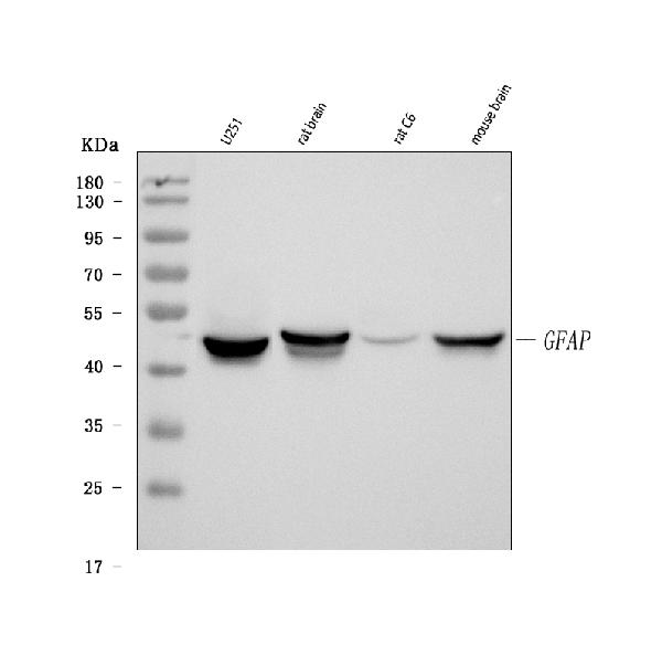
Figure 1. Western blot analysis of GFAP using anti-GFAP antibody (M00213-8).
Electrophoresis was performed on a 5-20% SDS-PAGE gel at 70V (Stacking gel) / 90V (Resolving gel) for 2-3 hours. The sample well of each lane was loaded with 50ug of sample under reducing conditions.
Lane 1: rat brain tissue lysates,
Lane 2: mouse brain tissue lysates.
After Electrophoresis, proteins were transferred to a Nitrocellulose membrane at 150mA for 50-90 minutes. Blocked the membrane with 5% Non-fat Milk/ TBS for 1.5 hour at RT. The membrane was incubated with mouse anti-GFAP antigen affinity purified monoclonal antibody (Catalog # M00213-8) at 0.5 μg/mL overnight at 4°C, then washed with TBS-0.1%Tween 3 times with 5 minutes each and probed with a goat anti-mouse IgG-HRP secondary antibody at a dilution of 1:10000 for 1.5 hour at RT. The signal is developed using an Enhanced Chemiluminescent detection (ECL) kit (Catalog # EK1001) with Tanon 5200 system. A specific band was detected for GFAP at approximately 50KD. The expected band size for GFAP is at 50KD.

Click image to see more details
Figure 2. IHC analysis of GFAP using anti-GFAP antibody (M00213-8).
GFAP was detected in paraffin-embedded section of human glioma tissue. Heat mediated antigen retrieval was performed in EDTA buffer (pH8.0, epitope retrieval solution). The tissue section was blocked with 10% goat serum. The tissue section was then incubated with 1μg/ml mouse anti-GFAP Antibody (M00213-8) overnight at 4°C. Biotinylated goat anti-mouse IgG was used as secondary antibody and incubated for 30 minutes at 37°C. The tissue section was developed using Strepavidin-Biotin-Complex (SABC) (Catalog # SA1021) with DAB as the chromogen.

Click image to see more details
Figure 3. IHC analysis of GFAP using anti-GFAP antibody (M00213-8).
GFAP was detected in paraffin-embedded section of rat brain tissue. Heat mediated antigen retrieval was performed in EDTA buffer (pH8.0, epitope retrieval solution). The tissue section was blocked with 10% goat serum. The tissue section was then incubated with 1μg/ml mouse anti-GFAP Antibody (M00213-8) overnight at 4°C. Biotinylated goat anti-mouse IgG was used as secondary antibody and incubated for 30 minutes at 37°C. The tissue section was developed using Strepavidin-Biotin-Complex (SABC) (Catalog # SA1021) with DAB as the chromogen.

Click image to see more details
Figure 4. IF analysis of Histone H3 and GFAP using anti-Histone H3 antibody (A12477-2) and anti-GFAP antibody (M00213-8).
Histone H3 and GFAP was detected in a paraffin-embedded section of rat brain tissue. Heat mediated antigen retrieval was performed in EDTA buffer (pH 8.0, epitope retrieval solution). The tissue section was blocked with 10% goat serum. The tissue section was then incubated with 5 μg/mL rabbit anti-Histone H3 antibody (A12477-2) and mouse anti-GFAP antibody (M00213-8) overnight at 4°C. DyLight®488 Conjugated Goat Anti-Rabbit IgG (BA1127), Cy3 Conjugated Goat Anti-Mouse IgG (BA1031) was used as secondary antibody at 1:100 dilution and incubated for 30 minutes at 37°C. The section was counterstained with DAPI. Visualize using a fluorescence microscope and filter sets appropriate for the label used.
Protein Target Info & Infographic
Gene/Protein Information For GFAP (Source: Uniprot.org, NCBI)
Gene Name
GFAP
Full Name
Glial fibrillary acidic protein
Weight
49.88kDa
Superfamily
intermediate filament family
Alternative Names
FLJ45472; GFAP astrocytes; GFAP immunohistochemistry; GFAP mouse; GFAP rabbit; GFAP stain; GFAP; glial fibrillary acidic protein GFAP ALXDRD glial fibrillary acidic protein glial fibrillary acidic protein
*If product is indicated to react with multiple species, protein info is based on the gene entry specified above in "Species".For more info on GFAP, check out the GFAP Infographic

We have 30,000+ of these available, one for each gene! Check them out.
In this infographic, you will see the following information for GFAP: database IDs, superfamily, protein function, synonyms, molecular weight, chromosomal locations, tissues of expression, subcellular locations, post-translational modifications, and related diseases, research areas & pathways. If you want to see more information included, or would like to contribute to it and be acknowledged, please contact [email protected].
Specific Publications For Anti-GFAP Antibody Picoband™ (monoclonal, 3F2) (M00213-8)
Hello CJ!
M00213-8 has been cited in 44 publications:
*The publications in this section are manually curated by our staff scientists. They may differ from Bioz's machine gathered results. Both are accurate. If you find a publication citing this product but is missing from this list, please let us know we will issue you a thank-you coupon.
β-elemene inhibits stemness, promotes differentiation and impairs chemoresistance to temozolomide in glioblastoma stem-like cells
Effect of Corilagin on the Proliferation and NF-κB in U251 Glioblastoma Cells and U251 Glioblastoma Stem-Like Cells
3D bioprinted glioma stem cells for brain tumor model and applications of drug susceptibility
NDGA-P21, a novel derivative of nordihydroguaiaretic acid, inhibits glioma cell proliferation and stemness
Increased expression of calponin-3 in epileptic patients and experimental rats
Decreased expression of Ras-GRF1 in the brain tissue of the intractable epilepsy patients and experimental rats
Lentiviral Vector-Induced Overexpression of RGMa in the Hippocampus Suppresses Seizures and Mossy Fiber Sprouting
Elevated Expression of the Delta-Subunit of Epithelial Sodium Channel in Temporal Lobe Epilepsy Patients and Rat Model
Decreased expression of Gab2 in patients with temporal lobe epilepsy and pilocarpine‐induced rat model
Liu Y,Li L,Qiu M,Tan L,Zhang M,Li J,Zhu H,Jiang S,Su X,Li A.Renal and cerebral RAS interaction contributes to diabetic kidney disease.Am J Transl Res.2019 May 15;11(5):2925-2939.PMID:31217864;PMCID:PMC6556645.
Species: Rat
M00213-8 usage in article: APP:IF, SAMPLE:BRAIN TISSUE, DILUTION:NA
Recommended Resources
Here are featured tools and databases that you might find useful.
- Boster's Pathways Library
- Protein Databases
- Bioscience Research Protocol Resources
- Data Processing & Analysis Software
- Photo Editing Software
- Scientific Literature Resources
- Research Paper Management Tools
- Molecular Biology Software
- Primer Design Tools
- Bioinformatics Tools
- Phylogenetic Tree Analysis
Customer Reviews
Have you used Anti-GFAP Antibody Picoband™ (monoclonal, 3F2)?
Submit a review and receive an Amazon gift card.
- $30 for a review with an image
Be the first to review Anti-GFAP Antibody Picoband™ (monoclonal, 3F2)
*The first user to submit a review for a product is eligible for Boster's Innovating Scientists Reward, which gives product credits. This is in addition to the gift card reward.
Customer Q&As
Have a question?
Find answers in Q&As, reviews.
Can't find your answer?
Submit your question
4 Customer Q&As for Anti-GFAP Antibody Picoband™ (monoclonal, 3F2)
Question
We are currently using anti-GFAP in antibody (monoclonal, 3F2) M00213-8 for rat tissue, and we are well pleased with the IHC-P results. The species of reactivity given in the datasheet says human, mouse, rat. Is it likely that the antibody can work on pig tissues as well?
Verified Customer
Verified customer
Asked: 2020-03-04
Answer
The anti-GFAP in antibody (monoclonal, 3F2) (M00213-8) has not been validated for cross reactivity specifically with pig tissues, though there is a good chance of cross reactivity. We have an innovator award program that if you test this antibody and show it works in pig you can get your next antibody for free. Please contact me if I can help you with anything.
Boster Scientific Support
Answered: 2020-03-04
Question
My team were content with the WB result of your anti-GFAP in antibody (monoclonal, 3F2). However we have observed positive staining in blood cytoplasm. using this antibody. Is that expected? Could you tell me where is GFAP supposed to be expressed?
Verified Customer
Verified customer
Asked: 2019-11-28
Answer
From what I have seen in literature, blood does express GFAP. Generally GFAP expresses in cytoplasm. Regarding which tissues have GFAP expression, here are a few articles citing expression in various tissues:
Blood, Pubmed ID: 12837269
Brain, Pubmed ID: 15489334
Brain, and Thalamus, Pubmed ID: 14702039
Fetal brain, Pubmed ID: 12058025
Fetal brain cortex, Pubmed ID: 2780570
Kidney, Pubmed ID: 17974005
Boster Scientific Support
Answered: 2019-11-28
Question
We have tried in the past anti-GFAP in antibody (monoclonal, 3F2) for IHC-P on dorsal motor nucleus of vagus nerve in a previous project. I am using rat, and We are going to use the antibody for WB next. Our lab want to know about examining dorsal motor nucleus of vagus nerve as well as brain thalamus in our next experiment. Could you please give me some suggestion on which antibody would work the best for WB?
B. Krishna
Verified customer
Asked: 2017-02-23
Answer
I viewed the website and datasheets of our anti-GFAP in antibody (monoclonal, 3F2) and I see that M00213-8 has been tested on rat in both IHC-P and WB. Thus M00213-8 should work for your application. Our Boster satisfaction guarantee will cover this product for WB in rat even if the specific tissue type has not been validated. We do have a comprehensive range of products for WB detection and you can check out our website bosterbio.com to find out more information about them.
Boster Scientific Support
Answered: 2017-02-23
Question
We have seen staining in rat fetal brain. Any tips? Is anti-GFAP in antibody (monoclonal, 3F2) supposed to stain fetal brain positively?
E. Wu
Verified customer
Asked: 2016-07-27
Answer
According to literature fetal brain does express GFAP. According to Uniprot.org, GFAP is expressed in dorsal motor nucleus of vagus nerve, brain thalamus, brain, kidney, fetal brain cortex, fetal brain, blood, among other tissues. Regarding which tissues have GFAP expression, here are a few articles citing expression in various tissues:
Blood, Pubmed ID: 12837269
Brain, Pubmed ID: 15489334
Brain, and Thalamus, Pubmed ID: 14702039
Fetal brain, Pubmed ID: 12058025
Fetal brain cortex, Pubmed ID: 2780570
Kidney, Pubmed ID: 17974005
Boster Scientific Support
Answered: 2016-07-27
