Click image to see more details
-

-
-
-
-
+6
Product Info Summary
| SKU: | M00066-1 |
|---|---|
| Size: | 100 μl |
| Reactive Species: | Human, Mouse, Rat |
| Host: | Rabbit |
| Application: | Flow Cytometry, IF, IHC, ICC, WB |
Customers Who Bought This Also Bought


Product info
Product Name
Anti-HMGB1/Hmg 1 Rabbit Monoclonal Antibody
View all HMGB1/HMG-1 Antibodies
SKU/Catalog Number
M00066-1
Size
100 μl
Form
Liquid
Description
Boster Bio Anti-HMGB1/Hmg 1 Rabbit Monoclonal Antibody catalog # M00066-1. Tested in WB, IHC, ICC/IF, Flow Cytometry applications. This antibody reacts with Human, Mouse, Rat.
Storage & Handling
Store at -20°C for one year. For short term storage and frequent use, store at 4°C for up to one month. Avoid repeated freeze-thaw cycles.
Cite This Product
Anti-HMGB1/Hmg 1 Rabbit Monoclonal Antibody (Boster Biological Technology, Pleasanton CA, USA, Catalog # M00066-1)
Host
Rabbit
Contents
Rabbit IgG in phosphate buffered saline, pH 7.4, 150mM NaCl, 0.02% sodium azide and 50% glycerol, 0.4-0.5mg/ml BSA.
Clonality
Monoclonal
Clone Number
AAC-8
Isotype
Rabbit IgG
Immunogen
A synthesized peptide derived from human HMGB1
*Blocking peptide can be purchased. Costs vary based on immunogen length. Contact us for pricing.
Reactive Species
M00066-1 is reactive to HMGB1 in Human, Mouse, Rat
Applications
M00066-1 is guaranteed for Flow Cytometry, IF, IHC, ICC, WB Boster Guarantee
Observed Molecular Weight
25 kDa
Calculated molecular weight
24.894kDa
Background of HMGB1/HMG-1
Phosphoinositide-3-kinase (PI3K) that phosphorylates PtdIns (Phosphatidylinositol), PtdIns4P (Phosphatidylinositol 4-phosphate) and PtdIns (4,5) P2 (Phosphatidylinositol 4,5-bisphosphate) to generate phosphatidylinositol 3,4,5-trisphosphate (PIP3) . PIP3 plays a key role by recruiting PH domain-containing proteins to the membrane, including AKT1 and PDPK1, activating signaling cascades involved in cell growth, survival, proliferation, motility and morphology. Participates in cellular signaling in response to various growth factors.
Antibody Validation
Boster validates all antibodies on WB, IHC, ICC, Immunofluorescence, and ELISA with known positive control and negative samples to ensure specificity and high affinity, including thorough antibody incubations.
Innovating Scientists Reward
If you are the first to review this product, or if you have results for a special sample, species or application this product is not validated in, share your results with us and receive product credits you can use towards any Boster products! Applicable to all scientists worldwide.
Submit A Review
Assay dilution & Images
Reconsitution
Restore with deionized water (or equivalent) for reconstitution volume of 1.0 mL
Assay Dilutions Recommendation
The recommendations below provide a starting point for assay optimization. The actual working concentration varies and should be decided by the user.
WB 1:1000-1:2000
IHC 1:50-1:100
ICC/IF 1:50-1:100
FC 1:30
Validation Images & Assay Conditions
Click image to see more details
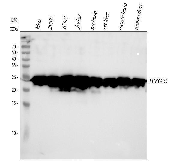
Figure 1. Western blot analysis of HMGB1 using anti-HMGB1 antibody (M00066-1).
Electrophoresis was performed on a 5-20% SDS-PAGE gel at 70V (Stacking gel) / 90V (Resolving gel) for 2-3 hours. The sample well of each lane was loaded with 30 ug of sample under reducing conditions.
Lane 1: human Hela whole cell lysates,
Lane 2: human 293T whole cell lysates,
Lane 3: human K562 whole cell lysates,
Lane 4: human Jurkat whole cell lysates,
Lane 5: rat brain tissue lysates,
Lane 6: rat liver tissue lysates,
Lane 7: mouse brain tissue lysates,
Lane 8: mouse liver tissue lysates.
After electrophoresis, proteins were transferred to a nitrocellulose membrane at 150 mA for 50-90 minutes. Blocked the membrane with 5% non-fat milk/TBS for 1.5 hour at RT. The membrane was incubated with rabbit anti-HMGB1 antigen affinity purified monoclonal antibody (Catalog # M00066-1) at 1:1000 overnight at 4°C, then washed with TBS-0.1%Tween 3 times with 5 minutes each and probed with a goat anti-rabbit IgG-HRP secondary antibody at a dilution of 1:1000 for 1.5 hour at RT. The signal is developed using an Enhanced Chemiluminescent detection (ECL) kit (Catalog # EK1002) with Tanon 5200 system. A specific band was detected for HMGB1 at approximately 25 kDa. The expected band size for HMGB1 is at 25 kDa.

Click image to see more details
Figure 2. IHC analysis of HMGB1 using anti-HMGB1 antibody (M00066-1).
HMGB1 was detected in a paraffin-embedded section of human colorectal adenocarcinoma tissue. Heat mediated antigen retrieval was performed in EDTA buffer (pH 8.0, epitope retrieval solution). The tissue section was blocked with 10% goat serum. The tissue section was then incubated with 1:50 rabbit anti-HMGB1 Antibody (M00066-1) overnight at 4°C. Peroxidase Conjugated Goat Anti-rabbit IgG was used as secondary antibody and incubated for 30 minutes at 37°C. The tissue section was developed using HRP Conjugated Rabbit IgG Super Vision Assay Kit (Catalog # SV0002) with DAB as the chromogen.

Click image to see more details
Figure 3. IHC analysis of HMGB1 using anti-HMGB1 antibody (M00066-1).
HMGB1 was detected in a paraffin-embedded section of human lung adenocarcinoma tissue. Heat mediated antigen retrieval was performed in EDTA buffer (pH 8.0, epitope retrieval solution). The tissue section was blocked with 10% goat serum. The tissue section was then incubated with 1:50 rabbit anti-HMGB1 Antibody (M00066-1) overnight at 4°C. Peroxidase Conjugated Goat Anti-rabbit IgG was used as secondary antibody and incubated for 30 minutes at 37°C. The tissue section was developed using HRP Conjugated Rabbit IgG Super Vision Assay Kit (Catalog # SV0002) with DAB as the chromogen.

Click image to see more details
Figure 4. IHC analysis of HMGB1 using anti-HMGB1 antibody (M00066-1).
HMGB1 was detected in a paraffin-embedded section of human spleen tissue. Heat mediated antigen retrieval was performed in EDTA buffer (pH 8.0, epitope retrieval solution). The tissue section was blocked with 10% goat serum. The tissue section was then incubated with 1:50 rabbit anti-HMGB1 Antibody (M00066-1) overnight at 4°C. Peroxidase Conjugated Goat Anti-rabbit IgG was used as secondary antibody and incubated for 30 minutes at 37°C. The tissue section was developed using HRP Conjugated Rabbit IgG Super Vision Assay Kit (Catalog # SV0002) with DAB as the chromogen.

Click image to see more details
Figure 5. IHC analysis of HMGB1 using anti-HMGB1 antibody (M00066-1).
HMGB1 was detected in a paraffin-embedded section of human prostatic acinar adenocarcinoma tissue. Heat mediated antigen retrieval was performed in EDTA buffer (pH 8.0, epitope retrieval solution). The tissue section was blocked with 10% goat serum. The tissue section was then incubated with 1:50 rabbit anti-HMGB1 Antibody (M00066-1) overnight at 4°C. Peroxidase Conjugated Goat Anti-rabbit IgG was used as secondary antibody and incubated for 30 minutes at 37°C. The tissue section was developed using HRP Conjugated Rabbit IgG Super Vision Assay Kit (Catalog # SV0002) with DAB as the chromogen.

Click image to see more details
Figure 6. IHC analysis of HMGB1 using anti-HMGB1 antibody (M00066-1).
HMGB1 was detected in a paraffin-embedded section of human cervical squamous carcinoma tissue. Heat mediated antigen retrieval was performed in EDTA buffer (pH 8.0, epitope retrieval solution). The tissue section was blocked with 10% goat serum. The tissue section was then incubated with 1:50 rabbit anti-HMGB1 Antibody (M00066-1) overnight at 4°C. Peroxidase Conjugated Goat Anti-rabbit IgG was used as secondary antibody and incubated for 30 minutes at 37°C. The tissue section was developed using HRP Conjugated Rabbit IgG Super Vision Assay Kit (Catalog # SV0002) with DAB as the chromogen.

Click image to see more details
Figure 7. IF analysis of HMGB1 using anti-HMGB1 antibody (M00066-1) and anti-Beta Tubulin antibody (M01857-3).
HMGB1 was detected in immunocytochemical section of Hela cell. Enzyme antigen retrieval was performed using IHC enzyme antigen retrieval reagent (AR0022) for 15 mins. The cells were blocked with 10% goat serum. And then incubated at 1:50 with rabbit anti-HMGB1 Antibody (M00066-1) and mouse anti-Beta Tubulin antibody (M01857-3) overnight at 4°C. DyLight®488 Conjugated Goat Anti-Rabbit IgG (BA1127) and Cy3 Conjugated Goat Anti-Mouse IgG (BA1031) were used as secondary antibody at 1:500 dilution and incubated for 30 minutes at 37°C. Visualize using a fluorescence microscope and filter sets appropriate for the label used.

Click image to see more details
Immunohistochemical analysis of paraffin-embedded Rat liver, using the Antibody at 1:300 dilution.

Click image to see more details
Immunohistochemical analysis of paraffin-embedded Rat kidney, using the Antibody at 1:300 dilution.

Click image to see more details
Immunohistochemical analysis of paraffin-embedded Mouse skeletal muscle - gastrocnemius , using the Antibody at 1:300 dilution.
Protein Target Info & Infographic
Gene/Protein Information For HMGB1 (Source: Uniprot.org, NCBI)
Gene Name
HMGB1
Full Name
High mobility group protein B1
Weight
24.894kDa
Superfamily
HMGB family
Alternative Names
Amphoterin; high mobility group box 1; High mobility group protein 1; high mobility group protein B1; high-mobility group (nonhistone chromosomal) protein 1; high-mobility group box 1; HMG1; HMG-1; HMG1DKFZp686A04236; HMG3; HMGB1; SBP-1; Sulfoglucuronyl carbohydrate binding protein HMGB1 HMG-1, HMG1, HMG3, SBP-1 high mobility group box 1 high mobility group protein B1|Amphoterin|Sulfoglucuronyl carbohydrate binding protein|high-mobility group (nonhistone chromosomal) protein 1
*If product is indicated to react with multiple species, protein info is based on the gene entry specified above in "Species".For more info on HMGB1, check out the HMGB1 Infographic

We have 30,000+ of these available, one for each gene! Check them out.
In this infographic, you will see the following information for HMGB1: database IDs, superfamily, protein function, synonyms, molecular weight, chromosomal locations, tissues of expression, subcellular locations, post-translational modifications, and related diseases, research areas & pathways. If you want to see more information included, or would like to contribute to it and be acknowledged, please contact [email protected].
Specific Publications For Anti-HMGB1/Hmg 1 Rabbit Monoclonal Antibody (M00066-1)
Hello CJ!
M00066-1 has been cited in 5 publications:
*The publications in this section are manually curated by our staff scientists. They may differ from Bioz's machine gathered results. Both are accurate. If you find a publication citing this product but is missing from this list, please let us know we will issue you a thank-you coupon.
Wen Y,Sun HY,Tan Z,Liu RH,Huang SQ,Chen GY,Qi H,Tang LJ.Abdominal paracentesis drainage ameliorates myocardial injury in severe experimental pancreatitis rats through suppressing oxidative stress.World J Gastroenterol.2020 Jan 7;26(1):35-54.doi:10.3748/wj
Species: Rat
M00066-1 usage in article: APP:WB, SAMPLE:PAAF CELLS, DILUTION:NA
MicroRNA-205%u20115b inhibits HMGB1 expression in LPS-induced sepsis
Galantamine protects against lipopolysaccharide-induced acute lung injury in rats
Association of Upregulated HMGB1 and c-IAP2 Proteins With Hepatocellular Carcinoma Development and Progression
Yang B, Gao P, Wu X, Yu J, Li Y, Meng R, Li Y, Yan J, Jin X. Exp Ther Med. 2017 Sep;14(3):1975-1982. doi: 10.3892/etm.2017.4774. Epub 2017 Jul 11. Epigallocatechin-3-gallate attenuates neointimal hyperplasia in a rat model of carotid artery injury...
Recommended Resources
Here are featured tools and databases that you might find useful.
- Boster's Pathways Library
- Protein Databases
- Bioscience Research Protocol Resources
- Data Processing & Analysis Software
- Photo Editing Software
- Scientific Literature Resources
- Research Paper Management Tools
- Molecular Biology Software
- Primer Design Tools
- Bioinformatics Tools
- Phylogenetic Tree Analysis
Customer Reviews
Have you used Anti-HMGB1/Hmg 1 Rabbit Monoclonal Antibody?
Submit a review and receive an Amazon gift card.
- $30 for a review with an image
Be the first to review Anti-HMGB1/Hmg 1 Rabbit Monoclonal Antibody
*The first user to submit a review for a product is eligible for Boster's Innovating Scientists Reward, which gives product credits. This is in addition to the gift card reward.
Customer Q&As
Have a question?
Find answers in Q&As, reviews.
Can't find your answer?
Submit your question
16 Customer Q&As for Anti-HMGB1/Hmg 1 Rabbit Monoclonal Antibody
Question
We have seen staining in mouse brain. Any tips? Is anti-HMGB1/Hmg 1 Rabbit Monoclonal antibody supposed to stain brain positively?
Verified Customer
Verified customer
Asked: 2020-04-23
Answer
Based on literature brain does express HMGB1. Based on Uniprot.org, HMGB1 is expressed in kidney, cerebellum, small intestine, brain, cervix testis, mammary carcinoma, cervix carcinoma, cervix carcinoma erythroleukemia, liver, among other tissues. Regarding which tissues have HMGB1 expression, here are a few articles citing expression in various tissues:
Brain, Cervix, and Testis, Pubmed ID: 15489334
Cerebellum, Pubmed ID: 14702039
Cervix carcinoma, Pubmed ID: 18669648, 20068231
Cervix carcinoma, and Erythroleukemia, Pubmed ID: 23186163
Liver, Pubmed ID: 24275569
Mammary carcinoma, Pubmed ID: 9150946
Small intestine, Pubmed ID: 17974005
Boster Scientific Support
Answered: 2020-04-23
Question
Is there a BSA free version of anti-HMGB1/Hmg 1 Rabbit Monoclonal antibody M00066-1 available?
Verified Customer
Verified customer
Asked: 2020-02-20
Answer
I appreciate your recent telephone inquiry. I can confirm that some lots of this anti-HMGB1/Hmg 1 Rabbit Monoclonal antibody M00066-1 are BSA free. For now, these lots are available and we can make a BSA free formula for you free of charge. It will take 3 extra days to prepare. If you require this antibody BSA free again in future, please do not hesitate to contact me and I will be pleased to check which lots we have in stock that are BSA free.
Boster Scientific Support
Answered: 2020-02-20
Question
Does anti-HMGB1/Hmg 1 Rabbit Monoclonal antibody M00066-1 work for ICC with brain?
Verified Customer
Verified customer
Asked: 2020-02-10
Answer
According to the expression profile of brain, HMGB1 is highly expressed in brain. So, it is likely that anti-HMGB1/Hmg 1 Rabbit Monoclonal antibody M00066-1 will work for ICC with brain.
Boster Scientific Support
Answered: 2020-02-10
Question
Our lab used your anti-HMGB1/Hmg 1 Rabbit Monoclonal antibody for Flow Cytometry on cerebellum a few months ago. I am using human, and I plan to use the antibody for IF next. I am interested in examining cerebellum as well as cervix carcinoma in our next experiment. Could you please give me some suggestion on which antibody would work the best for IF?
Verified Customer
Verified customer
Asked: 2020-01-03
Answer
I have checked the website and datasheets of our anti-HMGB1/Hmg 1 Rabbit Monoclonal antibody and I see that M00066-1 has been validated on human in both Flow Cytometry and IF. Thus M00066-1 should work for your application. Our Boster satisfaction guarantee will cover this product for IF in human even if the specific tissue type has not been validated. We do have a comprehensive range of products for IF detection and you can check out our website bosterbio.com to find out more information about them.
Boster Scientific Support
Answered: 2020-01-03
Question
Is a blocking peptide available for product anti-HMGB1/Hmg 1 Rabbit Monoclonal antibody (M00066-1)?
Verified Customer
Verified customer
Asked: 2020-01-02
Answer
We do provide the blocking peptide for product anti-HMGB1/Hmg 1 Rabbit Monoclonal antibody (M00066-1). If you would like to place an order for it please contact [email protected] and make a special request.
Boster Scientific Support
Answered: 2020-01-02
Question
Thanks for helping with my inquiry over the phone. Here are the WB image, lot number and protocol we used for brain using anti-HMGB1/Hmg 1 Rabbit Monoclonal antibody M00066-1. Let me know if you need anything else.
Verified Customer
Verified customer
Asked: 2019-07-16
Answer
Thank you for the data. You have provided everything we needed. Our lab team are working to resolve your inquiry as quickly as possible, and we appreciate your patience and understanding! Please let me know if there is anything you need in the meantime.
Boster Scientific Support
Answered: 2019-07-16
Question
We need to test anti-HMGB1/Hmg 1 Rabbit Monoclonal antibody M00066-1 on rat brain for research purposes, then I may be interested in using anti-HMGB1/Hmg 1 Rabbit Monoclonal antibody M00066-1 for diagnostic purposes as well. Is the antibody suitable for diagnostic purposes?
Verified Customer
Verified customer
Asked: 2019-07-04
Answer
The products we sell, including anti-HMGB1/Hmg 1 Rabbit Monoclonal antibody M00066-1, are only intended for research use. They would not be suitable for use in diagnostic work. If you have the means to develop a product into diagnostic use, and are interested in collaborating with us and develop our product into an IVD product, please contact us for more discussions.
Boster Scientific Support
Answered: 2019-07-04
Question
Is this M00066-1 anti-HMGB1/Hmg 1 Rabbit Monoclonal antibody reactive to the isotypes of HMGB1?
Verified Customer
Verified customer
Asked: 2019-05-16
Answer
The immunogen of M00066-1 anti-HMGB1/Hmg 1 Rabbit Monoclonal antibody is A synthesized peptide derived from human HMGB1. Could you tell me which isotype you are interested in so I can help see if the immunogen is part of this isotype?
Boster Scientific Support
Answered: 2019-05-16
Question
I see that the anti-HMGB1/Hmg 1 Rabbit Monoclonal antibody M00066-1 works with ICC, what is the protocol used to produce the result images on the product page?
Verified Customer
Verified customer
Asked: 2019-04-01
Answer
You can find protocols for ICC on the "support/technical resources" section of our navigation menu. If you have any further questions, please send an email to [email protected]
Boster Scientific Support
Answered: 2019-04-01
Question
Will M00066-1 anti-HMGB1/Hmg 1 Rabbit Monoclonal antibody work on parafin embedded sections? If so, which fixation method do you recommend we use (PFA, paraformaldehyde, other)?
Verified Customer
Verified customer
Asked: 2018-10-31
Answer
As indicated on the product datasheet, M00066-1 anti-HMGB1/Hmg 1 Rabbit Monoclonal antibody as been tested on ICC. It is best to use PFA for fixation because it has better tissue penetration ability. PFA needs to be prepared fresh before use. Long term stored PFA turns into formalin, as the PFA molecules congregate and become formalin.
Boster Scientific Support
Answered: 2018-10-31
Question
My question regarding product M00066-1, anti-HMGB1/Hmg 1 Rabbit Monoclonal antibody. I was wondering if it would be possible to conjugate this antibody with biotin. I would need it to be without BSA or sodium azide. I am planning on using a buffer exchange of sodium azide with PBS only. Would there be problems for me to conjugate the antibody and store it in -20 degrees in small aliquots?
Verified Customer
Verified customer
Asked: 2017-12-18
Answer
We do not recommend storing this antibody with PBS buffer only in -20 degrees. If you want to store it in -20 degrees it is best to add some cryoprotectant like glycerol. If you want carrier free M00066-1 anti-HMGB1/Hmg 1 Rabbit Monoclonal antibody, we can provide it to you in a special formula with trehalose and/or glycerol. These molecules will not interfere with conjugation chemistry and provide a good level of protection for the antibody from degradation. Please be sure to specify this in your purchase order.
Boster Scientific Support
Answered: 2017-12-18
Question
We were satisfied with the WB result of your anti-HMGB1/Hmg 1 Rabbit Monoclonal antibody. However we have seen positive staining in brain nucleus using this antibody. Is that expected? Could you tell me where is HMGB1 supposed to be expressed?
Verified Customer
Verified customer
Asked: 2017-10-19
Answer
Based on literature, brain does express HMGB1. Generally HMGB1 expresses in nucleus. Regarding which tissues have HMGB1 expression, here are a few articles citing expression in various tissues:
Brain, Cervix, and Testis, Pubmed ID: 15489334
Cerebellum, Pubmed ID: 14702039
Cervix carcinoma, Pubmed ID: 18669648, 20068231
Cervix carcinoma, and Erythroleukemia, Pubmed ID: 23186163
Liver, Pubmed ID: 24275569
Mammary carcinoma, Pubmed ID: 9150946
Small intestine, Pubmed ID: 17974005
Boster Scientific Support
Answered: 2017-10-19
Question
Would anti-HMGB1/Hmg 1 Rabbit Monoclonal antibody M00066-1 work on horse IF with brain?
B. Huang
Verified customer
Asked: 2016-06-29
Answer
Our lab technicians have not tested anti-HMGB1/Hmg 1 Rabbit Monoclonal antibody M00066-1 on horse. You can run a BLAST between horse and the immunogen sequence of anti-HMGB1/Hmg 1 Rabbit Monoclonal antibody M00066-1 to see if they may cross-react. If the sequence homology is close, then you can perform a pilot test. Keep in mind that since we have not validated horse samples, this use of the antibody is not covered by our guarantee. However we have an innovator award program that if you test this antibody and show it works in horse brain in IF, you can get your next antibody for free.
Boster Scientific Support
Answered: 2016-06-29
Question
I was wanting to use your anti-HMGB1/Hmg 1 Rabbit Monoclonal antibody for ICC for rat brain on frozen tissues, but I want to know if it has been validated for this particular application. Has this antibody been validated and is this antibody a good choice for rat brain identification?
R. Yang
Verified customer
Asked: 2016-01-08
Answer
You can see on the product datasheet, M00066-1 anti-HMGB1/Hmg 1 Rabbit Monoclonal antibody has been validated for Flow Cytometry, IF, IHC, ICC, WB on human, mouse, rat tissues. We have an innovator award program that if you test this antibody and show it works in rat brain in IHC-frozen, you can get your next antibody for free.
Boster Scientific Support
Answered: 2016-01-08
Question
We are currently using anti-HMGB1/Hmg 1 Rabbit Monoclonal antibody M00066-1 for mouse tissue, and we are well pleased with the Flow Cytometry results. The species of reactivity given in the datasheet says human, mouse, rat. Is it true that the antibody can work on primate tissues as well?
P. Lewis
Verified customer
Asked: 2013-12-12
Answer
The anti-HMGB1/Hmg 1 Rabbit Monoclonal antibody (M00066-1) has not been validated for cross reactivity specifically with primate tissues, but there is a good chance of cross reactivity. We have an innovator award program that if you test this antibody and show it works in primate you can get your next antibody for free. Please contact me if I can help you with anything.
Boster Scientific Support
Answered: 2013-12-12
Question
See attached the WB image, lot number and protocol we used for brain using anti-HMGB1/Hmg 1 Rabbit Monoclonal antibody M00066-1. Please let me know if you require anything else.
L. Patel
Verified customer
Asked: 2013-12-04
Answer
Thank you very much for the data. Our lab team are working to resolve this as quickly as possible, and we appreciate your patience and understanding! You have provided everything we needed. Please let me know if there is anything you need in the meantime.
Boster Scientific Support
Answered: 2013-12-04
