Click image to see more details
-

-
-
-
-
+4
Product Info Summary
| SKU: | M04168-1 |
|---|---|
| Size: | 100 μg/vial |
| Reactive Species: | Human |
| Host: | Mouse |
| Application: | Flow Cytometry, IF, IHC, ICC, WB |
Customers Who Bought This Also Bought

Product info
Product Name
Anti-Hsp105/HSPH1 Antibody Picoband® (monoclonal, 3D10)
SKU/Catalog Number
M04168-1
Size
100 μg/vial
Form
Lyophilized
Description
Boster Bio Anti-Hsp105/HSPH1 Antibody Picoband® (monoclonal, 3D10) catalog # M04168-1. Tested in Flow Cytometry, IF, IHC, ICC, WB applications. This antibody reacts with Human. The brand Picoband indicates this is a premium antibody that guarantees superior quality, high affinity, and strong signals with minimal background in Western blot applications. Only our best-performing antibodies are designated as Picoband, ensuring unmatched performance.
Storage & Handling
Store at -20˚C for one year from date of receipt. After reconstitution, at 4˚C for one month. It can also be aliquotted and stored frozen at -20˚C for six months. Avoid repeated freeze-thaw cycles.
Cite This Product
Anti-Hsp105/HSPH1 Antibody Picoband® (monoclonal, 3D10) (Boster Biological Technology, Pleasanton CA, USA, Catalog # M04168-1)
Host
Mouse
Contents
Each vial contains 4mg Trehalose, 0.9mg NaCl, 0.2mg Na2HPO4, 0.05mg NaN3.
Clonality
Monoclonal
Clone Number
3D10
Isotype
Mouse IgG1
Immunogen
E. coli-derived human Hsp105 recombinant protein (Position: Y653-D858).
Cross-reactivity
No cross-reactivity with other proteins.
Reactive Species
M04168-1 is reactive to HSPH1 in Human
Observed Molecular Weight
105 kDa
Calculated molecular weight
96.9 kDa
Background of HSPH1
HSP105 (HEAT-SHOCK 105/110-KD PROTEIN 1), also called HSPH1 or HSP110, is a protein that in humans is encoded by the HSPH1 gene. Immunohistochemical analysis localizes HSP105 mainly in the cytoplasm. Database analysis indicates that both HSP105 isoforms are highly conserved during evolution. By analysis of radiation hybrids and human/rodent hybrid cell lines, the HSPH1 gene is mapped to chromosome 13. Both HSP105-alpha and HSP105-beta are upregulated in HeLa cells exposed to heat shock. HSP105-alpha, but not HSP105-beta, is also upregulate in response to other cell stresses. Following heat shock, HSP105 relocalizes from a cytoplasmic to perinuclear position. Besides, HSP110 may thus constitute a major determinant for both prognosis and treatment response in colorectal cancer.
Antibody Validation
Boster validates all antibodies on WB, IHC, ICC, Immunofluorescence, and ELISA with known positive control and negative samples to ensure specificity and high affinity, including thorough antibody incubations.
Application & Images
Applications
M04168-1 is guaranteed for Flow Cytometry, IF, IHC, ICC, WB Boster Guarantee
Assay Dilutions Recommendation
The recommendations below provide a starting point for assay optimization. The actual working concentration varies and should be decided by the user.
Western blot, 0.1-0.5μg/ml, Human
Immunohistochemistry (Paraffin-embedded Section), 0.5-1μg/ml, Human
Immunocytochemistry/Immunofluorescence, 2μg/ml, Human
Flow Cytometry (Fixed), 1-3μg/1x106 cells, Human
Positive Control
WB: human Caco-2 whole cell, human K562 whole cell, human A549 whole cell, human HepG2 whole cell, human PANC-1 whole cell, human SGC-7901 whole cell,
IHC: human lung cancer tissue, human mammary cancer tissue, human intestinal cancer tissue
ICC/IF: A431 cell, MCF7 cell
FCM: A431 cell, HepG2 cell
Validation Images & Assay Conditions
Click image to see more details
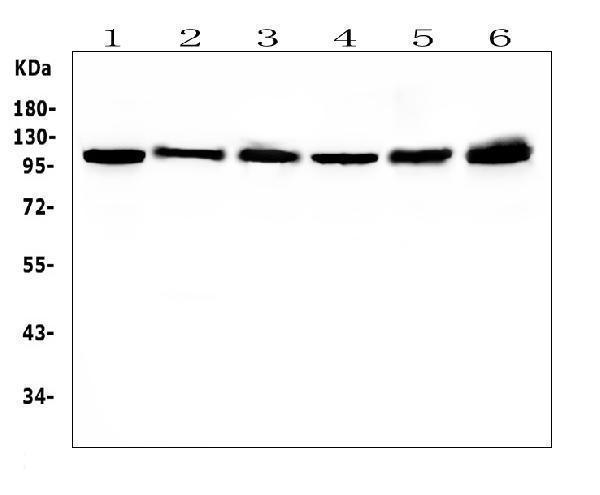
Western blot analysis of HSPH1 using anti-HSPH1 antibody (M04168-1).
Electrophoresis was performed on a 5-20% SDS-PAGE gel at 70V (Stacking gel) / 90V (Resolving gel) for 2-3 hours. The sample well of each lane was loaded with 50ug of sample under reducing conditions.
Lane 1: human Caco-2 whole cell lysates
Lane 2: human K562 whole cell lysates
Lane 3: human A549 whole cell lysates
Lane 4: human HepG2 whole cell lysates
Lane 5: human PANC-1 whole cell lysates
Lane 6: human SGC-7901 whole cell lysates
After Electrophoresis, proteins were transferred to a Nitrocellulose membrane at 150mA for 50-90 minutes. Blocked the membrane with 5% Non-fat Milk/ TBS for 1.5 hour at RT. The membrane was incubated with mouse anti-HSPH1 antigen affinity purified monoclonal antibody (Catalog # M04168-1) at 0.5 μg/mL overnight at 4°C, then washed with TBS-0.1%Tween 3 times with 5 minutes each and probed with a goat anti-mouse IgG-HRP secondary antibody at a dilution of 1:10000 for 1.5 hour at RT. The signal is developed using an Enhanced Chemiluminescent detection (ECL) kit (Catalog # EK1001) with Tanon 5200 system. A specific band was detected for HSPH1 at approximately 105KD. The expected band size for HSPH1 is at 105KD.

Click image to see more details
IHC analysis of HSPH1 using anti-HSPH1 antibody (M04168-1).
HSPH1 was detected in paraffin-embedded section of human lung cancer tissues. Heat mediated antigen retrieval was performed in citrate buffer (pH6, epitope retrieval solution) for 20 mins. The tissue section was blocked with 10% goat serum. The tissue section was then incubated with 1μg/ml mouse anti-HSPH1 Antibody (M04168-1) overnight at 4°C. Biotinylated goat anti-mouse IgG was used as secondary antibody and incubated for 30 minutes at 37°C. The tissue section was developed using Strepavidin-Biotin-Complex (SABC)(Catalog # SA1021) with DAB as the chromogen.

Click image to see more details
IHC analysis of HSPH1 using anti-HSPH1 antibody (M04168-1).
HSPH1 was detected in paraffin-embedded section of human mammary cancer tissues. Heat mediated antigen retrieval was performed in citrate buffer (pH6, epitope retrieval solution) for 20 mins. The tissue section was blocked with 10% goat serum. The tissue section was then incubated with 1μg/ml mouse anti-HSPH1 Antibody (M04168-1) overnight at 4°C. Biotinylated goat anti-mouse IgG was used as secondary antibody and incubated for 30 minutes at 37°C. The tissue section was developed using Strepavidin-Biotin-Complex (SABC)(Catalog # SA1021) with DAB as the chromogen.

Click image to see more details
IHC analysis of HSPH1 using anti-HSPH1 antibody (M04168-1).
HSPH1 was detected in paraffin-embedded section of human intestinal cancer tissues. Heat mediated antigen retrieval was performed in citrate buffer (pH6, epitope retrieval solution) for 20 mins. The tissue section was blocked with 10% goat serum. The tissue section was then incubated with 1μg/ml mouse anti-HSPH1 Antibody (M04168-1) overnight at 4°C. Biotinylated goat anti-mouse IgG was used as secondary antibody and incubated for 30 minutes at 37°C. The tissue section was developed using Strepavidin-Biotin-Complex (SABC)(Catalog # SA1021) with DAB as the chromogen.

Click image to see more details
IF analysis of HSPH1 using anti-HSPH1 antibody (M04168-1).
HSPH1 was detected in immunocytochemical section of A431 cell. Enzyme antigen retrieval was performed using IHC enzyme antigen retrieval reagent (AR0022) for 15 mins. The cells were blocked with 10% goat serum. And then incubated with 2μg/mL mouse anti-HSPH1 Antibody (M04168-1) overnight at 4°C. DyLight®488 Conjugated Goat Anti-Mouse IgG (BA1126) was used as secondary antibody at 1:100 dilution and incubated for 30 minutes at 37°C. The section was counterstained with DAPI. Visualize using a fluorescence microscope and filter sets appropriate for the label used.

Click image to see more details
Flow Cytometry analysis of A431 cells using anti-HSPH1 antibody (M04168-1).
Overlay histogram showing A431 cells stained with M04168-1 (Blue line). To facilitate intracellular staining, cells were fixed with 4% paraformaldehyde and permeabilized with permeabilization buffer. The cells were blocked with 10% normal goat serum. And then incubated with mouse anti-HSPH1 Antibody (M04168-1,1μg/1x106 cells) for 30 min at 20°C. DyLight®488 conjugated goat anti-mouse IgG (BA1126, 5-10μg/1x106 cells) was used as secondary antibody for 30 minutes at 20°C. Isotype control antibody (Green line) was mouse IgG (1μg/1x106) used under the same conditions. Unlabelled sample without incubation with primary antibody and secondary antibody (Red line) was used as a blank control.

Click image to see more details
Flow Cytometry analysis of HepG2 cells using anti-HSPH1 antibody (M04168-1).
Overlay histogram showing HepG2 cells stained with M04168-1 (Blue line). To facilitate intracellular staining, cells were fixed with 4% paraformaldehyde and permeabilized with permeabilization buffer. The cells were blocked with 10% normal goat serum. And then incubated with mouse anti-HSPH1 Antibody (M04168-1,1μg/1x106 cells) for 30 min at 20°C. DyLight®488 conjugated goat anti-mouse IgG (BA1126, 5-10μg/1x106 cells) was used as secondary antibody for 30 minutes at 20°C. Isotype control antibody (Green line) was mouse IgG (1μg/1x106) used under the same conditions. Unlabelled sample without incubation with primary antibody and secondary antibody (Red line) was used as a blank control.

Click image to see more details
IF analysis of HSPH1 using anti-HSPH1 antibody (M04168-1).
HSPH1 was detected in immunocytochemical section of MCF7 cells. Enzyme antigen retrieval was performed using IHC enzyme antigen retrieval reagent (AR0022) for 15 mins. The cells were blocked with 10% goat serum. And then incubated with 2μg/mL mouse anti-HSPH1 Antibody (M04168-1) overnight at 4°C. DyLight®488 Conjugated Goat Anti-Mouse IgG (BA1126) was used as secondary antibody at 1:100 dilution and incubated for 30 minutes at 37°C. The section was counterstained with DAPI. Visualize using a fluorescence microscope and filter sets appropriate for the label used.
Specific Publications For Anti-Hsp105/HSPH1 Antibody Picoband® (monoclonal, 3D10) (M04168-1)
Loading publications
Recommended Resources
Here are featured tools and databases that you might find useful.
- Boster's Pathways Library
- Protein Databases
- Bioscience Research Protocol Resources
- Data Processing & Analysis Software
- Photo Editing Software
- Scientific Literature Resources
- Research Paper Management Tools
- Molecular Biology Software
- Primer Design Tools
- Bioinformatics Tools
- Phylogenetic Tree Analysis
Customer Reviews
Have you used Anti-Hsp105/HSPH1 Antibody Picoband® (monoclonal, 3D10)?
Share your experimental results or join a short interview to earn up to $1,000 in product credits or other rewards.
0 Reviews For Anti-Hsp105/HSPH1 Antibody Picoband® (monoclonal, 3D10)
Customer Q&As
Have a question?
Find answers in Q&As, reviews.
Can't find your answer?
Submit your question
