Click image to see more details
-

-
-
-
-
+10
Product Info Summary
| SKU: | PB9611 |
|---|---|
| Size: | 100 μg/vial |
| Reactive Species: | Human, Mouse, Rat |
| Host: | Rabbit |
| Application: | Flow Cytometry, IP, IF, IHC, ICC, WB |
Customers Who Bought This Also Bought

Product info
Product Name
Anti-Lamin B1/LMNB1 Antibody Picoband®
SKU/Catalog Number
PB9611
Size
100 μg/vial
Form
Lyophilized
Description
Boster Bio Anti-Lamin B1/LMNB1 Antibody Picoband® catalog # PB9611. Tested in Flow Cytometry, IF, IHC, IHC-F, ICC, WB applications. This antibody reacts with Human, Mouse, Rat. The brand Picoband indicates this is a premium antibody that guarantees superior quality, high affinity, and strong signals with minimal background in Western blot applications. Only our best-performing antibodies are designated as Picoband, ensuring unmatched performance.
Storage & Handling
Store at -20˚C for one year from date of receipt. After reconstitution, at 4˚C for one month. It can also be aliquotted and stored frozen at -20˚C for six months. Avoid repeated freeze-thaw cycles.
Cite This Product
Anti-Lamin B1/LMNB1 Antibody Picoband® (Boster Biological Technology, Pleasanton CA, USA, Catalog # PB9611)
Host
Rabbit
Contents
Each vial contains 4 mg Trehalose, 0.9 mg NaCl and 0.2 mg Na2HPO4.
Clonality
Polyclonal
Isotype
Rabbit IgG
Immunogen
E.coli-derived human Lamin B1 recombinant protein (Position: Q266-C583). Human Lamin B1 shares 95.9% and 95% amino acid (aa) sequence identity with mouse and rat Lamin B1, respectively.
Cross-reactivity
No cross-reactivity with other proteins
Reactive Species
PB9611 is reactive to LMNB1 in Human, Mouse, Rat
Observed Molecular Weight
66 kDa
Calculated molecular weight
66.4 kDa
Background of LMNB1
Lamin-B1 is a protein that in humans is encoded by the LMNB1 gene. The nuclear lamina consists of a two-dimensional matrix of proteins located next to the inner nuclear membrane. The lamin family of proteins make up the matrix and are highly conserved in evolution. During mitosis, the lamina matrix is reversibly disassembled as the lamin proteins are phosphorylated. Lamin proteins are thought to be involved in nuclear stability, chromatin structure and gene expression. Vertebrate lamins consist of two types, A and B. This gene encodes one of the two B type proteins, B1.
Antibody Validation
Boster validates all antibodies on WB, IHC, ICC, Immunofluorescence, and ELISA with known positive control and negative samples to ensure specificity and high affinity, including thorough antibody incubations.
Application & Images
Applications
PB9611 is guaranteed for Flow Cytometry, IP, IF, IHC, ICC, WB Boster Guarantee
Recommend Dilution
| Application | Dilution | Species |
|---|---|---|
| Western blot | 0.1-0.5 μg/ml | |
| Immunohistochemistry (Paraffin-embedded Section) | 2-5 μg/ml | |
| Immunocytochemistry/Immunofluorescence | 5 μg/ml | |
| Immunoprecipitation | 0.5-2 μg/ml | |
| Flow Cytometry (Fixed) | 1-3 μg/1x106 cells |
Tested application
Suggested blocking solution with 5% non-fat milk or BSA; (*)Recommended protein loading: 20-40 µg per lane
Use TE buffer pH 9.0 for antigen retrieval; (*) citrate buffer pH 6.0 is an alternative.
Validation Images & Assay Conditions
Click image to see more details
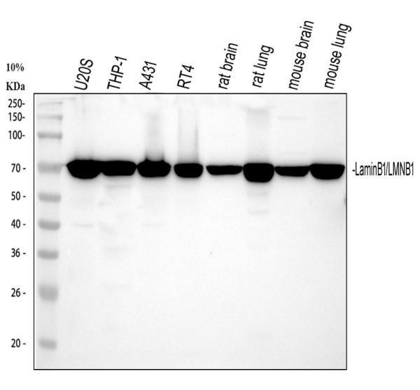
Western blot analysis of Lamin B1 using anti-Lamin B1 antibody (PB9611).
Electrophoresis was performed on a 5-20% SDS-PAGE gel at 70V (Stacking gel) / 90V (Resolving gel) for 2-3 hours. The sample well of each lane was loaded with 30 ug of sample under reducing conditions.
Lane 1: human U2OS whole cell lysates,
Lane 2: human THP-1 whole cell lysates,
Lane 3: human A431 whole cell lysates,
Lane 4: human RT4 whole cell lysates,
Lane 5: rat brain tissue lysates,
Lane 6: rat lung tissue lysates,
Lane 7: mouse brain tissue lysates,
Lane 8: mouse lung tissue lysates.
After electrophoresis, proteins were transferred to a nitrocellulose membrane at 150 mA for 50-90 minutes. Blocked the membrane with 5% non-fat milk/TBS for 1.5 hour at RT. The membrane was incubated with rabbit anti-Lamin B1 antigen affinity purified polyclonal antibody (Catalog # PB9611) at 0.5 μg/mL overnight at 4°C, then washed with TBS-0.1%Tween 3 times with 5 minutes each and probed with a goat anti-rabbit IgG-HRP secondary antibody at a dilution of 1:5000 for 1.5 hour at RT. The signal is developed using an Enhanced Chemiluminescent detection (ECL) kit (Catalog # EK1002) with Tanon 5200 system. A specific band was detected for Lamin B1 at approximately 72 kDa. The expected band size for Lamin B1 is at 66 kDa.

Click image to see more details
IHC analysis of Lamin B1 using anti-Lamin B1 antibody (PB9611).
Lamin B1 was detected in a paraffin-embedded section of human stomach cancer tissue. Heat mediated antigen retrieval was performed in EDTA buffer (pH 8.0, epitope retrieval solution). The tissue section was blocked with 10% goat serum. The tissue section was then incubated with 2 μg/ml rabbit anti-Lamin B1 Antibody (PB9611) overnight at 4°C. Peroxidase Conjugated Goat Anti-rabbit IgG was used as secondary antibody and incubated for 30 minutes at 37°C. The tissue section was developed using HRP Conjugated Rabbit IgG Super Vision Assay Kit (Catalog # SV0002) with DAB as the chromogen.

Click image to see more details
IHC analysis of Lamin B1 using anti-Lamin B1 antibody (PB9611).
Lamin B1 was detected in a paraffin-embedded section of human glioblastoma tissue. Heat mediated antigen retrieval was performed in EDTA buffer (pH 8.0, epitope retrieval solution). The tissue section was blocked with 10% goat serum. The tissue section was then incubated with 2 μg/ml rabbit anti-Lamin B1 Antibody (PB9611) overnight at 4°C. Peroxidase Conjugated Goat Anti-rabbit IgG was used as secondary antibody and incubated for 30 minutes at 37°C. The tissue section was developed using HRP Conjugated Rabbit IgG Super Vision Assay Kit (Catalog # SV0002) with DAB as the chromogen.

Click image to see more details
IHC analysis of Lamin B1 using anti-Lamin B1 antibody (PB9611).
Lamin B1 was detected in a paraffin-embedded section of human colorectal adenocarcinoma tissue. Heat mediated antigen retrieval was performed in EDTA buffer (pH 8.0, epitope retrieval solution). The tissue section was blocked with 10% goat serum. The tissue section was then incubated with 2 μg/ml rabbit anti-Lamin B1 Antibody (PB9611) overnight at 4°C. Peroxidase Conjugated Goat Anti-rabbit IgG was used as secondary antibody and incubated for 30 minutes at 37°C. The tissue section was developed using HRP Conjugated Rabbit IgG Super Vision Assay Kit (Catalog # SV0002) with DAB as the chromogen.

Click image to see more details
IHC analysis of Lamin B1 using anti-Lamin B1 antibody (PB9611).
Lamin B1 was detected in a paraffin-embedded section of human breast cancer tissue. Heat mediated antigen retrieval was performed in EDTA buffer (pH 8.0, epitope retrieval solution). The tissue section was blocked with 10% goat serum. The tissue section was then incubated with 2 μg/ml rabbit anti-Lamin B1 Antibody (PB9611) overnight at 4°C. Peroxidase Conjugated Goat Anti-rabbit IgG was used as secondary antibody and incubated for 30 minutes at 37°C. The tissue section was developed using HRP Conjugated Rabbit IgG Super Vision Assay Kit (Catalog # SV0002) with DAB as the chromogen.

Click image to see more details
IHC analysis of Lamin B1 using anti-Lamin B1 antibody (PB9611).
Lamin B1 was detected in a paraffin-embedded section of human urothelial carcinoma of the bladder with squamous differentiation tissue. Heat mediated antigen retrieval was performed in EDTA buffer (pH 8.0, epitope retrieval solution). The tissue section was blocked with 10% goat serum. The tissue section was then incubated with 2 μg/ml rabbit anti-Lamin B1 Antibody (PB9611) overnight at 4°C. Peroxidase Conjugated Goat Anti-rabbit IgG was used as secondary antibody and incubated for 30 minutes at 37°C. The tissue section was developed using HRP Conjugated Rabbit IgG Super Vision Assay Kit (Catalog # SV0002) with DAB as the chromogen.

Click image to see more details
IHC analysis of Lamin B1 using anti-Lamin B1 antibody (PB9611).
Lamin B1 was detected in a paraffin-embedded section of mouse cerebellum tissue. Heat mediated antigen retrieval was performed in EDTA buffer (pH 8.0, epitope retrieval solution). The tissue section was blocked with 10% goat serum. The tissue section was then incubated with 2 μg/ml rabbit anti-Lamin B1 Antibody (PB9611) overnight at 4°C. Peroxidase Conjugated Goat Anti-rabbit IgG was used as secondary antibody and incubated for 30 minutes at 37°C. The tissue section was developed using HRP Conjugated Rabbit IgG Super Vision Assay Kit (Catalog # SV0002) with DAB as the chromogen.

Click image to see more details
IHC analysis of Lamin B1 using anti-Lamin B1 antibody (PB9611).
Lamin B1 was detected in a paraffin-embedded section of rat cerebellum tissue. Heat mediated antigen retrieval was performed in EDTA buffer (pH 8.0, epitope retrieval solution). The tissue section was blocked with 10% goat serum. The tissue section was then incubated with 2 μg/ml rabbit anti-Lamin B1 Antibody (PB9611) overnight at 4°C. Peroxidase Conjugated Goat Anti-rabbit IgG was used as secondary antibody and incubated for 30 minutes at 37°C. The tissue section was developed using HRP Conjugated Rabbit IgG Super Vision Assay Kit (Catalog # SV0002) with DAB as the chromogen.

Click image to see more details
IF analysis of Lamin B1 using anti-Lamin B1 antibody (PB9611).
Lamin B1 was detected in immunocytochemical section of U2OS cells. Enzyme antigen retrieval was performed using IHC enzyme antigen retrieval reagent (AR0022) for 15 mins. The cells were blocked with 10% goat serum. And then incubated with 5μg/mL rabbit anti-Lamin B1 Antibody (PB9611) overnight at 4°C. DyLight®488 Conjugated Goat Anti-Rabbit IgG (BA1127) was used as secondary antibody at 1:500 dilution and incubated for 30 minutes at 37°C. The section was counterstained with DAPI. Visualize using a fluorescence microscope and filter sets appropriate for the label used.

Click image to see more details
Flow Cytometry analysis of A431 cells using anti-Lamin B1 antibody (PB9611).
Overlay histogram showing A431 cells stained with PB9611 (Blue line). To facilitate intracellular staining, cells were fixed with 4% paraformaldehyde and permeabilized with permeabilization buffer. The cells were blocked with 10% normal goat serum. And then incubated with rabbit anti-Lamin B1 Antibody (PB9611, 1 μg/1x106 cells) for 30 min at 20°C. DyLight®488 conjugated goat anti-rabbit IgG (BA1127, 5-10 μg/1x106 cells) was used as secondary antibody for 30 minutes at 20°C. Isotype control antibody (Green line) was rabbit IgG (1 μg/1x106) used under the same conditions. Unlabelled sample without incubation with primary antibody and secondary antibody (Red line) was used as a blank control.

Click image to see more details
Immunoprecipitating Lamin B1 in Hela whole cell lysate.
Western blot analysis of Lamin B1 using anti-Lamin B1 antibody (PB9611).
Lane 1: Hela whole cell lysates (30ug),
Lane 2: Rabbit control IgG instead of anti-Lamin B1 antibody in Hela whole cell lysate,
Lane 3: anti-Lamin B1 antibody (2μg) + Hela whole cell lysate (500μg).
After electrophoresis, proteins were transferred to a membrane. Then the membrane was incubated with rabbit anti-Lamin B1 antigen affinity purified polyclonal antibody (PB9611) at a dilution of 0.5 μg/mL and probed with a mouse anti-rabbit IgG-HRP secondary antibody (Catalog # BA1054). The signal is developed using ECL Plus Western Blotting Substrate (Catalog # AR1196-200). A specific band was detected for Lamin B1 at approximately 66 kDa. The expected band size for Lamin B1 is at 72 kDa.

Click image to see more details
Effects of deguelin on TNF-α induced NF-κB activation in BEAS-2B cells. BEAS-2B cells were pretreated with 10 μM deguelin for 24 h and then exposed to 10 ng/ml TNF-α for the indicated times. (A) Expressions of NF-κB p65, phospho-p65, IκBα, phospho-IκBα and nuclear p65 were analyzed by Western Blotting analysis. β-actin and Lamin B were used as internal controls. (B, C, D, E, F) Grey values of the indicated proteins were measured by Quantity One software. Values are shown as mean ± SEM of three independent experiments. # P <0.05, ## P <0.01 vs. Medium plus TNF-α-10 min group; * P <0.05, ** P <0.01 vs. Medium plus TNF-α-30 min group.
Index in PubMed under a CC BY license. PMID: 28529457

Click image to see more details
Deguelin suppressed OVA-induced NF-κB activation in lung tissues. Total protein, cytosol protein and nuclear protein were separately extracted from lung tissues 24 h after the last OVA challenge. (A) Expressions of NF-κB p65, phospho-p65, IκBα, and phosph-IκBα were analyzed by western blotting analysis. β-actin was used as an internal control. (E) Expressions of cytosol p65 and nuclear p65 were analyzed by western blotting. β-actin and Lamin B were used as internal controls. (B, C, D, F, G, H) Grey values of the indicated proteins were measured by Quantity One software. Values are shown as mean ± SEM (n = 6 for each group). DXM = dexamethasone. # P <0.05, ## P <0.01 vs. Control group; * P <0.05, ** P <0.01 vs. OVA-challenged group.
Index in PubMed under a CC BY license. PMID: 28529457

Click image to see more details
Effects of Klotho overexpression on neuroinflammatory responses in the ischemic brain induced by 2VO in mice. Four weeks after the intracerebroventricular injection of a lentivirus that encoded Klotho (LV-KL) or GFP (LV-GFP), the mice were exposed to 20 min 2VO and 72 h reperfusion. Western blot and qPCR were performed 72 h after surgery. (A) Representative Western blots and quantitative analysis of the expression of RIG-1 and the nuclear translocation of NF-κB. (B) The amount of RIG-I expression and NF-κB that translocated to the nucleus was normalized to β-actin or Lamin B, respectively. The results were expressed as each normalized value relative to LV-GFP-treated sham controls. (C) The mRNA levels of TNF-α and IL-6 in the brain. The results were normalized to the corresponding reporter gene GAPDH and are presented as a fold change relative to the sham-operated group. The data are expressed as mean ± SEM. One-way ANOVA followed by Dunnett’s test. n = 3–4 per group. ∗ p < 0.05, ∗∗ p < 0.01, vs. LV-GFP-treated 2VO group; # p < 0.05, vs. LV-GFP-treated sham group.
Index in PubMed under a CC BY license. PMID: 29403373
Specific Publications For Anti-Lamin B1/LMNB1 Antibody Picoband® (PB9611)
Loading publications
Recommended Resources
Here are featured tools and databases that you might find useful.
- Boster's Pathways Library
- Protein Databases
- Bioscience Research Protocol Resources
- Data Processing & Analysis Software
- Photo Editing Software
- Scientific Literature Resources
- Research Paper Management Tools
- Molecular Biology Software
- Primer Design Tools
- Bioinformatics Tools
- Phylogenetic Tree Analysis
Customer Reviews
Have you used Anti-Lamin B1/LMNB1 Antibody Picoband®?
Share your experimental results or join a short interview to earn up to $1,000 in product credits or other rewards.
0 Reviews For Anti-Lamin B1/LMNB1 Antibody Picoband®
Customer Q&As
Have a question?
Find answers in Q&As, reviews.
Can't find your answer?
Submit your question
1 Customer Q&As for Anti-Lamin B1/LMNB1 Antibody Picoband®
Question
We are currently using anti-Lamin B1/LMNB1 antibody PB9611 for rat tissue, and we are content with the IHC results. The species of reactivity given in the datasheet says human, mouse, rat. Is it likely that the antibody can work on primate tissues as well?
Verified Customer
Verified customer
Asked: 2017-05-29
Answer
The anti-Lamin B1/LMNB1 antibody (PB9611) has not been tested for cross reactivity specifically with primate tissues, though there is a good chance of cross reactivity. We have an innovator award program that if you test this antibody and show it works in primate you can get your next antibody for free. Please contact me if I can help you with anything.
Boster Scientific Support
Answered: 2017-05-29
