Click image to see more details
-

-
-
-
-
+14
Product Info Summary
| SKU: | PA1328 |
|---|---|
| Size: | 100 μg/vial |
| Reactive Species: | Human, Mouse, Rat |
| Host: | Rabbit |
| Application: | Flow Cytometry, IF, IHC, WB |
Customers Who Bought This Also Bought

Product info
Product Name
Anti-N-Cadherin-2 CDH2 CD325-Antibody Picoband®
SKU/Catalog Number
PA1328
BA0673 is an alternative SKU for this antibody, used in previous lots.
Size
100 μg/vial
Form
Lyophilized
Description
Boster Bio Anti-N-Cadherin-2 CDH2 CD325-Antibody catalog # PA1328. Tested in Flow Cytometry, IF, IHC, WB applications. This antibody reacts with Human, Mouse, Rat. The brand Picoband indicates this is a premium antibody that guarantees superior quality, high affinity, and strong signals with minimal background in Western blot applications. Only our best-performing antibodies are designated as Picoband, ensuring unmatched performance.
Storage & Handling
Store at -20˚C for one year from date of receipt. After reconstitution, at 4˚C for one month. It can also be aliquotted and stored frozen at -20˚C for six months. Avoid repeated freeze-thaw cycles.
Cite This Product
Anti-N-Cadherin-2 CDH2 CD325-Antibody Picoband® (Boster Biological Technology, Pleasanton CA, USA, Catalog # PA1328)
Host
Rabbit
Contents
Each vial contains 4 mg Trehalose, 0.9 mg NaCl and 0.2 mg Na2HPO4.
Clonality
Polyclonal
Isotype
Rabbit IgG
Immunogen
A synthetic peptide corresponding to a sequence in the middle region of human N Cadherin, identical to the related rat and mouse sequences.
Cross-reactivity
No cross-reactivity with other proteins
Reactive Species
PA1328 is reactive to CDH2 in Human, Mouse, Rat
Observed Molecular Weight
140 kDa
Calculated molecular weight
99.8 kDa
Background of CDH2
N-cadherin (NCAD) is a member of the cadherin cell-cell adhesion receptor family that includes P-, E-, and R-cadherin and liver cell adhesion molecule (L-CAM). N-Cadherin,, also known as Cadherin-2, encodes a 907-amino acid protein that includes a 159-amino acid signal sequence. Human and mouse nucleotide sequences are 96% identical. Mouse Ncad gene consists of 16 exons dispersed over more than 200 kb of genomic DNA. Human N-cadherin gene contains 16 exons and its sequence is highly similar to both the mouse NCAD gene (including the large first and second introns) and other cadherin genes. N-cadherin is mapped to 18q11.2. Cadherin regulates dendritic spine morphogenesis.
Antibody Validation
Boster validates all antibodies on WB, IHC, ICC, Immunofluorescence, and ELISA with known positive control and negative samples to ensure specificity and high affinity, including thorough antibody incubations.
Application & Images
Applications
PA1328 is guaranteed for Flow Cytometry, IF, IHC, WB Boster Guarantee
Assay Dilutions Recommendation
The recommendations below provide a starting point for assay optimization. The actual working concentration varies and should be decided by the user.
Western blot, 0.1-0.5μg/ml, Human, Mouse, Rat
Immunohistochemistry (Paraffin-embedded Section), 2-5μg/ml, Mouse, Rat
Immunofluorescence, 5 μg/ml, Rat
Flow Cytometry (Fixed), 1-3μg/1x106 cells, Human
Positive Control
WB: human 293T whole cell, human Hela whole cell, rat brain tissue, mouse brain tissue
IHC: mouse heart tissue, rat heart tissue
IF: rat heart tissue
FCM: A549 cell
Validation Images & Assay Conditions
Click image to see more details
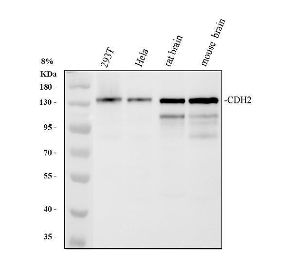
Western blot analysis of CDH2 using anti-CDH2 antibody (PA1328).
Electrophoresis was performed on a 8% SDS-PAGE gel at 80V (Stacking gel) / 120V (Resolving gel) for 2 hours. The sample well of each lane was loaded with 30 ug of sample under reducing conditions.
Lane 1: human 293T whole cell lysates,
Lane 2: human Hela whole cell lysates,
Lane 3: rat brain tissue lysates,
Lane 4: mouse brain tissue lysates.
After electrophoresis, proteins were transferred to a nitrocellulose membrane at 150 mA for 50-90 minutes. Blocked the membrane with 5% non-fat milk/TBS for 1.5 hour at RT. The membrane was incubated with rabbit anti-CDH2 antigen affinity purified polyclonal antibody (PA1328) at 0.5 μg/mL overnight at 4°C, then washed with TBS-0.1%Tween 3 times with 5 minutes each and probed with a goat anti-rabbit IgG-HRP secondary antibody (Catalog # BA1054) at a dilution of 1:5000 for 1.5 hour at RT. The signal is developed using an ECL Plus Western Blotting Substrate (Catalog # AR1196-200) with Tanon 5200 system. A specific band was detected for CDH2 at approximately 140 kDa. The expected band size for CDH2 is at 100 kDa.

Click image to see more details
IHC analysis of CDH2 using anti-CDH2 antibody (PA1328).
CDH2 was detected in a paraffin-embedded section of mouse heart tissue. Heat mediated antigen retrieval was performed in EDTA buffer (pH 8.0, epitope retrieval solution). The tissue section was blocked with 10% goat serum. The tissue section was then incubated with 2 μg/ml rabbit anti-CDH2 Antibody (PA1328) overnight at 4°C. Peroxidase Conjugated Goat Anti-rabbit IgG was used as secondary antibody and incubated for 30 minutes at 37°C. The tissue section was developed using HRP Conjugated Rabbit IgG Super Vision Assay Kit (Catalog # SV0002) with DAB as the chromogen.

Click image to see more details
IHC analysis of CDH2 using anti-CDH2 antibody (PA1328).
CDH2 was detected in a paraffin-embedded section of rat heart tissue. Heat mediated antigen retrieval was performed in EDTA buffer (pH 8.0, epitope retrieval solution). The tissue section was blocked with 10% goat serum. The tissue section was then incubated with 2 μg/ml rabbit anti-CDH2 Antibody (PA1328) overnight at 4°C. Peroxidase Conjugated Goat Anti-rabbit IgG was used as secondary antibody and incubated for 30 minutes at 37°C. The tissue section was developed using HRP Conjugated Rabbit IgG Super Vision Assay Kit (Catalog # SV0002) with DAB as the chromogen.

Click image to see more details
IHC analysis of CDH2 using anti-CDH2 antibody (PA1328).
CDH2 was detected in a paraffin-embedded section of human liver cancer tissue. Heat mediated antigen retrieval was performed in EDTA buffer (pH 8.0, epitope retrieval solution). The tissue section was blocked with 10% goat serum. The tissue section was then incubated with 5 μg/ml rabbit anti-CDH2 Antibody (PA1328) overnight at 4°C. HRP-AffiniPure Goat Anti-Rabbit IgG was used as secondary antibody and incubated for 30 minutes at 37°C. The tissue section was developed using HRP Conjugated Rabbit IgG Super Vision Assay Kit (Catalog # SV0002) with DAB as the chromogen.

Click image to see more details
IF analysis of CDH2 using anti-CDH2 antibody (PA1328).
CDH2 was detected in a paraffin-embedded section of rat heart tissue. Heat mediated antigen retrieval was performed in EDTA buffer (pH 8.0, epitope retrieval solution). The tissue section was blocked with 10% goat serum. The tissue section was then incubated with 5 μg/mL rabbit anti-CDH2 Antibody (PA1328) overnight at 4°C. DyLight®488 Conjugated Goat Anti-Rabbit IgG (BA1127) was used as secondary antibody at 1:500 dilution and incubated for 30 minutes at 37°C. The section was counterstained with DAPI. Visualize using a fluorescence microscope and filter sets appropriate for the label used.

Click image to see more details
IF analysis of CDH2 using anti-CDH2 antibody (PA1328).
CDH2 was detected in a paraffin-embedded section of human thyroid cancer tissue. Heat mediated antigen retrieval was performed in EDTA buffer (pH 8.0, epitope retrieval solution). The tissue section was blocked with 10% goat serum. The tissue section was then incubated with 5 μg/mL rabbit anti-CDH2 Antibody (PA1328) overnight at 4°C. DyLight®594 Conjugated Goat Anti-Rabbit IgG (BA1142) was used as secondary antibody at 1:100 dilution and incubated for 30 minutes at 37°C. The section was counterstained with DAPI. Visualize using a fluorescence microscope and filter sets appropriate for the label used.

Click image to see more details
IF analysis of CDH2 using anti-CDH2 antibody (PA1328).
CDH2 was detected in a paraffin-embedded section of human endometrial cancer tissue. Heat mediated antigen retrieval was performed in EDTA buffer (pH 8.0, epitope retrieval solution). The tissue section was blocked with 10% goat serum. The tissue section was then incubated with 5 μg/mL rabbit anti-CDH2 Antibody (PA1328) overnight at 4°C. DyLight®594 Conjugated Goat Anti-Rabbit IgG (BA1142) was used as secondary antibody at 1:100 dilution and incubated for 30 minutes at 37°C. The section was counterstained with DAPI. Visualize using a fluorescence microscope and filter sets appropriate for the label used.

Click image to see more details
IF analysis of CDH2 using anti-CDH2 antibody (PA1328).
CDH2 was detected in a paraffin-embedded section of human liver cancer tissue. Heat mediated antigen retrieval was performed in EDTA buffer (pH 8.0, epitope retrieval solution). The tissue section was blocked with 10% goat serum. The tissue section was then incubated with 5 μg/mL rabbit anti-CDH2 Antibody (PA1328) overnight at 4°C. DyLight®594 Conjugated Goat Anti-Rabbit IgG (BA1142) was used as secondary antibody at 1:100 dilution and incubated for 30 minutes at 37°C. The section was counterstained with DAPI. Visualize using a fluorescence microscope and filter sets appropriate for the label used.

Click image to see more details
IF analysis of CDH2 using anti-CDH2 antibody (PA1328).
CDH2 was detected in a paraffin-embedded section of human lung cancer tissue. Heat mediated antigen retrieval was performed in EDTA buffer (pH 8.0, epitope retrieval solution). The tissue section was blocked with 10% goat serum. The tissue section was then incubated with 5 μg/mL rabbit anti-CDH2 Antibody (PA1328) overnight at 4°C. DyLight®594 Conjugated Goat Anti-Rabbit IgG (BA1142) was used as secondary antibody at 1:100 dilution and incubated for 30 minutes at 37°C. The section was counterstained with DAPI. Visualize using a fluorescence microscope and filter sets appropriate for the label used.

Click image to see more details
IF analysis of CDH2 using anti-CDH2 antibody (PA1328).
CDH2 was detected in a paraffin-embedded section of mouse testis tissue. Heat mediated antigen retrieval was performed in EDTA buffer (pH 8.0, epitope retrieval solution). The tissue section was blocked with 10% goat serum. The tissue section was then incubated with 5 μg/mL rabbit anti-CDH2 Antibody (PA1328) overnight at 4°C. DyLight®594 Conjugated Goat Anti-Rabbit IgG (BA1142) was used as secondary antibody at 1:100 dilution and incubated for 30 minutes at 37°C. The section was counterstained with DAPI. Visualize using a fluorescence microscope and filter sets appropriate for the label used.

Click image to see more details
IF analysis of CDH2 using anti-CDH2 antibody (PA1328).
CDH2 was detected in a paraffin-embedded section of human ovarian cancer tissue. Heat mediated antigen retrieval was performed in EDTA buffer (pH 8.0, epitope retrieval solution). The tissue section was blocked with 10% goat serum. The tissue section was then incubated with 5 μg/mL rabbit anti-CDH2 Antibody (PA1328) overnight at 4°C. DyLight®594 Conjugated Goat Anti-Rabbit IgG (BA1142) was used as secondary antibody at 1:100 dilution and incubated for 30 minutes at 37°C. The section was counterstained with DAPI. Visualize using a fluorescence microscope and filter sets appropriate for the label used.

Click image to see more details
IF analysis of CDH2 using anti-CDH2 antibody (PA1328).
CDH2 was detected in a paraffin-embedded section of human pancreas cancer tissue. Heat mediated antigen retrieval was performed in EDTA buffer (pH 8.0, epitope retrieval solution). The tissue section was blocked with 10% goat serum. The tissue section was then incubated with 5 μg/mL rabbit anti-CDH2 Antibody (PA1328) overnight at 4°C. DyLight®594 Conjugated Goat Anti-Rabbit IgG (BA1142) was used as secondary antibody at 1:100 dilution and incubated for 30 minutes at 37°C. The section was counterstained with DAPI. Visualize using a fluorescence microscope and filter sets appropriate for the label used.

Click image to see more details
IF analysis of CDH2 using anti-CDH2 antibody (PA1328).
CDH2 was detected in a paraffin-embedded section of rat cardiac tissue. Heat mediated antigen retrieval was performed in EDTA buffer (pH 8.0, epitope retrieval solution). The tissue section was blocked with 10% goat serum. The tissue section was then incubated with 5 μg/mL rabbit anti-CDH2 Antibody (PA1328) overnight at 4°C. DyLight®594 Conjugated Goat Anti-Rabbit IgG (BA1142) was used as secondary antibody at 1:100 dilution and incubated for 30 minutes at 37°C. The section was counterstained with DAPI. Visualize using a fluorescence microscope and filter sets appropriate for the label used.

Click image to see more details
Flow Cytometry analysis of A549 cells using anti-CDH2 antibody (PA1328).
Overlay histogram showing A549 cells stained with PA1328 (Blue line). The cells were fixed with 4% paraformaldehyde and blocked with 10% normal goat serum. And then incubated with rabbit anti-CDH2 Antibody (PA1328, 1 μg/1x106 cells) for 30 min at 20°C. DyLight®488 conjugated goat anti-rabbit IgG (BA1127, 5-10 μg/1x106 cells) was used as secondary antibody for 30 minutes at 20°C. Isotype control antibody (Green line) was rabbit IgG (1 μg/1x106) used under the same conditions. Unlabelled sample without incubation with primary antibody and secondary antibody (Red line) was used as a blank control.

Click image to see more details
The effects of Ube2v1 on epithelial mesenchymal transition and autophagy program in colorectal cancer. a , b Expressions of E-cadherin, β-catenin, Vimentin, Fibronectin, N-cadherin, Twist1, and Snai1were detected after Ube2v1 was overexpressed ( a ) or knocked down ( b ) in DLD-1 and SW480 cells. c Tumor exacted from xenograft model were used to detect expressions of E-cadherin and LC3-II in both DLD-1 and SW480 cells with Ube2v1 stable overexpression by western blotting. d Endogenous LC3 puncta and expressions of E-cadherin, β-catenin, and Vimentin were observed after Ube2v1 was knocked down in SW480cells by immunofluorescent analysis. e Expressions of Ube2v1, ATG5, ATG7,LC3-II, E-cadherin, β-catenin, Vimentin, Fibronectin, and N-cadherin were observed after Ube2v1 was knocked down in HCT116 and SW480 cells with or without knockdown of ATG5 or ATG7 using RNA interference
Index in PubMed under a CC BY license. PMID: 30016968

Click image to see more details
Interfering with DLGAP4 inhibits the PPARβ/δ signalling pathway and the expression of proliferation- and metastasis-related proteins. Western blotting was performed to measure the protein expression of DLGAP4, PPARβ/δ, CyclinD1, E-cadherin and N-cadherin in shNC or shDLGAP4 HepG2 and HCCLM3 cells. The data were obtained from the average of three independent experiments. *P < 0.05.
Index in PubMed under a CC BY license. PMID: 36396671

Click image to see more details
Specific Publications For Anti-N-Cadherin-2 CDH2 CD325-Antibody Picoband® (PA1328)
Loading publications
Recommended Resources
Here are featured tools and databases that you might find useful.
- Boster's Pathways Library
- Protein Databases
- Bioscience Research Protocol Resources
- Data Processing & Analysis Software
- Photo Editing Software
- Scientific Literature Resources
- Research Paper Management Tools
- Molecular Biology Software
- Primer Design Tools
- Bioinformatics Tools
- Phylogenetic Tree Analysis
Customer Reviews
Have you used Anti-N-Cadherin-2 CDH2 CD325-Antibody Picoband®?
Share your experimental results or join a short interview to earn up to $1,000 in product credits or other rewards.
0 Reviews For Anti-N-Cadherin-2 CDH2 CD325-Antibody Picoband®
Customer Q&As
Have a question?
Find answers in Q&As, reviews.
Can't find your answer?
Submit your question
7 Customer Q&As for Anti-N-Cadherin-2 CDH2 CD325-Antibody Picoband®
Question
We are currently using anti-N Cadherin/CDH2 antibody PA1328 for rat tissue, and we are well pleased with the IHC results. The species of reactivity given in the datasheet says human, mouse, rat. Is it true that the antibody can work on monkey tissues as well?
Verified Customer
Verified customer
Asked: 2020-03-06
Answer
The anti-N Cadherin/CDH2 antibody (PA1328) has not been validated for cross reactivity specifically with monkey tissues, though there is a good chance of cross reactivity. We have an innovator award program that if you test this antibody and show it works in monkey you can get your next antibody for free. Please contact me if I can help you with anything.
Boster Scientific Support
Answered: 2020-03-06
Question
I was wanting to use your anti-N Cadherin/CDH2 antibody for WB for rat testis on frozen tissues, but I want to know if it has been validated for this particular application. Has this antibody been validated and is this antibody a good choice for rat testis identification?
Verified Customer
Verified customer
Asked: 2020-02-25
Answer
You can see on the product datasheet, PA1328 anti-N Cadherin/CDH2 antibody has been tested for IHC, WB on human, mouse, rat tissues. We have an innovator award program that if you test this antibody and show it works in rat testis in IHC-frozen, you can get your next antibody for free.
Boster Scientific Support
Answered: 2020-02-25
Question
I see that the anti-N Cadherin/CDH2 antibody PA1328 works with WB, what is the protocol used to produce the result images on the product page?
Verified Customer
Verified customer
Asked: 2019-11-14
Answer
You can find protocols for WB on the "support/technical resources" section of our navigation menu. If you have any further questions, please send an email to support@bosterbio.com
Boster Scientific Support
Answered: 2019-11-14
Question
I have a question about product PA1328, anti-N Cadherin/CDH2 antibody. I was wondering if it would be possible to conjugate this antibody with biotin. I would need it to be without BSA or sodium azide. I am planning on using a buffer exchange of sodium azide with PBS only. Would there be problems for me to conjugate the antibody and store it in -20 degrees in small aliquots?
B. Singh
Verified customer
Asked: 2019-02-04
Answer
We do not recommend storing this antibody with PBS buffer only in -20 degrees. If you want to store it in -20 degrees it is best to add some cryoprotectant like glycerol. If you want carrier free PA1328 anti-N Cadherin/CDH2 antibody, we can provide it to you in a special formula with trehalose and/or glycerol. These molecules will not interfere with conjugation chemistry and provide a good level of protection for the antibody from degradation. Please be sure to specify this in your purchase order.
Boster Scientific Support
Answered: 2019-02-04
Question
Would anti-N Cadherin/CDH2 antibody PA1328 work on bovine IHC with liver?
Verified Customer
Verified customer
Asked: 2017-12-15
Answer
Our lab technicians have not tested anti-N Cadherin/CDH2 antibody PA1328 on bovine. You can run a BLAST between bovine and the immunogen sequence of anti-N Cadherin/CDH2 antibody PA1328 to see if they may cross-react. If the sequence homology is close, then you can perform a pilot test. Keep in mind that since we have not validated bovine samples, this use of the antibody is not covered by our guarantee. However we have an innovator award program that if you test this antibody and show it works in bovine liver in IHC, you can get your next antibody for free.
Boster Scientific Support
Answered: 2017-12-15
Question
you antibody to test anti-N Cadherin/CDH2 antibody PA1328 on rat testis for research purposes, then I may be interested in using anti-N Cadherin/CDH2 antibody PA1328 for diagnostic purposes as well. Is the antibody suitable for diagnostic purposes?
C. Zhang
Verified customer
Asked: 2015-07-30
Answer
The products we sell, including anti-N Cadherin/CDH2 antibody PA1328, are only intended for research use. They would not be suitable for use in diagnostic work. If you have the means to develop a product into diagnostic use, and are interested in collaborating with us and develop our product into an IVD product, please contact us for more discussions.
Boster Scientific Support
Answered: 2015-07-30
Question
Is a blocking peptide available for product anti-N Cadherin/CDH2 antibody (PA1328)?
A. Brown
Verified customer
Asked: 2015-01-07
Answer
We do provide the blocking peptide for product anti-N Cadherin/CDH2 antibody (PA1328). If you would like to place an order for it please contact support@bosterbio.com and make a special request.
Boster Scientific Support
Answered: 2015-01-07
