
Click image to see more details
-

-
-
-
-
+7
Product Info Summary
| SKU: | PB9402 |
|---|---|
| Size: | 100 μg/vial |
| Reactive Species: | Human, Mouse, Rat |
| Host: | Rabbit |
| Application: | Flow Cytometry, IF, IHC, ICC, WB |
Customers Who Bought This Also Bought

Product info
Product Name
Anti-Superoxide Dismutase 1/SOD1 Antibody Picoband™
View all SOD1/Cu-Zn SOD Antibodies
SKU/Catalog Number
PB9402
Size
100 μg/vial
Form
Lyophilized
Description
Boster Bio Anti-Superoxide Dismutase 1/SOD1 Antibody Picoband™ catalog # PB9402. Tested in Flow Cytometry, IF, IHC, ICC, WB applications. This antibody reacts with Human, Mouse, Rat.
Storage & Handling
Store at -20˚C for one year from date of receipt. After reconstitution, at 4˚C for one month. It can also be aliquotted and stored frozen at -20˚C for six months. Avoid repeated freeze-thaw cycles.
Cite This Product
Anti-Superoxide Dismutase 1/SOD1 Antibody Picoband™ (Boster Biological Technology, Pleasanton CA, USA, Catalog # PB9402)
Host
Rabbit
Contents
Each vial contains 5mg BSA, 0.9mg NaCl, 0.2mg Na2HPO4, 0.05mg NaN3.
Clonality
Polyclonal
Isotype
Rabbit IgG
Immunogen
A synthetic peptide corresponding to a sequence at the C-terminus of Human SOD1, different from the related mouse and rat sequences by two amino acids.
*Blocking peptide can be purchased. Costs vary based on immunogen length. Contact us for pricing.
Cross-reactivity
No cross-reactivity with other proteins.
Reactive Species
PB9402 is reactive to SOD1 in Human, Mouse, Rat
Applications
PB9402 is guaranteed for Flow Cytometry, IF, IHC, ICC, WB Boster Guarantee
Observed Molecular Weight
16-18 kDa
Calculated molecular weight
15.936kDa
Background of SOD1/Cu-Zn SOD
Superoxide dismutases (SOD) are a class of enzymes that catalyze the dismutation of superoxide into oxygen and hydrogen peroxide. As such, they are an important antioxidant defense in nearly all cells exposed to oxygen. One of the exceedingly rare exceptions is Lactobacillus plantarum and related lactobacilli, which use a different mechanism.Cu,Zn-SOD was found widely distributed in the cell cytosol and in the cell nucleus, consistent with it being a soluble cytosolic protein. Mitochondria and secretory compartments did not label for this protein. In human cells, peroxisomes showed a labeling density slightly less than that of cytoplasm.
Antibody Validation
Boster validates all antibodies on WB, IHC, ICC, Immunofluorescence, and ELISA with known positive control and negative samples to ensure specificity and high affinity, including thorough antibody incubations.
Innovating Scientists Reward
If you are the first to review this product, or if you have results for a special sample, species or application this product is not validated in, share your results with us and receive product credits you can use towards any Boster products! Applicable to all scientists worldwide.
Submit A Review
Assay dilution & Images
Reconsitution
Add 0.2ml of distilled water will yield a concentration of 500ug/ml.
Assay Dilutions Recommendation
The recommendations below provide a starting point for assay optimization. The actual working concentration varies and should be decided by the user.
Western blot, 0.1-0.5μg/ml, Human, Mouse, Rat
Immunohistochemistry(Paraffin-embedded Section), 0.5-1 μg/ml, Human, Mouse, Rat, By Heat
Immunocytochemistry/Immunofluorescence, 2 μg/ml, Human
Flow Cytometry, 1-3 μg/1x106 cells, Human
Validation Images & Assay Conditions
Click image to see more details
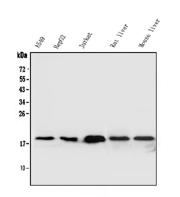
Figure 1. Western blot analysis of SOD1 using anti-SOD1 antibody (PB9402).
Electrophoresis was performed on a 5-20% SDS-PAGE gel at 70V (Stacking gel) / 90V (Resolving gel) for 2-3 hours. The sample well of each lane was loaded with 30 ug of sample under reducing conditions.
Lane 1: human Hela whole cell lysates,
Lane 2: human Jurkat whole cell lysates,
Lane 3: rat brain tissue lysates,
Lane 4: rat spleen tissue lysates,
Lane 5: mouse brain tissue lysates,
Lane 6: mouse spleen tissue lysates.
After electrophoresis, proteins were transferred to a nitrocellulose membrane at 150 mA for 50-90 minutes. Blocked the membrane with 5% non-fat milk/TBS for 1.5 hour at RT. The membrane was incubated with rabbit anti-SOD1 antigen affinity purified polyclonal antibody (Catalog # PB9402) at 0.5 μg/mL overnight at 4°C, then washed with TBS-0.1%Tween 3 times with 5 minutes each and probed with a goat anti-rabbit IgG-HRP secondary antibody at a dilution of 1:5000 for 1.5 hour at RT. The signal is developed using an Enhanced Chemiluminescent detection (ECL) kit (Catalog # EK1002) with Tanon 5200 system. A specific band was detected for SOD1 at approximately 16-18 kDa. The expected band size for SOD1 is at 16 kDa.

Click image to see more details
Figure 2. IHC analysis of SOD1 using anti-SOD1 antibody (PB9402).
SOD1 was detected in paraffin-embedded section of mouse liver tissue. Heat mediated antigen retrieval was performed in EDTA buffer (pH8.0, epitope retrieval solution). The tissue section was blocked with 10% goat serum. The tissue section was then incubated with 1μg/ml rabbit anti-SOD1 Antibody (PB9402) overnight at 4°C. Biotinylated goat anti-rabbit IgG was used as secondary antibody and incubated for 30 minutes at 37°C. The tissue section was developed using Strepavidin-Biotin-Complex (SABC) (Catalog # SA1022) with DAB as the chromogen.

Click image to see more details
Figure 3. IHC analysis of SOD1 using anti-SOD1 antibody (PB9402).
SOD1 was detected in paraffin-embedded section of human intestinal cancer tissue. Heat mediated antigen retrieval was performed in EDTA buffer (pH8.0, epitope retrieval solution). The tissue section was blocked with 10% goat serum. The tissue section was then incubated with 1μg/ml rabbit anti-SOD1 Antibody (PB9402) overnight at 4°C. Biotinylated goat anti-rabbit IgG was used as secondary antibody and incubated for 30 minutes at 37°C. The tissue section was developed using Strepavidin-Biotin-Complex (SABC) (Catalog # SA1022) with DAB as the chromogen.

Click image to see more details
Figure 4. IHC analysis of SOD1 using anti-SOD1 antibody (PB9402).
SOD1 was detected in paraffin-embedded section of human intestinal cancer tissue. Heat mediated antigen retrieval was performed in EDTA buffer (pH8.0, epitope retrieval solution). The tissue section was blocked with 10% goat serum. The tissue section was then incubated with 1μg/ml rabbit anti-SOD1 Antibody (PB9402) overnight at 4°C. Biotinylated goat anti-rabbit IgG was used as secondary antibody and incubated for 30 minutes at 37°C. The tissue section was developed using Strepavidin-Biotin-Complex (SABC) (Catalog # SA1022) with DAB as the chromogen.

Click image to see more details
Figure 5. IHC analysis of SOD1 using anti-SOD1 antibody (PB9402).
SOD1 was detected in paraffin-embedded section of human lung cancer tissue. Heat mediated antigen retrieval was performed in EDTA buffer (pH8.0, epitope retrieval solution). The tissue section was blocked with 10% goat serum. The tissue section was then incubated with 1μg/ml rabbit anti-SOD1 Antibody (PB9402) overnight at 4°C. Biotinylated goat anti-rabbit IgG was used as secondary antibody and incubated for 30 minutes at 37°C. The tissue section was developed using Strepavidin-Biotin-Complex (SABC) (Catalog # SA1022) with DAB as the chromogen.

Click image to see more details
Figure 6. IHC analysis of SOD1 using anti-SOD1 antibody (PB9402).
SOD1 was detected in paraffin-embedded section of human lung cancer tissue. Heat mediated antigen retrieval was performed in EDTA buffer (pH8.0, epitope retrieval solution). The tissue section was blocked with 10% goat serum. The tissue section was then incubated with 1μg/ml rabbit anti-SOD1 Antibody (PB9402) overnight at 4°C. Biotinylated goat anti-rabbit IgG was used as secondary antibody and incubated for 30 minutes at 37°C. The tissue section was developed using Strepavidin-Biotin-Complex (SABC) (Catalog # SA1022) with DAB as the chromogen.

Click image to see more details
Figure 7. IHC analysis of SOD1 using anti-SOD1 antibody (PB9402).
SOD1 was detected in paraffin-embedded section of human mammary cancer tissue. Heat mediated antigen retrieval was performed in EDTA buffer (pH8.0, epitope retrieval solution). The tissue section was blocked with 10% goat serum. The tissue section was then incubated with 1μg/ml rabbit anti-SOD1 Antibody (PB9402) overnight at 4°C. Biotinylated goat anti-rabbit IgG was used as secondary antibody and incubated for 30 minutes at 37°C. The tissue section was developed using Strepavidin-Biotin-Complex (SABC) (Catalog # SA1022) with DAB as the chromogen.

Click image to see more details
Figure 8. IHC analysis of SOD1 using anti-SOD1 antibody (PB9402).
SOD1 was detected in paraffin-embedded section of human mammary cancer tissue. Heat mediated antigen retrieval was performed in EDTA buffer (pH8.0, epitope retrieval solution). The tissue section was blocked with 10% goat serum. The tissue section was then incubated with 1μg/ml rabbit anti-SOD1 Antibody (PB9402) overnight at 4°C. Biotinylated goat anti-rabbit IgG was used as secondary antibody and incubated for 30 minutes at 37°C. The tissue section was developed using Strepavidin-Biotin-Complex (SABC) (Catalog # SA1022) with DAB as the chromogen.

Click image to see more details
Figure 9. IHC analysis of SOD1 using anti-SOD1 antibody (PB9402).
SOD1 was detected in paraffin-embedded section of rat liver tissue. Heat mediated antigen retrieval was performed in EDTA buffer (pH8.0, epitope retrieval solution). The tissue section was blocked with 10% goat serum. The tissue section was then incubated with 1μg/ml rabbit anti-SOD1 Antibody (PB9402) overnight at 4°C. Biotinylated goat anti-rabbit IgG was used as secondary antibody and incubated for 30 minutes at 37°C. The tissue section was developed using Strepavidin-Biotin-Complex (SABC) (Catalog # SA1022) with DAB as the chromogen.

Click image to see more details
Figure 10. IF analysis of SOD1 using anti-SOD1 antibody (PB9402).
SOD1 was detected in immunocytochemical section of Hep2 cells. Enzyme antigen retrieval was performed using IHC enzyme antigen retrieval reagent (AR0022) for 15 mins. The cells were blocked with 10% goat serum. And then incubated with 2μg/mL rabbit anti-SOD1 Antibody (PB9402) overnight at 4°C. DyLight®488 Conjugated Goat Anti-Rabbit IgG (BA1127) was used as secondary antibody at 1:100 dilution and incubated for 30 minutes at 37°C. The section was counterstained with DAPI. Visualize using a fluorescence microscope and filter sets appropriate for the label used.

Click image to see more details
Figure 11. Flow Cytometry analysis of A549 cells using anti-SOD1 antibody (PB9402).
Overlay histogram showing A549 cells stained with PB9402 (Blue line).The cells were blocked with 10% normal goat serum. And then incubated with rabbit anti-SOD1Antibody (PB9402, 1μg/1x106 cells) for 30 min at 20°C. DyLight®488 conjugated goat anti-rabbit IgG (BA1127, 5-10μg/1x106 cells) was used as secondary antibody for 30 minutes at 20°C. Isotype control antibody (Green line) was rabbit IgG (1μg/1x106) used under the same conditions. Unlabelled sample (Red line) was also used as a control.
Protein Target Info & Infographic
Gene/Protein Information For SOD1 (Source: Uniprot.org, NCBI)
Gene Name
SOD1
Full Name
Superoxide dismutase [Cu-Zn]
Weight
15.936kDa
Superfamily
Cu-Zn superoxide dismutase family
Alternative Names
ALS; ALS1; amyotrophic lateral sclerosis 1 (adult); Cu; Cu/Zn superoxide dismutase; CuZn SOD; Cu-Zn SOD; EC 1.15.1.1; homodimer; hSod1; indophenoloxidase A; Ipo1; IPOA; SOD; SOD, cytosolic; SOD, Soluble; SOD1; superoxide dismutase [Cu-Zn]; Superoxide dismutase 1; superoxide dismutase 1, soluble; Zn superoxide dismutase, EC 1.15.1.110superoxide dismutase, cystolic SOD1 ALS, ALS1, HEL-S-44, IPOA, SOD, STAHP, hSod1, homodimer superoxide dismutase 1 superoxide dismutase [Cu-Zn]|Cu/Zn superoxide dismutase|SOD, soluble|epididymis secretory protein Li 44|indophenoloxidase A|superoxide dismutase 1, soluble|superoxide dismutase, cystolic
*If product is indicated to react with multiple species, protein info is based on the gene entry specified above in "Species".For more info on SOD1, check out the SOD1 Infographic

We have 30,000+ of these available, one for each gene! Check them out.
In this infographic, you will see the following information for SOD1: database IDs, superfamily, protein function, synonyms, molecular weight, chromosomal locations, tissues of expression, subcellular locations, post-translational modifications, and related diseases, research areas & pathways. If you want to see more information included, or would like to contribute to it and be acknowledged, please contact [email protected].
Specific Publications For Anti-Superoxide Dismutase 1/SOD1 Antibody Picoband™ (PB9402)
Hello CJ!
PB9402 has been cited in 8 publications:
*The publications in this section are manually curated by our staff scientists. They may differ from Bioz's machine gathered results. Both are accurate. If you find a publication citing this product but is missing from this list, please let us know we will issue you a thank-you coupon.
Akt specific activator SC79 protects against early brain injury following subarachnoid hemorrhage
The Natural Flavone Acacetin Confers Cardiomyocyte Protection Against Hypoxia/Reoxygenation Injury via AMPK-Mediated Activation of Nrf2 Signaling Pathway
Protective Role of Antioxidant Huskless Barley Extracts on TNF-?-Induced Endothelial Dysfunction in Human Vascular Endothelial Cells
Maternal inflammation activated ROS-p38 MAPK predisposes offspring to heart damages caused by isoproterenol via augmenting ROS generation
Zhu X, Chen Y, Chen Q, Yang H, Xie X. J Diabetes Res. 2018 Mar 21;2018:6730315. doi: 10.1155/2018/6730315. eCollection 2018. Astaxanthin Promotes Nrf2/ARE Signaling to Alleviate Renal Fibronectin and Collagen IV Accumulation in Diabetic Rats
Guo Z, Qi W, Yu Y, Du S, Wu J, Liu J. Diabetol Metab Syndr. 2014 Feb 28;6(1):29. Doi: 10.1186/1758-5996-6-29. Effect Of Exenatide On The Cardiac Expression Of Adiponectin Receptor 1 And Nadph Oxidase Subunits And Heart Function In Streptozotocin-I...
Hou S, Zheng F, Li Y, Gao L, Zhang J. Int J Mol Sci. 2014 Aug 26;15(9):15026-43. Doi: 10.3390/Ijms150915026. The Protective Effect Of Glycyrrhizic Acid On Renal Tubular Epithelial Cell Injury Induced By High Glucose.
Long Sh, Yu Zq, Shuai L, Guo Yl, Duan Dl, Xu Xy, Li Xd. Int J Mol Sci. 2012;13(3):3354-65. Doi: 10.3390/Ijms13033354. Epub 2012 Mar 12. The Hypoglycemic Effect Of The Kelp On Diabetes Mellitus Model Induced By Alloxan In Rats.
Recommended Resources
Here are featured tools and databases that you might find useful.
- Boster's Pathways Library
- Protein Databases
- Bioscience Research Protocol Resources
- Data Processing & Analysis Software
- Photo Editing Software
- Scientific Literature Resources
- Research Paper Management Tools
- Molecular Biology Software
- Primer Design Tools
- Bioinformatics Tools
- Phylogenetic Tree Analysis
Customer Reviews
Have you used Anti-Superoxide Dismutase 1/SOD1 Antibody Picoband™?
Submit a review and receive an Amazon gift card.
- $30 for a review with an image
Be the first to review Anti-Superoxide Dismutase 1/SOD1 Antibody Picoband™
*The first user to submit a review for a product is eligible for Boster's Innovating Scientists Reward, which gives product credits. This is in addition to the gift card reward.
Customer Q&As
Have a question?
Find answers in Q&As, reviews.
Can't find your answer?
Submit your question
16 Customer Q&As for Anti-Superoxide Dismutase 1/SOD1 Antibody Picoband™
Question
I was wanting to use your anti-Superoxide Dismutase 1/SOD1 antibody for IF for rat liver on frozen tissues, but I want to know if it has been validated for this particular application. Has this antibody been validated and is this antibody a good choice for rat liver identification?
Verified Customer
Verified customer
Asked: 2020-03-31
Answer
As indicated on the product datasheet, PB9402 anti-Superoxide Dismutase 1/SOD1 antibody has been tested for Flow Cytometry, IF, IHC-P, ICC, WB on human, mouse, rat tissues. We have an innovator award program that if you test this antibody and show it works in rat liver in IHC-frozen, you can get your next antibody for free.
Boster Scientific Support
Answered: 2020-03-31
Question
Is this PB9402 anti-Superoxide Dismutase 1/SOD1 antibody reactive to the isotypes of SOD1?
Verified Customer
Verified customer
Asked: 2020-02-11
Answer
The immunogen of PB9402 anti-Superoxide Dismutase 1/SOD1 antibody is A synthetic peptide corresponding to a sequence at the C-terminus of Human SOD1 (116-146aa RTLVVHEKADDLGKGGNEESTKTGNAGSRLA), different from the related mouse and rat sequences by two amino acids. Could you tell me which isotype you are interested in so I can help see if the immunogen is part of this isotype?
Boster Scientific Support
Answered: 2020-02-11
Question
My question regarding product PB9402, anti-Superoxide Dismutase 1/SOD1 antibody. I was wondering if it would be possible to conjugate this antibody with biotin. I would need it to be without BSA or sodium azide. I am planning on using a buffer exchange of sodium azide with PBS only. Would there be problems for me to conjugate the antibody and store it in -20 degrees in small aliquots?
Verified Customer
Verified customer
Asked: 2020-01-22
Answer
It is not recommended storing this antibody with PBS buffer only in -20 degrees. If you want to store it in -20 degrees it is best to add some cryoprotectant like glycerol. If you want carrier free PB9402 anti-Superoxide Dismutase 1/SOD1 antibody, we can provide it to you in a special formula with trehalose and/or glycerol. These molecules will not interfere with conjugation chemistry and provide a good level of protection for the antibody from degradation. Please be sure to specify this in your purchase order.
Boster Scientific Support
Answered: 2020-01-22
Question
I am looking for using your anti-Superoxide Dismutase 1/SOD1 antibody for auditory receptor cell stereocilium organization studies. Has this antibody been tested with western blotting on mammary cancer tissue? We would like to see some validation images before ordering.
Verified Customer
Verified customer
Asked: 2019-12-25
Answer
We appreciate your inquiry. This PB9402 anti-Superoxide Dismutase 1/SOD1 antibody is tested on human placenta tissue, hela whole cell lysates, sw620 whole cell lysates, a549 whole cell lysates, hepg2 whole cell lysates, jurkat whole cell lysates, k562 whole cell lysates, rat liver tissue, brain tissue, spleen tissue, kidney tissue, mouse liver tissue, intestinal cancer tissue, lung cancer tissue, mammary cancer tissue. It is guaranteed to work for Flow Cytometry, IF, IHC-P, ICC, WB in human, mouse, rat. Our Boster guarantee will cover your intended experiment even if the sample type has not been be directly tested.
Boster Scientific Support
Answered: 2019-12-25
Question
We have tried in the past anti-Superoxide Dismutase 1/SOD1 antibody for ICC on placenta a few months ago. I am using mouse, and We are going to use the antibody for IF next. Our lab want to know about examining placenta as well as cervix carcinoma in our next experiment. Could you please give me some suggestion on which antibody would work the best for IF?
K. Moore
Verified customer
Asked: 2019-11-12
Answer
I looked at the website and datasheets of our anti-Superoxide Dismutase 1/SOD1 antibody and I see that PB9402 has been validated on mouse in both ICC and IF. Thus PB9402 should work for your application. Our Boster satisfaction guarantee will cover this product for IF in mouse even if the specific tissue type has not been validated. We do have a comprehensive range of products for IF detection and you can check out our website bosterbio.com to find out more information about them.
Boster Scientific Support
Answered: 2019-11-12
Question
My colleagues were happy with the WB result of your anti-Superoxide Dismutase 1/SOD1 antibody. However we have observed positive staining in pons cytoplasm. using this antibody. Is that expected? Could you tell me where is SOD1 supposed to be expressed?
Verified Customer
Verified customer
Asked: 2019-10-01
Answer
From what I have seen in literature, pons does express SOD1. Generally SOD1 expresses in cytoplasm. Regarding which tissues have SOD1 expression, here are a few articles citing expression in various tissues:
Cervix carcinoma, Pubmed ID: 18669648, 20068231
Colon, Pubmed ID: 14702039
Fetal brain cortex, Pubmed ID: 8528216
Liver, Pubmed ID: 24275569
Placenta, Pubmed ID: 15489334
Boster Scientific Support
Answered: 2019-10-01
Question
I see that the anti-Superoxide Dismutase 1/SOD1 antibody PB9402 works with IF, what is the protocol used to produce the result images on the product page?
Verified Customer
Verified customer
Asked: 2019-09-09
Answer
You can find protocols for IF on the "support/technical resources" section of our navigation menu. If you have any further questions, please send an email to [email protected]
Boster Scientific Support
Answered: 2019-09-09
Question
We have observed staining in rat fetal brain cortex. Do you have any suggestions? Is anti-Superoxide Dismutase 1/SOD1 antibody supposed to stain fetal brain cortex positively?
Verified Customer
Verified customer
Asked: 2019-08-05
Answer
From literature fetal brain cortex does express SOD1. From Uniprot.org, SOD1 is expressed in pons, colon, placenta, fetal brain cortex, cervix carcinoma, liver, among other tissues. Regarding which tissues have SOD1 expression, here are a few articles citing expression in various tissues:
Cervix carcinoma, Pubmed ID: 18669648, 20068231
Colon, Pubmed ID: 14702039
Fetal brain cortex, Pubmed ID: 8528216
Liver, Pubmed ID: 24275569
Placenta, Pubmed ID: 15489334
Boster Scientific Support
Answered: 2019-08-05
Question
Please see the WB image, lot number and protocol we used for liver using anti-Superoxide Dismutase 1/SOD1 antibody PB9402. Please let me know if you require anything else.
Verified Customer
Verified customer
Asked: 2019-07-26
Answer
Thank you very much for the data. Our lab team are working to resolve this as quickly as possible, and we appreciate your patience and understanding! You have provided everything we needed. Please let me know if there is anything you need in the meantime.
Boster Scientific Support
Answered: 2019-07-26
Question
My lab would like to test anti-Superoxide Dismutase 1/SOD1 antibody PB9402 on rat liver for research purposes, then I may be interested in using anti-Superoxide Dismutase 1/SOD1 antibody PB9402 for diagnostic purposes as well. Is the antibody suitable for diagnostic purposes?
N. Wu
Verified customer
Asked: 2019-01-14
Answer
The products we sell, including anti-Superoxide Dismutase 1/SOD1 antibody PB9402, are only intended for research use. They would not be suitable for use in diagnostic work. If you have the means to develop a product into diagnostic use, and are interested in collaborating with us and develop our product into an IVD product, please contact us for more discussions.
Boster Scientific Support
Answered: 2019-01-14
Question
Do you have a BSA free version of anti-Superoxide Dismutase 1/SOD1 antibody PB9402 available?
A. Evans
Verified customer
Asked: 2018-08-01
Answer
I appreciate your recent telephone inquiry. I can confirm that some lots of this anti-Superoxide Dismutase 1/SOD1 antibody PB9402 are BSA free. For now, these lots are available and we can make a BSA free formula for you free of charge. It will take 3 extra days to prepare. If you require this antibody BSA free again in future, please do not hesitate to contact me and I will be pleased to check which lots we have in stock that are BSA free.
Boster Scientific Support
Answered: 2018-08-01
Question
Is a blocking peptide available for product anti-Superoxide Dismutase 1/SOD1 antibody (PB9402)?
Z. Carter
Verified customer
Asked: 2018-07-05
Answer
We do provide the blocking peptide for product anti-Superoxide Dismutase 1/SOD1 antibody (PB9402). If you would like to place an order for it please contact [email protected] and make a special request.
Boster Scientific Support
Answered: 2018-07-05
Question
Will PB9402 anti-Superoxide Dismutase 1/SOD1 antibody work on parafin embedded sections? If so, which fixation method do you recommend we use (PFA, paraformaldehyde, other)?
T. Dhar
Verified customer
Asked: 2018-05-15
Answer
You can see on the product datasheet, PB9402 anti-Superoxide Dismutase 1/SOD1 antibody as been tested on IF. It is best to use PFA for fixation because it has better tissue penetration ability. PFA needs to be prepared fresh before use. Long term stored PFA turns into formalin, as the PFA molecules congregate and become formalin.
Boster Scientific Support
Answered: 2018-05-15
Question
Thank you for helping with my inquiry over the phone. Here are the WB image, lot number and protocol we used for liver using anti-Superoxide Dismutase 1/SOD1 antibody PB9402. Let me know if you need anything else.
J. Moore
Verified customer
Asked: 2016-06-01
Answer
Thank you for the data. You have provided everything we needed. Our lab team are working to resolve your inquiry as quickly as possible, and we appreciate your patience and understanding! Please let me know if there is anything you need in the meantime.
Boster Scientific Support
Answered: 2016-06-01
Question
Will anti-Superoxide Dismutase 1/SOD1 antibody PB9402 work for IF with liver?
S. Edwards
Verified customer
Asked: 2015-09-14
Answer
According to the expression profile of liver, SOD1 is highly expressed in liver. So, it is likely that anti-Superoxide Dismutase 1/SOD1 antibody PB9402 will work for IF with liver.
Boster Scientific Support
Answered: 2015-09-14
Question
We are currently using anti-Superoxide Dismutase 1/SOD1 antibody PB9402 for rat tissue, and we are satisfied with the ICC results. The species of reactivity given in the datasheet says human, mouse, rat. Is it possible that the antibody can work on bovine tissues as well?
L. Mitchell
Verified customer
Asked: 2014-10-06
Answer
The anti-Superoxide Dismutase 1/SOD1 antibody (PB9402) has not been validated for cross reactivity specifically with bovine tissues, though there is a good chance of cross reactivity. We have an innovator award program that if you test this antibody and show it works in bovine you can get your next antibody for free. Please contact me if I can help you with anything.
Boster Scientific Support
Answered: 2014-10-06
