
Click image to see more details
Product Info Summary
| SKU: | M05049-3 |
|---|---|
| Size: | 100 μg/vial |
| Reactive Species: | Mouse, Rat |
| Host: | Mouse |
| Application: | WB |
Customers Who Bought This Also Bought

Product info
Product Name
Anti-SYP/Synaptophysin Antibody Picoband™ (monoclonal, 3G12)
View all Synaptophysin Antibodies
SKU/Catalog Number
M05049-3
Size
100 μg/vial
Form
Lyophilized
Description
Boster Bio Anti-SYP/Synaptophysin Antibody Picoband™ (monoclonal, 3G12) catalog # M05049-3. Tested in WB applications. This antibody reacts with Mouse, Rat.
Storage & Handling
Store at -20˚C for one year from date of receipt. After reconstitution, at 4˚C for one month. It can also be aliquotted and stored frozen at -20˚C for six months. Avoid repeated freeze-thaw cycles.
Cite This Product
Anti-SYP/Synaptophysin Antibody Picoband™ (monoclonal, 3G12) (Boster Biological Technology, Pleasanton CA, USA, Catalog # M05049-3)
Host
Mouse
Contents
Each vial contains 4mg Trehalose, 0.9mg NaCl, 0.2mg Na2HPO4, 0.05mg NaN3.
Clonality
Monoclonal
Clone Number
3G12
Isotype
Mouse IgG2b
Immunogen
A synthetic peptide corresponding to a sequence at the N-terminus of human Synaptophysin, identical to the related mouse and rat sequences.
*Blocking peptide can be purchased. Costs vary based on immunogen length. Contact us for pricing.
Cross-reactivity
No cross-reactivity with other proteins.
Reactive Species
M05049-3 is reactive to SYP in Mouse, Rat
Applications
M05049-3 is guaranteed for WB Boster Guarantee
Observed Molecular Weight
38 kDa
Calculated molecular weight
33.845kDa
Background of Synaptophysin
SYP (Synaptophysin) is located on the short arm of X chromosome (Xp11.23-p11.22). The protein is a synaptic vesicle glycoprotein with four transmembrane domains weighing 38kDa. It is present inneuroendocrine cells and in virtually all neurons in the brain and spinal cord that participate in synaptic transmission. It acts as a marker for neuroendocrine tumors, and its ubiquity at the synapse has led to the use of synaptophysin immunostaining for quantification of synapses. Also, the protein binds cholesterol and is thought to direct targeting of vesicle-associated membrane protein 2 (synaptobrevin) to intracellular compartments. Mutations in this gene are associated with X-linked mental retardation (XLMR).
Antibody Validation
Boster validates all antibodies on WB, IHC, ICC, Immunofluorescence, and ELISA with known positive control and negative samples to ensure specificity and high affinity, including thorough antibody incubations.
Innovating Scientists Reward
If you are the first to review this product, or if you have results for a special sample, species or application this product is not validated in, share your results with us and receive product credits you can use towards any Boster products! Applicable to all scientists worldwide.
Submit A Review
Assay dilution & Images
Reconsitution
Add 0.2ml of distilled water will yield a concentration of 500μg/ml.
Assay Dilutions Recommendation
The recommendations below provide a starting point for assay optimization. The actual working concentration varies and should be decided by the user.
Western blot, 0.1-0.5μg/ml, Mouse, Rat
Validation Images & Assay Conditions
Click image to see more details
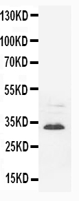
Figure 1. Western blot analysis of SYP using anti-SYP antibody (M05049-3).
Electrophoresis was performed on a 5-20% SDS-PAGE gel at 70V (Stacking gel) / 90V (Resolving gel) for 2-3 hours. The sample well of each lane was loaded with 50ug of sample under reducing conditions.
Lane 1: rat brain tissue lysates,
Lane 2: rat brain whole cell lysates,
Lane 3: mouse brain whole cell lysates,
Lane 4: mouse brain whole cell lysates,
After Electrophoresis, proteins were transferred to a Nitrocellulose membrane at 150mA for 50-90 minutes. Blocked the membrane with 5% Non-fat Milk/ TBS for 1.5 hour at RT. The membrane was incubated with mouse anti-SYP antigen affinity purified polyclonal antibody (Catalog # M05049-3) at 0.5 g/mL overnight at 4C, then washed with TBS-0.1%Tween 3 times with 5 minutes each and probed with a goat anti-mouse IgG-HRP secondary antibody at a dilution of 1:10000 for 1.5 hour at RT. The signal is developed using an Enhanced Chemiluminescent detection (ECL) kit (Catalog # EK1001) with Tanon 5200 system. A specific band was detected for SYP at approximately 38KD. The expected band size for SYP is at 34KD.
Protein Target Info & Infographic
Gene/Protein Information For SYP (Source: Uniprot.org, NCBI)
Gene Name
SYP
Full Name
Synaptophysin
Weight
33.845kDa
Superfamily
synaptophysin/synaptobrevin family
Alternative Names
Major synaptic vesicle protein p38; MRX; MRXSYP; Synaptophysin; SYP SYP MRX96, MRXSYP synaptophysin synaptophysin|major synaptic vesicle protein P38
*If product is indicated to react with multiple species, protein info is based on the gene entry specified above in "Species".For more info on SYP, check out the SYP Infographic

We have 30,000+ of these available, one for each gene! Check them out.
In this infographic, you will see the following information for SYP: database IDs, superfamily, protein function, synonyms, molecular weight, chromosomal locations, tissues of expression, subcellular locations, post-translational modifications, and related diseases, research areas & pathways. If you want to see more information included, or would like to contribute to it and be acknowledged, please contact [email protected].
Specific Publications For Anti-SYP/Synaptophysin Antibody Picoband™ (monoclonal, 3G12) (M05049-3)
Hello CJ!
M05049-3 has been cited in 2 publications:
*The publications in this section are manually curated by our staff scientists. They may differ from Bioz's machine gathered results. Both are accurate. If you find a publication citing this product but is missing from this list, please let us know we will issue you a thank-you coupon.
Abnormality of synaptic vesicular associated proteins in cerebral cortex and hippocampus after microwave exposure
Lin J,Hao C,Gong Y,Zhang Y,Li Y,Feng Z,Xu X,Huang H,Liao W. Effect of Tetramethylpyrazine on Neuroplasticity after Transient Focal Cerebral Ischemia Reperfusion in Rats. Evid Based Complement Alternat Med. 2021 Jan 18;2021:1587241.doi:10.1155/2021/1587241
Species: Rat
M05049-3 usage in article: APP:IHC, SAMPLE:BRAIN TISSUE, DILUTION:1:200
Recommended Resources
Here are featured tools and databases that you might find useful.
- Boster's Pathways Library
- Protein Databases
- Bioscience Research Protocol Resources
- Data Processing & Analysis Software
- Photo Editing Software
- Scientific Literature Resources
- Research Paper Management Tools
- Molecular Biology Software
- Primer Design Tools
- Bioinformatics Tools
- Phylogenetic Tree Analysis
Customer Reviews
Have you used Anti-SYP/Synaptophysin Antibody Picoband™ (monoclonal, 3G12)?
Submit a review and receive an Amazon gift card.
- $30 for a review with an image
Be the first to review Anti-SYP/Synaptophysin Antibody Picoband™ (monoclonal, 3G12)
*The first user to submit a review for a product is eligible for Boster's Innovating Scientists Reward, which gives product credits. This is in addition to the gift card reward.
Customer Q&As
Have a question?
Find answers in Q&As, reviews.
Can't find your answer?
Submit your question
4 Customer Q&As for Anti-SYP/Synaptophysin Antibody Picoband™ (monoclonal, 3G12)
Question
We are currently using anti-SYP/Synaptophysin antibody (monoclonal, 3G12) M05049-3 for rat tissue, and we are well pleased with the ICC results. The species of reactivity given in the datasheet says mouse, rat. Is it true that the antibody can work on goat tissues as well?
Verified Customer
Verified customer
Asked: 2019-12-13
Answer
The anti-SYP/Synaptophysin antibody (monoclonal, 3G12) (M05049-3) has not been validated for cross reactivity specifically with goat tissues, but there is a good chance of cross reactivity. We have an innovator award program that if you test this antibody and show it works in goat you can get your next antibody for free. Please contact me if I can help you with anything.
Boster Scientific Support
Answered: 2019-12-13
Question
We have seen staining in mouse amygdala. Any tips? Is anti-SYP/Synaptophysin antibody (monoclonal, 3G12) supposed to stain amygdala positively?
Verified Customer
Verified customer
Asked: 2019-09-17
Answer
From literature amygdala does express SYP. From Uniprot.org, SYP is expressed in anterior cingulate cortex, amygdala, brain, among other tissues. Regarding which tissues have SYP expression, here are a few articles citing expression in various tissues:
Amygdala, Pubmed ID: 14702039
Brain, Pubmed ID: 15489334
Boster Scientific Support
Answered: 2019-09-17
Question
We ordered your anti-SYP/Synaptophysin antibody (monoclonal, 3G12) for Flow Cytometry on brain in a previous experiment. I am using rat, and We want to use the antibody for ICC next. We want examining brain as well as anterior cingulate cortex in our next experiment. Do you have any suggestion on which antibody would work the best for ICC?
Verified Customer
Verified customer
Asked: 2019-06-11
Answer
I viewed the website and datasheets of our anti-SYP/Synaptophysin antibody (monoclonal, 3G12) and I see that M05049-3 has been validated on rat in both Flow Cytometry and ICC. Thus M05049-3 should work for your application. Our Boster satisfaction guarantee will cover this product for ICC in rat even if the specific tissue type has not been validated. We do have a comprehensive range of products for ICC detection and you can check out our website bosterbio.com to find out more information about them.
Boster Scientific Support
Answered: 2019-06-11
Question
Our team were happy with the WB result of your anti-SYP/Synaptophysin antibody (monoclonal, 3G12). However we have observed positive staining in anterior cingulate cortex secretory vesicle, using this antibody. Is that expected? Could you tell me where is SYP supposed to be expressed?
T. Wu
Verified customer
Asked: 2014-03-25
Answer
According to literature, anterior cingulate cortex does express SYP. Generally SYP expresses in cytoplasmic vesicle, secretory vesicle,. Regarding which tissues have SYP expression, here are a few articles citing expression in various tissues:
Amygdala, Pubmed ID: 14702039
Brain, Pubmed ID: 15489334
Boster Scientific Support
Answered: 2014-03-25
