
Click image to see more details
-

-
-
-
-
+1
Product Info Summary
| SKU: | PB9519 |
|---|---|
| Size: | 100 μg/vial |
| Reactive Species: | Human, Mouse, Rat |
| Host: | Rabbit |
| Application: | Flow Cytometry, IHC, IHC-F, ICC, WB |
Customers Who Bought This Also Bought

Product info
Product Name
Anti-ULK3 Antibody Picoband™
SKU/Catalog Number
PB9519
Size
100 μg/vial
Form
Lyophilized
Description
Boster Bio Anti-ULK3 Antibody Picoband™ catalog # PB9519. Tested in Flow Cytometry, IHC, IHC-F, ICC, WB applications. This antibody reacts with Human, Mouse, Rat.
Storage & Handling
Store at -20˚C for one year from date of receipt. After reconstitution, at 4˚C for one month. It can also be aliquotted and stored frozen at -20˚C for six months. Avoid repeated freeze-thaw cycles.
Cite This Product
Anti-ULK3 Antibody Picoband™ (Boster Biological Technology, Pleasanton CA, USA, Catalog # PB9519)
Host
Rabbit
Contents
Each vial contains 5mg BSA, 0.9mg NaCl, 0.2mg Na2HPO4, 0.05mg NaN3.
Clonality
Polyclonal
Isotype
Rabbit IgG
Immunogen
A synthetic peptide corresponding to a sequence at C-terminus of human ULK3, different from the related mouse and rat sequences by four amino acids.
*Blocking peptide can be purchased. Costs vary based on immunogen length. Contact us for pricing.
Cross-reactivity
No cross-reactivity with other proteins
Reactive Species
PB9519 is reactive to ULK3 in Human, Mouse, Rat
Applications
PB9519 is guaranteed for Flow Cytometry, IHC, IHC-F, ICC, WB Boster Guarantee
Observed Molecular Weight
53 kDa
Calculated molecular weight
53.444kDa
Background of ULK3
Serine/threonine protein kinase that acts as a regulator of Sonic hedgehog (SHH) signaling and autophagy. In search of potential homologues to Drosophila and human Fu, people have cloned human serine/threonine kinase ULK3 and assessed its ability to regulate GLI transcription factors, mediators of SHH signaling. And ULK3 enhances endogenous and over-expressed GLI1 and GLI2 transcriptional activity in cultured cells, as assessed by GLI-luciferase reporter assay. Besides that, ULK3 alters subcellular localization of GLI1, as assessed by immunofluorescent staining and immunoblotting assays. It is showed that ULK3 is an autophosphorylated kinase and phosphorylates GLI proteins in vitro. Also, ULK3 catalytical activity is crucial for its function in SHH pathway. It is demonstrated that ULK3 is widely expressed and its expression is higher in a number of tissues where Shh signaling is known to be active. The data suggest that serine/threonine kinase ULK3 is involved in the SHH pathway as a positive regulator of GLI proteins.
Antibody Validation
Boster validates all antibodies on WB, IHC, ICC, Immunofluorescence, and ELISA with known positive control and negative samples to ensure specificity and high affinity, including thorough antibody incubations.
Innovating Scientists Reward
If you are the first to review this product, or if you have results for a special sample, species or application this product is not validated in, share your results with us and receive product credits you can use towards any Boster products! Applicable to all scientists worldwide.
Submit A Review
Assay dilution & Images
Reconsitution
Add 0.2ml of distilled water will yield a concentration of 500ug/ml.
Assay Dilutions Recommendation
The recommendations below provide a starting point for assay optimization. The actual working concentration varies and should be decided by the user.
Western blot, 0.1-0.5μg/ml, Human, Mouse, Rat
Immunohistochemistry (Paraffin-embedded Section), 0.5-1μg/ml, Human, Rat, By Heat
Immunohistochemistry (Frozen Section), 0.5-1μg/ml, Human
Immunocytochemistry, 0.5-1μg/ml, Human
Flow Cytometry, 1-3μg/1x106 cells, Human
Validation Images & Assay Conditions
Click image to see more details
Figure 1. Western blot analysis of ULK3 using anti-ULK3 antibody (PB9519).
Electrophoresis was performed on a 5-20% SDS-PAGE gel at 70V (Stacking gel) / 90V (Resolving gel) for 2-3 hours.
Lane 1: Rat Brain Tissue Lysate at 50ug,
Lane 2: Rat Testis Tissue Lysate at 50ug,
Lane 3: Mouse Brain Tissue Lysate at 50ug,
Lane 4: Human Placenta Tissue Lysate at 50ug,
Lane 5: 22RV1 Whole Cell Lysate at 40ug,
Lane 6: SMMC Whole Cell Lysate at 40ug.
After electrophoresis, proteins were transferred to a nitrocellulose membrane at 150 mA for 50-90 minutes. Blocked the membrane with 5% non-fat milk/TBS for 1.5 hour at RT. The membrane was incubated with rabbit anti-ULK3 antigen affinity purified polyclonal antibody (Catalog # PB9519) at 0.5 μg/mL overnight at 4°C, then washed with TBS-0.1%Tween 3 times with 5 minutes each and probed with a goat anti-rabbit IgG-HRP secondary antibody at a dilution of 1:5000 for 1.5 hour at RT. The signal is developed using an Enhanced Chemiluminescent detection (ECL) kit (Catalog # EK1002) with Tanon 5200 system. A specific band was detected for ULK3 at approximately 53 kDa. The expected band size for ULK3 is at 53 kDa.

Click image to see more details
Figure 2. IHC analysis of ULK3 using anti-ULK3 antibody (PB9519).
ULK3 was detected in a paraffin-embedded section of rat brain tissue. Heat mediated antigen retrieval was performed in EDTA buffer (pH 8.0, epitope retrieval solution). The tissue section was blocked with 10% goat serum. The tissue section was then incubated with 1 μg/ml rabbit anti-ULK3 Antibody (PB9519) overnight at 4°C. Biotinylated goat anti-rabbit IgG was used as secondary antibody and incubated for 30 minutes at 37°C. The tissue section was developed using Strepavidin-Biotin-Complex (SABC) (Catalog # SA1022) with DAB as the chromogen.

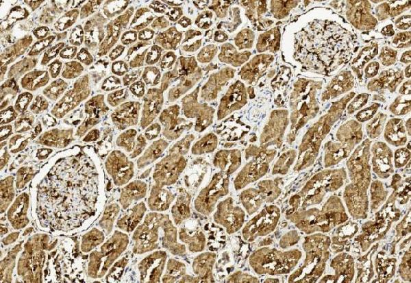
Click image to see more details
Figure 3. IHC analysis of ULK3 using anti-ULK3 antibody (PB9519).
ULK3 was detected in a paraffin-embedded section of human lung cancer tissue. Heat mediated antigen retrieval was performed in EDTA buffer (pH 8.0, epitope retrieval solution). The tissue section was blocked with 10% goat serum. The tissue section was then incubated with 1 μg/ml rabbit anti-ULK3 Antibody (PB9519) overnight at 4°C. Biotinylated goat anti-rabbit IgG was used as secondary antibody and incubated for 30 minutes at 37°C. The tissue section was developed using Strepavidin-Biotin-Complex (SABC) (Catalog # SA1022) with DAB as the chromogen.

Click image to see more details
Figure 4. Flow Cytometry analysis of U20S cells using anti-ULK3 antibody (PB9519).
Overlay histogram showing U20S cells stained with PB9519 (Blue line).The cells were blocked with 10% normal goat serum. And then incubated with rabbit anti-ULK3 Antibody (PB9519,1μg/1x106 cells) for 30 min at 20°C. DyLight®488 conjugated goat anti-rabbit IgG (BA1127, 5-10μg/1x106 cells) was used as secondary antibody for 30 minutes at 20°C. Isotype control antibody (Green line) was rabbit IgG (1μg/1x106) used under the same conditions. Unlabelled sample (Red line) was also used as a control.

Click image to see more details
Figure 5. Flow Cytometry analysis of THP-1 cells using anti-ULK3 antibody (PB9519).
Overlay histogram showing THP-1 cells stained with PB9519 (Blue line).The cells were blocked with 10% normal goat serum. And then incubated with rabbit anti-ULK3 Antibody (PB9519,1μg/1x106 cells) for 30 min at 20°C. DyLight488 conjugated goat anti-rabbit IgG (BA1127, 5-10μg/1x106 cells) was used as secondary antibody for 30 minutes at 20°C. Isotype control antibody (Green line) was rabbit IgG (1μg/1x106) used under the same conditions. Unlabelled sample (Red line) was also used as a control.
Protein Target Info & Infographic
Gene/Protein Information For ULK3 (Source: Uniprot.org, NCBI)
Gene Name
ULK3
Full Name
Serine/threonine-protein kinase ULK3
Weight
53.444kDa
Superfamily
protein kinase superfamily
Alternative Names
DKFZP434C131; EC 2.7.11.1; FLJ90566; serine/threonine-protein kinase ULK3; unc-51-like kinase 3 (C. elegans); Unc-51-like kinase 3 ULK3 unc-51 like kinase 3 serine/threonine-protein kinase ULK3
*If product is indicated to react with multiple species, protein info is based on the gene entry specified above in "Species".For more info on ULK3, check out the ULK3 Infographic

We have 30,000+ of these available, one for each gene! Check them out.
In this infographic, you will see the following information for ULK3: database IDs, superfamily, protein function, synonyms, molecular weight, chromosomal locations, tissues of expression, subcellular locations, post-translational modifications, and related diseases, research areas & pathways. If you want to see more information included, or would like to contribute to it and be acknowledged, please contact [email protected].
Specific Publications For Anti-ULK3 Antibody Picoband™ (PB9519)
Hello CJ!
No publications found for PB9519
*Do you have publications using this product? Share with us and receive a reward. Ask us for more details.
Recommended Resources
Here are featured tools and databases that you might find useful.
- Boster's Pathways Library
- Protein Databases
- Bioscience Research Protocol Resources
- Data Processing & Analysis Software
- Photo Editing Software
- Scientific Literature Resources
- Research Paper Management Tools
- Molecular Biology Software
- Primer Design Tools
- Bioinformatics Tools
- Phylogenetic Tree Analysis
Customer Reviews
Have you used Anti-ULK3 Antibody Picoband™?
Submit a review and receive an Amazon gift card.
- $30 for a review with an image
Be the first to review Anti-ULK3 Antibody Picoband™
*The first user to submit a review for a product is eligible for Boster's Innovating Scientists Reward, which gives product credits. This is in addition to the gift card reward.
Customer Q&As
Have a question?
Find answers in Q&As, reviews.
Can't find your answer?
Submit your question
1 Customer Q&As for Anti-ULK3 Antibody Picoband™
Question
We are currently using anti-ULK3 antibody PB9519 for human tissue, and we are well pleased with the Flow Cytometry results. The species of reactivity given in the datasheet says human, mouse, rat. Is it true that the antibody can work on goat tissues as well?
Verified Customer
Verified customer
Asked: 2020-01-02
Answer
The anti-ULK3 antibody (PB9519) has not been validated for cross reactivity specifically with goat tissues, though there is a good chance of cross reactivity. We have an innovator award program that if you test this antibody and show it works in goat you can get your next antibody for free. Please contact me if I can help you with anything.
Boster Scientific Support
Answered: 2020-01-02
