This website uses cookies to ensure you get the best experience on our website.
- Table of Contents
Boster takes pride in offering an annual scholarship aimed at supporting students pursuing or continuing a 2 to 4-year academic program related to Biology or Biotechnology. Our commitment to nurturing young scientists has led to the establishment of the Boster Bio Scholarships for Young Scientists of Excellence. Every year, this scholarship program grants $1,000 scholarships to exceptional graduating high school or college students to provide financial aid for their scientific research endeavors through their college education.
During the scholarship selection process, we consider the following in our criteria: proven interest in biomedical research, academic excellence, determination towards professional goals, and perseverance in challenging conditions.
As we carefully reviewed this year's exceptional and diverse pool of applicants, it was truly inspiring to witness the passion and dedication demonstrated by each and every one of them. Students from various academic backgrounds and research interests showcased their commitment to advancing the field of life sciences and making a positive impact on society.
After much discussion and a rigorous selection process, we are excited to announce the recipient of the 2023 Boster Bio Scholarship, whose dedication, hard work, and achievements have stood out from a highly competitive pool of applicants.
The 2023 Boster Bio Scholarship has been awarded to Divyathamizhini Kanakasabapathy!
We are delighted to recognize Kanakasabapathy’s exceptional abilities, academic excellence, and the potential to make a positive impact on the scientific community. Congratulations!
To all our applicants, thank you for sharing your passion for science with u...
Understanding the relative abundance of target proteins and effectively normalizing data are crucial aspects of Western blot analysis. These critical aspects are deeply rooted in the western blotting principle and procedure, which outline how proteins are separated...
We're delighted to bring you another issue of our Research Spotlight newsletter, showcasing the most recent papers our customers have published. These studies widen biological understanding and pave pathways for innovative therapies.
This month, we delve into a broad spectrum of biomedical research studies. We highlight research examining the intricate roles of the PD-L1/PD-1 pathway in neuroscience and the use of 3D bioprinting for biomimetic hydrogel scaffolds. Scientists demonstrate...

Compound screening is a service that enables rapid screening of thousands of compounds to identify a ‘hit’, or a compound that elicits a desired biochemical effect against a validated target or a phenotypic effect in cells. Compound screening can rapidly assess thousands of potential compounds and narrow down a list of ‘hits’ that can then be evaluated in more detail. Automated compound screening can complete this daunting task in a matter of hours which would otherwise take a team of researchers several days or months of laborious benchwork. There are a few types of compound screening, which we will briefly discuss below:
High-throughput screening is a highly automated process that allows for fast testing of hundreds of thousands of compounds in a library against a target or cell line for a particular biological or chemical effect [1]. The three general steps of high-throughput screening include: 1) Selecting a diverse, relevant library for testing, 2) setting up a suitable automated workflow with a robotics station, and 3) determining the method of acquiring and processing data. High-throughput screening usually occurs in a miniaturized format, such as in a 96-well, 384-well, or other plate format [1]. The miniaturization of the process enabled minute amounts of chemicals or drugs to be used in testing and the automation of the process allowed for a dramatic decrease in the time and labor needed to screen these compounds [1]. In fact, the process of quantitative high-throughput screening (qHTS) added an additional layer of complexity to high-throughput screening: testing varying concentrations of a single compound simultaneously to establish a dose curve [2]. By establishing a dose curve during screening, the rate of false negative and false positive hits has decreased [2]. Overall, high-throughput screening has dramatically improved the process of screening multitudes of compounds in a rapid fashion, helping researchers spend less time on labor-intensive screens and more time on validating intriguing hits.
While one of the advantages of high-throughput screening is the ability to screen through enormous libraries of compounds, focused screening, as the name suggests, narrows down the library to a smaller fraction of compounds [3]. If there is already some information on a target that suggests certain compounds would react with it in the desired fashion, focused screening allows for testing of a smaller list of compounds [3]. While this method may not work if there is not much data on a target or if the researcher wants to cast a wide net when searching for hits, it can certainly reduce the cost and the timeframe of screening if many compounds can already be eliminated from the initial screening process [3].
Virtual screening is a highly sophisticated process that considers the potential interactions of compounds and a target based on the current structural information of the target [3]. Similar to focused screening, this method can be used initially to narrow down a broad list of compounds to a more manageable list of potential compounds, all while conducting initial screens in silico [3].
Compound screening can be used in a variety of assays, from drug discovery to screening for compounds that target a receptor on cells to induce a signaling pathway. We will briefly discuss applications of compound screening in reporter cell lines, compound screening in drug discovery, as well as some practical considerations, such as timeline and cost for screening assays.
Many compound screens rely on examining the biochemical interaction between a target and a compound. However, other screens utilize cell culture in order to identify hits that affect receptors, ion channels on the cell surface, etc. [3]. Reporter cell lines in particular can prove useful when screening for hits. To support these assays, consider our curated Reporter cell lines — engineered for robust luciferase-based readouts in screening applications. For example, one study used cells overexpressing a validated target and a biosensor. When a compound in the screen would bind to the target receptor, a cascade of intracellular events resulted in luminescence, which could be measured and quantified in the assay [3,4]. Utilizing this reporter cell line in high-throughput screening enabled researchers to test numerous compounds quickly and simply through the evaluation of luminescence produced in vitro [3,4]. Other studies have used reporter cell lines in compound screening and identified hits by measuring luciferase in common murine and human cell lines [5]. To enhance delivery efficiency for transgene expression in reporter-based assays, AAV Packaging Service offers a reliable solution for generating high-titer viral vectors compatible with various reporter constructs.
For every drug that was discovered, there were countless potential compounds that were initially screened out. In the initial stages of drug discovery, compound screening is paramount in finding a few potential hits among thousands of compounds, especially since it already takes over a decade on average from the initial stages of research to the release of new drugs [3]. Compound screening, especially high-throughput screening, has improved the process of screening for potential drugs against targets. Quantitative high-throughput screening for compounds also improves the process, as it allows for screening of multiple concentrations of the drug and can decrease false positives and negatives in screens [2]. An ideal drug binds with high specificity to a target and works at a low concentration to minimize side effects. In this regard, high-throughput screening and quantitative high-throughput screening enable more rapid testing of compounds to find ‘hits’ that can be evaluated in vitro and in vivo.
In drug discovery, on average 200,000 to over 1 million compounds may be screened to narrow down a list of potential hits [3]. Once the list is narrowed after compound screening, select compounds are focused on and validated [3]. However, not every compound library contains over a million compounds to be tested. In fact, focused screening and physiological screening focus on a much smaller initial list, reducing the overall cost and time required for the screen.
Before a screen can begin, a target must first be identified and validated as biologically relevant to the disease or mechanism researchers aim to affect [3]. Through in vitro and in vivo work, the target should be validated in multiple ways that it plays an important role in the disease or mechanism of interest [3]. Once a target has been validated (or shown that it does play a crucial role in the disease or condition thr...
Cell lines are at the backbone of scientific research and have enabled countless discoveries and preclinical studies Although there are many cell lines available, researchers have many reasons to generate or modify their cell lines—perhaps to study a particular mutation in a cancer cell line or to determine the effects of knocking down a gene of interest.
To generate a stable cell line, there are three general steps:

Though there are different methods of stable cell line generation, each protocol follows these general steps. The most common methods of stable cell line generation include:
We will cover the specifics of each of these methods, as well as the advantages and disadvantages of each method in this article. When generating a cell line, it is important to consider if you require transient or stable expression of your gene of interest. Transient expression of a gene occurs when exogenous DNA introduced into a cell line is only temporarily expressed and does not integrate into the genome [1]. Stable expression of a gene means long-term, continued expression of a gene of interest [1]. Transient expression can be a better method when studying short-term effects in cells since this is a much faster method. However, if cells will be used over multiple experiments or studied for long-term changes, then stable expression is preferred [1].
Before generating a cell line, it is important to decide which type of cell is needed. First, let’s review the types of cells often used in research.
Primary cells are collected directly from patients (such as their blood, tumor, etc.), processed, and cultured. Compared to other cell types, primary cells are more heterogeneous and offer better biological relevance since they are derived directly from patients, though these cells cannot replicate as much and therefore cannot be cultured as long as other cell types [2].
Immortalized cells can grow indefinitely in culture [2]. Cells can be immortalized through the introduction of an immortalization gene (such as SV-40 large T antigen, hTERT, etc.), spontaneous immortalization through passaging, or due to an acquired ability to replicate indefinitely as seen in cancer cells [3-6]. The benefit of using an immortalized cell line is that they are much easier to work with in culture and have a much longer ‘lifespan’ as they can be cultured nearly indefinitely. However, immortalized cells often have less biological relevance compare...
Welcome to the June issue of Research Spotlight! We are happy to feature 10 selected publications from scientists whom Boster has served.
Recent neurological research has uncovered the impact of glioblastoma on human neural circuits, providing insights into decreased survival. In oncology, researchers revealed the role of hepatic stellate cell stearoyl co-A desaturase in promoting liver tumorigenesis through the leukotriene B4 receptor 2 - β-catenin cascade.
Exciting advancements...

Sodium citrate buffer, also known as citrate buffer, is a common reagent used for antigen retrieval in immunohistochemistry. Immunohistochemistry (IHC) is a technique that enables high-quality visualization of proteins in a variety of tissues. IHC is a multi-step process that comprises fixation, embedding, and sectioning of a target tissue followed by incubation with an antibody targeting a protein of interest.
Once the tissue is fixed, embedded in paraffin, and sectioned on slides, researchers can utilize antibodies to target and identify their protein of interest. However, fixation and embedding of tissues, often with 10% formalin and paraffin respectively, initiates cross-linking of proteins that can mask antigen epitopes. Antigen masking may reduce the sensitivity of antibodies and decrease the detection of certain proteins of interest. A process known as antigen retrieval breaks the bonds between formalin and proteins, removing the effects of cross-linking, thereby enabling better detection of proteins. Sodium citrate buffer is highly used in the process of antigen retrieval, specifically for heat-induced antigen retrieval. Boster Bio offers a simple-to-use Sodium Citrate Buffer (Catalog# AR0024) for heat-induced antigen retrieval to remove the effects of antigen masking in your IHC experiments.
Antigen retrieval is a process of breaking the bonds from cross-linking, enabling proteins to interact with antibodies during immunohistochemistry. The purpose of antigen retrieval in immunohistochemistry is to unmask antigens in fixed tissues, which can significantly improve the quality of staining and detection of proteins. While optimization of this method may add additional steps to an already multi-step, complex procedure, antigen retrieval has improved IHC...

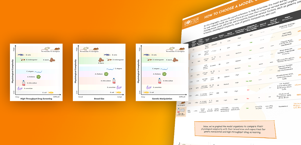
Download and share our “How to Choose a Model Organism” infographic! We offer an overview of model organisms in research to help you determine which one is most compatible with your research goals.
In the infographic, you will see a comparison of various model organisms, including mouse, yeast, zebrafish, dro...
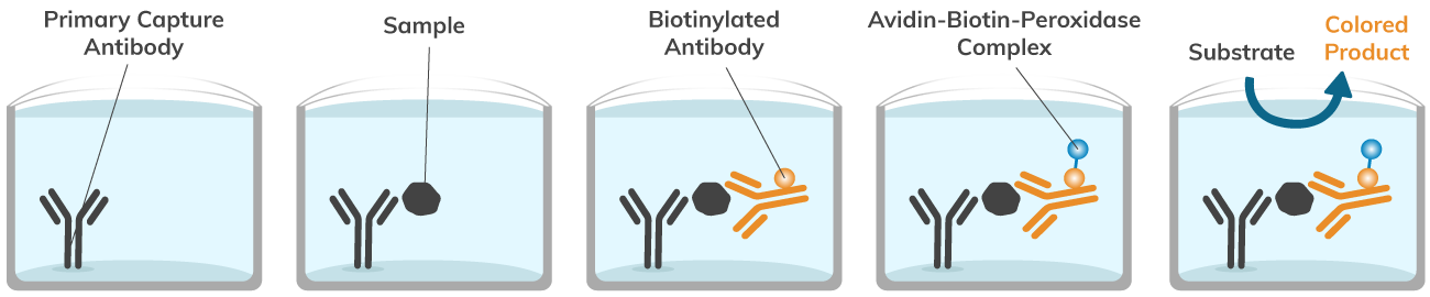
The avidin-biotin interaction remains one of the strongest non-covalent interactions between molecules, even surpassing the strength of antibody-antigen interactions. Interactions between these two molecules have been utilized in the laboratory for countless applications, such as immunohistochemistry (IHC), immunoprecipitation (IP), affinity purification, enzyme-linked immunosorbent assay (ELISA), and more—including advanced techniques like Multiplex Assay Services that allow simultaneous detection of multiple targets. The avidin-biotin-peroxidase complex has been used in countless assays for its simple and effective procedure, incomparable strength of binding between molecules, and ability to label and quantify lowly abundant proteins of interest in experiments. Boster Bio conveniently offers an Avidin-Biotin-Peroxidase Complex for ELISA kit (Catalog# AR1103), complete with necessary reagents, f...
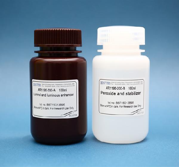
Western blotting (also called Protein Immunoblotting) is an analytical technique used to detect specific proteins in the given sample. A deep understanding of the western blot principle provides the essential foundation for applying this method successfully in protein detection. It uses SDS-polyacrylamide gel electrophoresis (SDS-PAGE) to separate various proteins contained in the sample. The separated proteins are then transferred or blotted onto a matrix, where they are stained with antibodies specific to the target protein. Expression details of the target proteins in the given cells or tissue homogenate can then be obtained through analyzing the location and intensity of the specific reaction. Western blotting analysis can detect target protein as low as 1 ng due to high resolution of the gel electrophoresis and strong specificity and high sensitivity of the immunoassay. This method is used in the fields of molecular biology, biochemistry, immunogenetics and other molecular biology disciplines for various experiments. Researchers who prefer standardized, high-sensitivity workflows often rely on a professional western blot service to ensure consistent and reproducible results.
Enhanced Chemiluminescence (ECL) Western Blot Substrate is a very sensitive, non-radioactive, enhanced luminol-based chemiluminescent substrate that allows for easy detection of horseradish peroxidase (HRP) on immunoblots. HRP is a common molecule conjugated to antibodies. ECL Western Blot Substrate has the capability...