This website uses cookies to ensure you get the best experience on our website.
- Table of Contents
Recombinant DNA technology is used to produce antibodies by assembling, expressing, and purifying antibody genes in vitro. This approach supports consistent, scalable antibody production without the need for animal immunogen preparation. It is widely applied in research, diagnostics, and therapeutic development, offering advantages in assay reliability and customization across the immune system.
Recombinant DNA technology involves combining DNA sequences from different sources to create new genetic constructs. In the context of antibody production, this means taking the genetic code that encodes an antibody’s variable and constant regions, inserting it into an expression vector, and producing the antibody in a host cell system such as mammalian cells, yeast, or bacteria.
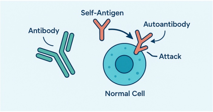
Roughly 10% of people worldwide live with an autoimmune condition, and more than 80 of these disorders trace at least part of their pathology to antibodies that have turned on the body. These rogue molecules—often referred to as autoantibodies—trigger inflammation, degrade healthy tis
Proteins are the workhorse molecules that drive virtually every biological system. With the increasing recognition of the role of proteins in various research and manufacturing activities, simply isolating them from their natural host cells cannot meet the escalating demand of the market. Chemical synthesis is also not a viable option for this endeavor due to the size and complexity of proteins. Instead...
The domestic chicken (Gallus gallus domesticus) has been a model organism for scientific research due to its accessibility, ease of breeding, and large-sized eggs, which are particularly useful for embryological studies.
In this blog, we describe a brief history and some key breakthroughs of chicken as a model organism. We discuss the research advantages and limitations of chickens, and describe some research areas where scientists have explored using chickens. For researchers interested in working with chickens, we have provided a list of resources and guiding questions. If you’re considering choosing chickens for your research studies or simply want to learn more about chickens, this blog is for you!
Feel free to jump to a specific section about chicken:
Gallus gallus domesticus, commonly known as the domestic chicken, is a bird species widely used in developmental biology and genetic research. As a subspecies of the red junglefowl, which is native to southern Asia, the domestic chicken is versatile and economically significant. Adult chickens typically measure between 40 to 60 centimeters in length from beak to tail and weigh around 2 to 4 kilograms. They are characterized by their feathered bodies, beaks, and a range of plumage colors and patterns.
Gallus gallus domesticus are chosen as research models due to its well-understood developmental processes and genetic makeup. Chickens have a relatively long incubation period of about 21 days from egg fertilization to hatching, during which the embryos can be observed and manipulated. This extended embryonic development allows for detailed studies of developmental biology, including organ formation and genetic regulation. Furthermore, the chicken genome was sequenced in 2004, presenting a comprehensive resource for genetic studies. This genome facilitates research into gene function, evolution, and disease mechanisms.
The chicken is examined in immunology and vaccine development due to its production of large amounts of antibodies. Additionally, its large eggs make it a favored model for studying early embryonic development and the effects of genetic and environmental factors on growth and differentiation. Overall, Gallus gallus domesticus offers valuable insights into vertebrate biology and developmental processes, bridging the gap between simpler model organisms and more complex mammalian systems.
Gallus gallus domesticus, the domestic chicken, has been instrumental in several key scientific breakthroughs, especially in embryology, virology, genetics, and developmental biology. We discuss some of the most notable milestones below.
In 1910, Peyton Rous discovered the Rous sarcoma virus (RSV) in chickens, marking one of the first demonstrations that viruses could cause cancer. This discovery crucially linked viruses and cancer, fundamentally changing our comprehension of carcinogenesis. The identification of RSV in chickens eventually led to the discovery of oncogenes, which are genes that can cause normal cells to become cancerous.1 Rous's work was so influential that he was awarded the Nobel Prize in Physiology or Medicine in 1966.2
The chicken embryo became a key model in embryology starting in the early 20th century. In the 1920s and 1930s, Viktor Hamburger’s work with chicken embryos was foundational in determining the stages of vertebrate development. His research, in collaboration with Howard L. Hamilton, led to the establishment of the Hamburger-Hamilton stages in 1951, a detailed series of 46 stages that describe the chronological development of chick embryos from the laying of the egg to hatching. This work became a standardized system for describing embryonic development in chickens and presented a vital framework for studying developmental processes across vertebrates.3,4
Chickens have played a critical role in immunology, notably in the development of vaccines. For instance, the use of chicken eggs in the production of vaccines for diseases such as influenza has been a significant breakthrough.5 The ability to produce large quantities of virus in chicken eggs has been crucial for the rapid development and distribution of vaccines, especially during pandemics.
In the 20th century, chickens gained prominence in genetics research. The establishment of inbred chicken strains empowered scientists to investigate genetic variation and inheritance patterns.6 Studies on chickens uncovered quantitative genetics related to traits such as disease resistance and growth.7,8
The sequencing of the chicken genome in 2004 was a major milestone in genetic research. The chicken was the first bird and the first agricultural animal to have its genome fully sequenced.9 This sequencing clarified the evolution of vertebrates, revealing how birds, including chickens, evolved from dinosaur ancestors. The chicken genome has also been used to examine gene function and genetic diseases, providing a comparative framework for understanding human genetics.
Chickens have been employed in genetic engineering and transgenics. The development of transgenic chickens, which carry foreign genes inserted into their genome, has allowed researchers to unravel gene function and regulation in a vertebrate model.10 This technology has implications for both basic research and the development of biopharmaceuticals.
The domestic chicken has been a valuable model organism, contributing to major scientific breakthroughs in various fields. From developmental biology to cancer research and vaccine production, the use of Gallus gallus domesticus has deepened our comprehension of fundamental biological processes and their applications in medicine and agriculture.
Gallus gal
...

Antibodies, also known as immunoglobulins, are specialized proteins produced by the immune system to identify and neutralize foreign invaders such as bacteria, viruses, and toxins. Their ability to recognize and bind to specific antigens make them play a critical role in the immune response and serve as key tools in biomedical research, including advanced applications like...

Antibodies do more than stick to germs. They block infection, label threats for cleanup, and spark immune system responses that keep the body safe.
The antibody action mechanism refers to the step-by-step process an antibody follows once it binds to a target antigen. The protein’s antibody structure enables each molecule to recognize a target with pinpoint precision and then trigger a robust immune response. Over the past several years, monoclonal antibodies have become a dominant format in therapeutic development, highlighting the importance of antibody engineering and antibody production in clinical applications. To understand why, we can examine four main mechanisms: neutralization, opsonization, complement-dependent cytotoxicity, and antibody-dependent cellular cytotoxicity (ADCC)
Binding of antibodies is only the first move. The antibody’s variable region grabs the antigen, while the Fc region faces outward—a process often studied using recombinant protein expression systems. That outward portion connects to immune receptors, starts signaling pathways, and guides the next steps. Signals can recruit other proteins, attract B cells or T cell populations, or alter the shape of the antibody itself. These downstream events decide whether the invader is blocked, swallowed, or destroyed. Such outcomes vary depending on the antibody class, the type of antigen, and the surrounding tissue environment.
Antibodies protect the body through the following mechanisms that block, tag, or destroy harmful targets:
Neutralization acts like closing a door before a burglar walks in. An antibody covers the exact part of a virus, toxin, or bacterium that would usually latch onto a host receptor. Viruses use viral proteins to attach to and fuse with tumor cells or human tissues. Toxins require antigen-binding site access to en...
Variable and constant regions give antibodies their precision and power. Explore how each component contributes to antigen recognition, immune signaling, and the development of therapeutic antibodies used in modern medicine.
The antibody structure enables each molecule to recognize a specific target. This target is usually a unique part of a foreign substance, such as a virus, bacterium, or toxin. Once identified, the antibody helps initiate the appropriate immune response to neutralize or eliminate the threat.
Precision and action work together because of the protein's shape. Each antibody has a variable fragment that binds specifically to its target. This precise binding ensures that the immune system responds only to harmful invaders, thereby preventing it from reacting to harmless substances. After binding, antibodies can block the target’s function, mark it for destruction, or activate other parts of the immune system. These actions help the body defend itself efficiently against a wide range of threats.
Monoclonal antibodies have become one of the most widely used formats in drug development over the past two decades. In recent years, regulatory agencies have continued to approve more mAbs each year, underscoring their importance in research and clinical practice. Many of these therapies perform better after scientists adjust their variable or constant domains. Comparing the two regions, we can see why antibodies bind so accurately and why this accuracy is crucial in both research and clinical care.
Antibodies, also known as immunoglobulins, are Y-shaped proteins built from two identical heavy and light chains. Disulfide bonds and disulfide bridges hold the chains together, giving the molecule stability and flexibility. Each chain contains a variable region at the tip and a constant region closer to the base. The variable region forms the antigen-binding site, whereas the constant region controls downstream immune activity.
Antigen recognition starts in the variable region, where sequence diversity produces unique binding surfaces.
Variable regions sit at the ends of both heavy and light chains. Within them, three complementarity-determining regions (CDRs) on each chain create the contact points for an antigen. Framework segments surround the CDRs, supporting the loops that enable binding. These hypervariable regions are critical for ensuring specificity.
When an antibody encounters its target, the six CDRs come together to form a grip on the antigen, much like a lock and key. That tight fit determines affinity, which influences how well the antibody can neutralize or flag the threat, an essential part of the antibody-antigen interaction.
Once the variable region locks onto a target, the constant region calls in reinforcements.
Constant regions extend from the hinge region of the antibody to the base of the heavy and light chains. In heavy chains, the constant segment determines the antibody isotypes, such as IgM antibodies IgG or IgA, each with distinct immune functions.
The constant region interacts with Fc fragment receptors on immune cells, activates the complement cascade, and influences serum half-life. For instance, IgG1 antibodies bind to Fcγ receptors on macrophages, guiding phagocytosis, while IgG4 has reduced effector activity, making it useful in chronic inflammatory settings.
Although neighbors within the same molecule, the two regions differ in almost every way.
| Feature | Variable Region | Constant Region |
|---|---|---|
| Primary role | Antigen recognition | Immune signalling, stability |
| Sequence variability | High | Low within an antibody class |
| Location | Tips for heavy chains and light chains | Lower arms and stem of the Y-shape |
| Flexibility | Greater to accommodate diverse targets | More rigid to maintain structural integrity |
| Engineering focus | Affinity tuning, specificity of antibody | Half-life, effector modulation |
Knowledge of the region function guides everything from laboratory assays to drug design.
Researchers often isolate variable regions to construct Fab fragments, single-chain variants, or nanobodies for use in imaging and diagnostic tests. Smaller antibody fragments retain antigen-binding activity specificity while reducing background noise.
Drug developers adjust constant regions to change how an antibody behaves in the body. For example, modifying Fc fragment domains of IgG molecules can extend half-life via neonatal Fc receptor recycling. Conversely, reducing Fc receptor binding can dampen inflammatory responses in therapeutic antibodies used to treat autoimmune diseases.
Tailoring both regions lets scientists create bispecific antibodies, antibody-drug conjugates, and chimeric antigen receptor constructs. Each relies on precise control of antigen binding and effector functional activity to improve safety and potency. Some designs also incorporate the J chain, especially in polymeric antibodies like IgM and IgA, influencing their quaternary structure and immune function.
Variable and constant regions work together, yet their tasks remain distinct. The variable portion seeks out the threat, while the constant portion signals for help. A clear understanding of both parts enables researchers to fine-tune antibodies for use in science, diagnostics, and treatment.
This includes optimizing the hinge region for structural flexibility, improving hypervariable regions for more accurate antigen recognition, and refining the amino acid sequence of polypeptide chains...
Antibody types form the foundation of adaptive immune system. In humans, IgG alone accounts for approximately 75% of circulating antibodies, while the other four classes—IgM antibodies, IgA, IgE, and IgD—fill specialized roles in blood, tissues, and secretions. Together, these Y-shaped glycoproteins create a flexible defense network that identifies, neutralizes, and clears pathogens with remarkable precision.
All antibodies share a common heterotetrametric structure consisting of heavy and lights connected by disulfide bonds in the hinge regions but variations in their constant regions define each antibody isotype. Understanding these differences is critical for selecting the right reagents for immunodiagnostic tests, designing effective vaccines, and developing therapeutic monoclonal antibodies. Each type plays a specific role in the adaptive immune response, inf...


