This website uses cookies to ensure you get the best experience on our website.


- Table of Contents
Plan chromogenic MAPK1 IHC in paraffin sections around the cytoplasmic and nuclear staining seen in most tissues (HPA tissue IHC). Use consistent fixation and score the two compartments separately, since phosphorylation can increase nuclear localization (UniProt).

Expected localisation, validated protocols, controls and antibodies — the at-a-glance facts below, then the full design guide.
| Expected localisation | Cytoplasmic and nuclear staining in most tissues (HPA tissue IHC) | |
| Staining pattern | Most tissues show cytoplasmic and nuclear staining (HPA tissue IHC) | |
| Antigen retrieval | Tris-EDTA pH 9.0 HIER, 95–98 °C, 20 min (rule: nuclear antigen) | |
| Positive control | Adipose tissue+4 more · see all | |
| Negative control | None in HPA (detected in all 45 tissues); use no-primary + isotype controls |
| Fixation | Keep fixation conditions consistent across paraffin sections. (standard IHC practice; not target-specific) | |
| Caveat | Phosphorylation can increase nuclear localization (UniProt) | |
| Regulation | Expression regulation is not annotated (UniProt) | |
| Isoform / epitope | 2 isoforms; check antibody epitope coverage (UniProt) |
The catalog antibody’s IHC-P protocol is followed by published MAPK1 IHC protocols for parathyroid adenoma, breast cancer, and mouse liver (PMC11279064; PMC5591392; PMC12587933).
| Sample | Paraffin-embedded Human melanoma tissue; fixative not specified (datasheet M00030) |
| Fixation | Image fixative and duration unreported (datasheet M00030); verify before use. |
| Sectioning | 4–5 µm sections on charged slides (standard) |
| Deparaffinisation | Xylene, graded ethanol series to water (standard) |
| Antigen retrieval | Heat-induced epitope retrieval in Tris-EDTA buffer, pH 9.0, 20 min at 95–98 °C (standard rule: nuclear antigen) |
| Peroxidase block | 3% H2O2, 10 min, room temperature (standard) |
| Blocking | 10% normal serum of the secondary host, 30 min, room temperature (standard) |
| Primary antibody | Rabbit monoclonal (clone II-13) anti-MAPK1, 1:50 recommended; image 1:100 (datasheet M00030) |
| Primary incubation | Overnight at 4 °C (standard) |
| Detection | HRP-polymer secondary, DAB chromogen 5–10 min (standard) |
| Counterstain | Hematoxylin, blue, dehydrate and mount (standard) |
| Expected result | MAPK1-positive staining in adipocytes of adipose tissue (HPA tissue IHC: High). HPA tissue profile: Cytoplasmic and nuclear expression in most tissues. No signal in the no-primary control. |
MAPK1/ERK2 is expected in the cytoplasm and nuclei of many cell types; HPA reports cytoplasmic and nuclear staining in most tissues, with high staining in selected adipocytes, epithelial cells, glia, and neurons (HPA: tissue IHC). Its lack of a transmembrane segment supports intracellular interpretation (UniProt P28482 topology). HPA rates the tissue profile Enhanced but reports only medium consistency between antibody staining and RNA expression (HPA: tissue IHC).
| Cytoplasmic and nuclear chromogen in adipocytes, bronchial respiratory epithelium, or cortical neurons (HPA: High in these cells). | This fits the broad tissue pattern (HPA: cytoplasmic and nuclear expression in most tissues). Judge intensity within identifiable cells and alongside controls; a nuclear-to-cytoplasmic difference alone does not establish a change in total MAPK1 abundance (UniProt P28482 subcellular location). |
| Predominantly nuclear or cytoplasmic staining within otherwise credible positive cells. | Either distribution can be plausible: MAPK1 occupies both compartments, and phosphorylation or binding partners can affect its localisation (UniProt P28482 subcellular location). Interpret a shift in the context of the antibody and specimen; total-MAPK1 staining alone does not identify the phosphorylated species (UniProt P28482 modified residues). |
| An isolated extracellular deposit or broad acellular chromogen is scored as MAPK1. | That is a compartment mismatch: MAPK1 has no transmembrane segment and is documented in intracellular locations, with additional caveola and focal-adhesion association (UniProt P28482 topology and subcellular location). Reassess deposits as possible IHC artefact before assigning a cellular score (general IHC practice). |
| Strong staining appears chiefly in cells reported as low, such as esophageal squamous epithelium or smooth muscle (HPA: Low). | Low means some staining is possible; it is not a negative-control designation (HPA: tissue IHC). An unexpected dominant signal warrants checks for antibody cross-reactivity, tissue pigment, or endogenous detection activity (general IHC practice). |
| No cellular signal appears in a well-preserved section containing bronchial respiratory epithelial cells (HPA: High). | This conflicts with the HPA reference pattern but does not by itself prove MAPK1 absence (HPA: High; tissue reliability Enhanced, medium RNA–staining consistency). Check assay controls and section handling before interpreting the specimen as negative (general IHC practice). |
| Reference cells and tissue range | HPA reports High staining in adipocytes, bronchial respiratory epithelium, cortical neurons, glia, and other listed cells; it reports Low staining in several other cell groups and lists no negatives (HPA: tissue IHC). Choose a documented high-staining cell population when judging whether the run produced the expected pattern. |
| Localisation and signalling state | MAPK1 is reported in cytoplasm and nucleus, with spindle, centrosome, caveola, and focal-adhesion associations; retention and phosphorylation can influence localisation (UniProt P28482 subcellular location). A nuclear fraction is therefore plausible, but distribution alone cannot establish pathway activation or the abundance of a particular phospho-form (UniProt P28482 modified residues). |
| Antibody and molecular scope | The HPA tissue profile is Enhanced, while individual listed IHC antibodies are Approved except CAB079933, which is Enhanced (HPA: antibodies; tissue IHC). MAPK1 has two isoforms and multiple modified residues; those facts do not establish which forms any given antibody detects (UniProt P28482 isoforms and modified residues). Check the selected antibody's stated specificity before comparing stains. |
| IF/ICC Q&A: Should its localisation set the IHC scoring rule? | No. HPA IF/ICC reports mainly nucleoplasmic MAPK1, plus cytosol and other locations, but cautions that its subcellular summary uses antibodies targeting proteins from multiple genes (HPA: subcellular ICC-IF). Use the cytoplasmic-and-nuclear tissue IHC profile for paraffin-section interpretation (HPA: tissue IHC). |
| Situation | Likely cause | Next action |
|---|---|---|
| The documented high-staining cells are blank (HPA: tissue IHC). | A failed run or weak detection is possible; a blank section alone does not identify the cause (general IHC practice). | Confirm that the chosen cells are present, then inspect the run controls, primary-antibody application, chromogen development, and counterstain (general IHC practice). HPA gives reference staining, not a target-specific retrieval requirement (HPA: tissue IHC). |
| The whole section has a brown haze that obscures cell borders. | Non-specific reagent retention or detection background can obscure a cellular pattern (general IHC practice). | Compare with an appropriate no-primary control; review blocking, washing, detection reagents, and development time (general IHC practice). Score MAPK1 only where cellular staining remains distinguishable (HPA: cytoplasmic and nuclear tissue pattern). |
| Pigmented or blood-rich areas seem positive despite weak cellular staining. | Intrinsic pigment or endogenous enzyme activity can mimic chromogenic signal (general IHC practice). | Inspect the no-primary control and tissue morphology; apply the detection system's endogenous-activity control where relevant (general IHC practice). Do not count acellular colour as cytoplasmic or nuclear MAPK1 (HPA: tissue IHC). |
| Only nuclei stain, or nuclei stain more strongly than cytoplasm. | A nuclear component is biologically plausible; localisation can vary with MAPK1 regulation (UniProt P28482 subcellular location). | Check that the signal is in intact cells and that controls are acceptable (general IHC practice). Record the compartmental pattern without calling activation unless the antibody is validated for the relevant phosphorylated form (UniProt P28482 modified residues). |
| A low-staining reference cell group gives the section's strongest signal (HPA: Low). | The pattern may reflect specimen variation, cross-reactivity, or detection background; HPA Low is not equivalent to absent (HPA: tissue IHC; general IHC practice). | Compare the same run with a documented HPA-high population and an appropriate control; review the selected antibody's validation before attributing the difference to MAPK1 (HPA: tissue IHC and antibodies; general IHC practice). |
| Two antibodies yield different nuclear-to-cytoplasmic patterns. | HPA assigns antibody-specific IHC validation levels, and MAPK1 has multiple isoforms and modified residues; neither fact alone explains the disagreement (HPA: antibodies; UniProt P28482 isoforms and modified residues). | Compare each antibody's stated target and validation, then inspect matched controls and cellular morphology (general IHC practice). Report the disagreement rather than merging the patterns into one MAPK1 score. |




Comprehensive Human Protein Atlas IHC scoring per tissue (reliability: Enhanced — Medium consistency between antibody staining and RNA expression data.). Rows are taken directly from the HPA tissue chart — click any row's HPA link to view the source.
| Tissue | Cell type | Level | Evidence | Source |
|---|---|---|---|---|
| Adipose tissue | Adipocytes | High | Protein (IHC) | HPA → |
| Adrenal gland | Glandular cells | High | Protein (IHC) | HPA → |
| Breast | Adipocytes | High | Protein (IHC) | HPA → |
| Bronchus | Respiratory epithelial cells | High | Protein (IHC) | HPA → |
| Caudate | Glial cells | High | Protein (IHC) | HPA → |
| Tissue | Cell type | Level | Evidence | Source |
|---|---|---|---|---|
| None in HPA: MAPK1 is detected in all 45 scored tissues. Use a no-primary (secondary-only) and an isotype control instead. | ||||
Troubleshoot MAPK1 staining in paraffin sections by checking retrieval, compartment pattern, controls and scoring before interpreting differences between samples.
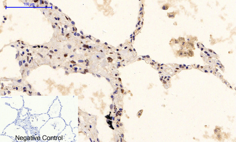
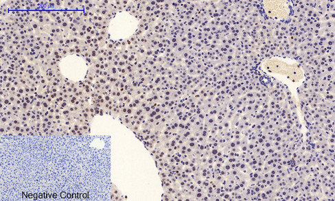
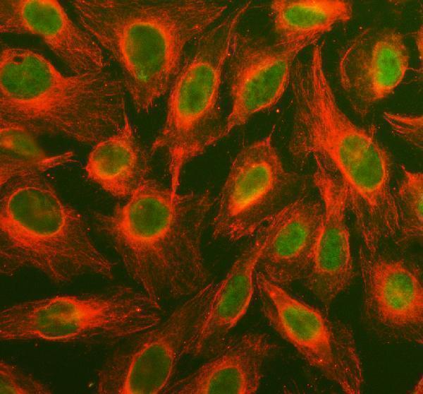
Catalog images show MAPK1/ERK2 IHC in human, mouse and rat paraffin sections (M00030 IHC captions) and MAPK1/3 IF in U2OS cells (PA1049 IF caption).
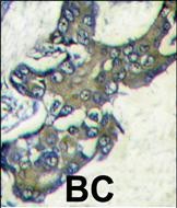
M00030 has IHC images from paraffin-embedded human melanoma and renal cancer, rat lung and mouse ovary (M00030 IHC captions); A00030Y222 has IHC images from paraffin-embedded human lung and appendix, rat lung and mouse liver (A00030Y222 IHC captions). A00030 has IHC-P data from formalin-fixed, paraffin-embedded human breast carcinoma (A00030 IHC caption); PA1049 has IF/ICC data from U2OS cells (PA1049 IF caption).
Which to pick: For tissue IHC across human, mouse and rat, consider M00030: it is monoclonal clone II-13 with IHC listed and paraffin-section images in all three species; its captions do not report a fixative (M00030 catalog and IHC captions). For human paraffin-section, paraffin-embedded tissue, A00030 has direct breast carcinoma DAB evidence; choose A00030Y222 when phospho ERK1/2 is the target, noting that its paraffin-section captions do not report a fixative (A00030 IHC caption; A00030Y222 title and IHC captions). For IF/ICC, PA1049 has a U2OS cell image and both applications listed, while M00030 also lists IF and ICC (PA1049 IF caption and applications; M00030 applications). The selected M00030 tissue-IHC caption documents paraffin sections, but does not specify the fixative (selected-SKU IHC image M00030).