This website uses cookies to ensure you get the best experience on our website.


- Table of Contents
Plan chromogenic RELA IHC on paraffin sections using PA1669 at 2–5 μg/mL (datasheet: PA1669). Assess the widespread cytoplasmic tissue pattern (HPA tissue IHC) while allowing for nuclear translocation after activation (UniProt).

Expected localisation, validated protocols, controls and antibodies — the at-a-glance facts below, then the full design guide.
| Expected localisation | Cytoplasmic staining in tissue sections (HPA tissue IHC) | |
| Staining pattern | Widespread cytoplasmic staining across cell types (HPA tissue IHC) | |
| Antigen retrieval | EDTA pH 8.0 HIER, heat-mediated (datasheet PA1669) | |
| Positive control | Appendix+4 more · see all | |
| Negative control | None in HPA (detected in all 45 tissues); use no-primary + isotype controls |
| Fixation | Keep fixation consistent across sections (standard IHC practice; not target-specific) | |
| Caveat | Activation can shift RELA into nuclei (UniProt) | |
| Regulation | Activation shifts nuclear staining (UniProt) | |
| Isoform / epitope | Four isoforms; verify epitope coverage (UniProt) |
The catalog antibody uses EDTA pH 8.0 retrieval (datasheet PA1669). Published RELA IHC protocols cover ovine thymus (PMC9587240) and mouse renal tissue (PMC9892181).
| Sample | Paraffin-embedded human breast cancer tissue; fixative not specified (datasheet PA1669) |
| Fixation | Image fixative and duration unreported (datasheet PA1669); verify before use. |
| Sectioning | 4–5 µm sections on charged slides (standard) |
| Deparaffinisation | Xylene, graded ethanol series to water (standard) |
| Antigen retrieval | Heat retrieval: EDTA pH 8.0 (datasheet PA1669); 20 min, 95–100 °C (standard) |
| Peroxidase block | 3% H2O2, 10 min, room temperature (standard) |
| Blocking | 10% goat serum (datasheet PA1669) |
| Primary antibody | Rabbit anti-RELA, 2-5μg/ml (datasheet PA1669) |
| Primary incubation | Overnight at 4 °C (datasheet PA1669) |
| Detection | HRP-conjugated secondary, DAB chromogen (datasheet PA1669) |
| Counterstain | Hematoxylin, blue, dehydrate and mount (standard) |
| Expected result | RELA-positive staining in lymphoid tissue of appendix (HPA tissue IHC: High). HPA tissue profile: Ubiquitous cytoplasmic expression. No signal in the no-primary control. |
RELA is mainly cytoplasmic in the tissue IHC profile, with nuclear localisation possible after activation (HPA: ubiquitous cytoplasmic expression; UniProt Q04206: nucleus and cytoplasm). Expect staining across many cell types, including strong staining in appendix lymphoid tissue, bone marrow hematopoietic cells, and several glandular epithelia (HPA: High in these cells). The tissue profile is Supported, with medium consistency between staining and RNA data (HPA: reliability Supported). RELA has no transmembrane segment (UniProt Q04206 topology).
| Cytoplasmic staining across many cells, with strong signal in appendix lymphoid tissue or bone marrow hematopoietic cells. | This fits the reported tissue pattern; compare intensity within the same run because staining can vary by cell type (HPA: ubiquitous cytoplasmic expression; High in these cells; standard IHC practice). |
| Nuclear staining appears in a subset of otherwise plausible positive cells. | Nuclear RELA can reflect activation and translocation; nuclear staining alone does not establish pathway activation without a suitable experimental comparison (UniProt Q04206: cytoplasmic inhibitor complex and nuclear translocation). |
| Signal is confined to cell membranes or extracellular material, with no convincing intracellular staining. | That distribution conflicts with the reported cytoplasmic and nuclear locations and warrants an artefact check; RELA has no transmembrane segment (HPA: cytoplasmic expression; UniProt Q04206 topology and subcellular location). |
| Strong staining appears in a cell population expected to be weak, while nearby expected-positive cells are unstained. | Consider cross-reactivity or endogenous chromogenic detection activity before assigning the signal to RELA; some populations have low reported staining, but HPA does not define them as negative controls (HPA: Low in selected cells; standard IHC practice). |
| Colour covers much of the section without clear cell boundaries, or a known high-staining tissue has no signal. | Diffuse colour suggests background, while absent signal calls for a control and detection check; neither observation alone establishes RELA absence (HPA: High in appendix lymphoid tissue and bone marrow hematopoietic cells; standard IHC practice). |
| Activation state and compartment | Inactive RELA can be retained in the cytoplasm by I-kappa-B; activation can release the complex for nuclear translocation, so record cytoplasmic and nuclear staining separately (UniProt Q04206 function and subcellular location). |
| Tissue and cell selection | Appendix lymphoid tissue, bone marrow hematopoietic cells, and breast or colon glandular cells are reported High; selected endothelial, glial, respiratory, and other cells are Low (HPA: tissue IHC). |
| Antibody evidence | CAB004264 and CAB005030 each have Supported IHC status; HPA calls the overall tissue profile Supported with medium staining–RNA consistency, so retain tissue controls when interpreting an unexpected pattern (HPA: antibody validation; tissue reliability). |
| Isoforms and epitope | Four RELA isoforms are listed, but this payload gives no antibody epitope or isoform coverage; an isoform-specific explanation for a staining difference is therefore unestablished (UniProt Q04206: four isoforms). |
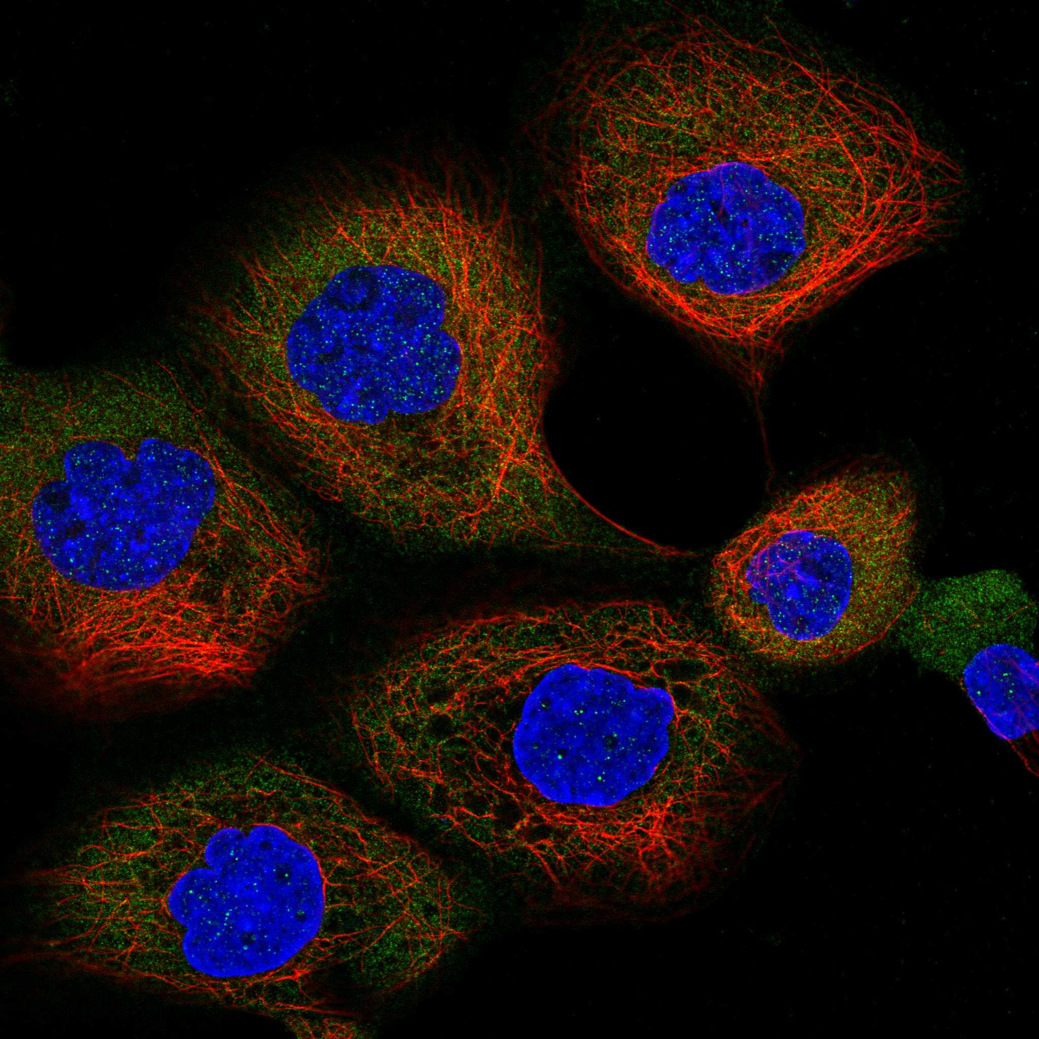
| IF/ICC Q: Where should RELA appear? | A: Cytosolic signal is the reported baseline, with nuclear localisation biologically possible after stimulation; assess IF/ICC on its own guide page (HPA: Cytosol, enhanced, in ICC-IF; UniProt Q04206: nuclear translocation). |
| Situation | Likely cause | Next action |
|---|---|---|
| Only membrane-like or extracellular colour is visible. | The distribution does not match reported RELA compartments; nonspecific deposition or detection artefact is possible (UniProt Q04206 topology and location; standard IHC practice). | Check the no-primary control and inspect whether colour follows cell interiors before scoring RELA (standard IHC practice). |
| Nuclear staining seems unexpectedly strong. | RELA can enter nuclei after activation, so the compartment alone cannot distinguish activation from an assay problem (UniProt Q04206 function). | Score nuclear and cytoplasmic compartments separately and compare matched conditions plus controls; avoid calling activation from one section alone (standard IHC practice; UniProt Q04206 function). |
| A nominally low-staining cell type shows strong colour. | Cell-type assignment may be wrong, or antibody cross-reactivity or endogenous detection activity may contribute (HPA: Low in selected cell types; standard IHC practice). | Recheck morphology and the HPA cell-level entry; examine no-primary and detection controls before attributing the colour to RELA (HPA: tissue IHC; standard IHC practice). |
| The whole section has diffuse chromogenic colour. | Background may arise from insufficient blocking, incomplete washing, or endogenous detection activity (standard IHC practice). | Inspect negative controls, then optimise blocking, washes, antibody dilution, and detection conditions using the assay's instructions (standard IHC practice). |
| Appendix lymphoid tissue or bone marrow hematopoietic cells show no convincing signal. | These are reported High, making an assay or specimen issue plausible; an absent result alone cannot identify its cause (HPA: High in these cells; standard IHC practice). | Verify positive-control staining and run records; review retrieval, antibody dilution, detection reagents, and counterstain without assuming a RELA-specific fixation effect (standard IHC practice). |
| Two sections give different proportions of nuclear and cytoplasmic staining. | Activation context can alter RELA location, while section and detection variation can complicate comparison (UniProt Q04206 function; standard IHC practice). | Compare equivalent cell populations under matched staining conditions, document each compartment, and interpret differences against experimental controls (standard IHC practice). |


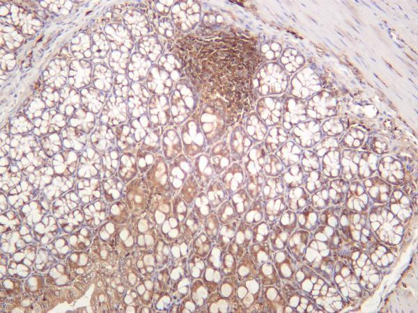
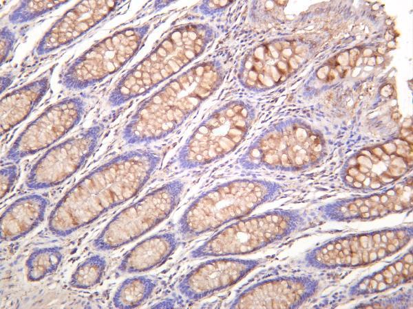
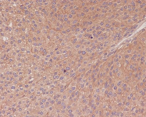
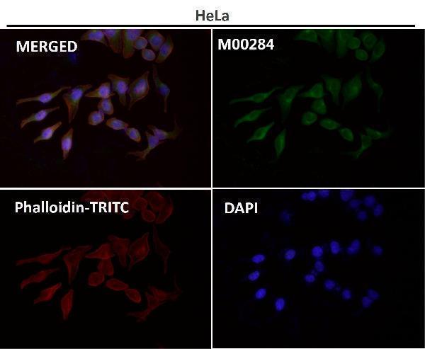


Comprehensive Human Protein Atlas IHC scoring per tissue (reliability: Supported — Medium consistency between antibody staining and RNA expression data.). Rows are taken directly from the HPA tissue chart — click any row's HPA link to view the source.
| Tissue | Cell type | Level | Evidence | Source |
|---|---|---|---|---|
| Appendix | Lymphoid tissue | High | Protein (IHC) | HPA → |
| Bone marrow | Hematopoietic cells | High | Protein (IHC) | HPA → |
| Breast | Glandular cells | High | Protein (IHC) | HPA → |
| Cervix | Glandular cells | High | Protein (IHC) | HPA → |
| Colon | Glandular cells | High | Protein (IHC) | HPA → |
| Tissue | Cell type | Level | Evidence | Source |
|---|---|---|---|---|
| None in HPA: RELA is detected in all 45 scored tissues. Use a no-primary (secondary-only) and an isotype control instead. | ||||
Use compartment-aware controls when optimizing chromogenic RELA IHC; nuclear staining and cytoplasmic staining answer different biological questions (UniProt Q04206; HPA tissue IHC).
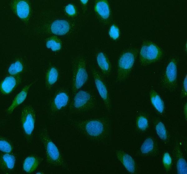
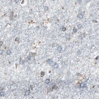
The catalog includes IHC images from human breast cancer, human bladder carcinoma, and mouse colon, plus an IF image from U2OS cells (catalog figure captions). Listed antibody reactivity spans human, mouse, and rat (catalog reactivity).
PA1669 has paraffin-section IHC data from human breast cancer, while M00284 has paraffin-section IHC data from human transitional cell carcinoma of the bladder (respective IHC figure captions). M00284-4 has mouse-colon IHC data, and A00284-1 has U2OS-cell IF data (respective figure captions).
Which to pick: For paraffin-section tissue IHC, choose PA1669 for the documented human breast cancer workflow or M00284 for the documented human bladder example; the fixative is unreported in both captions (respective IHC figure captions). For IF/ICC, choose A00284-1, which lists both applications and shows staining in U2OS cells (catalog applications; A00284-1 IF figure caption). For cross-species IHC planning, consider M00284-4: it is a monoclonal antibody listed as reactive with human, mouse, and rat, with IHC images from mouse colon, human colon, and human tonsil; those captions do not report the fixative (M00284-4 catalog description, reactivity, and IHC figure captions).