
Click image to see more details
Product Info Summary
| SKU: | M01047-3 |
|---|---|
| Size: | 100 μg/vial |
| Reactive Species: | Human |
| Host: | Mouse |
| Application: | IHC, WB |
Customers Who Bought This Also Bought

Product info
Product Name
Anti-CD79a Antibody Picoband™ (monoclonal, 4G4)
SKU/Catalog Number
M01047-3
Size
100 μg/vial
Form
Lyophilized
Description
Boster Bio Anti-CD79a Antibody Picoband™ (monoclonal, 4G4) catalog # M01047-3. Tested in IHC, WB applications. This antibody reacts with Human.
Storage & Handling
Store at -20˚C for one year from date of receipt. After reconstitution, at 4˚C for one month. It can also be aliquotted and stored frozen at -20˚C for six months. Avoid repeated freeze-thaw cycles.
Cite This Product
Anti-CD79a Antibody Picoband™ (monoclonal, 4G4) (Boster Biological Technology, Pleasanton CA, USA, Catalog # M01047-3)
Host
Mouse
Contents
Each vial contains 4mg Trehalose, 0.9mg NaCl, 0.2mg Na2HPO4, 0.05mg NaN3.
Clonality
Monoclonal
Clone Number
4G4
Isotype
Mouse IgG1
Immunogen
E.coli-derived human CD79a recombinant protein (Position: T121-P226). Human CD79a shares 91% amino acid (aa) sequence identity with mouse CD79a.
*Blocking peptide can be purchased. Costs vary based on immunogen length. Contact us for pricing.
Cross-reactivity
No cross-reactivity with other proteins.
Reactive Species
M01047-3 is reactive to CD79A in Human
Applications
M01047-3 is guaranteed for IHC, WB Boster Guarantee
Observed Molecular Weight
44 kDa
Calculated molecular weight
25.038kDa
Background of CD79A
Cluster of differentiation CD79A also known as B-cell antigen receptor complex-associated protein alpha chain and MB-1 membrane glycoprotein, is a protein that in humans is encoded by the CD79A gene. It is mapped to 19q13.2. CD79A is a membrane protein with an extracellular immunoglobulin domain, a single span transmembrane region and a short cytoplasmic domain. Genetic deletion of the transmembrane exon of CD79A results in loss of CD79A protein and a complete block of B cell development at the pro to pre B cell transition. Similarly, humans with homozygous splice variants in CD79A predicted to result in loss of the transmembrane region and a truncated or absent protein display agammaglobulinemia and no peripheral B cells.
Antibody Validation
Boster validates all antibodies on WB, IHC, ICC, Immunofluorescence, and ELISA with known positive control and negative samples to ensure specificity and high affinity, including thorough antibody incubations.
Innovating Scientists Reward
If you are the first to review this product, or if you have results for a special sample, species or application this product is not validated in, share your results with us and receive product credits you can use towards any Boster products! Applicable to all scientists worldwide.
Submit A Review
Assay dilution & Images
Reconsitution
Add 0.2ml of distilled water will yield a concentration of 500μg/ml.
Assay Dilutions Recommendation
The recommendations below provide a starting point for assay optimization. The actual working concentration varies and should be decided by the user.
Western blot, 0.1-0.5μg/ml, Human
Immunohistochemistry (Paraffin-embedded Section), 0.5-1μg/ml, Human, By Heat
Validation Images & Assay Conditions
Click image to see more details
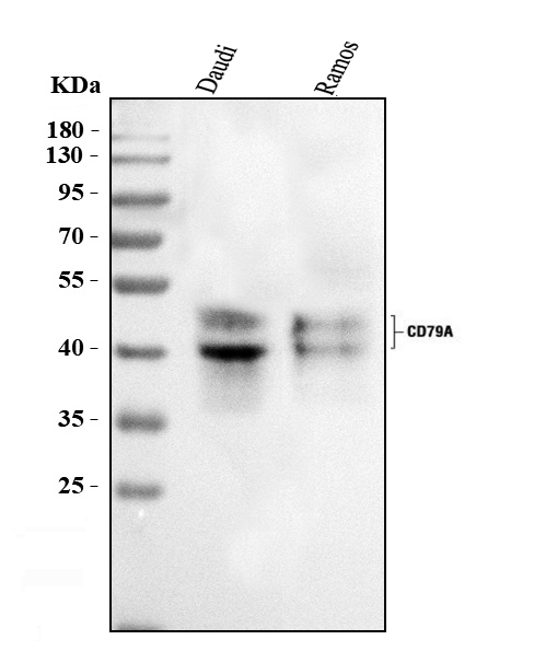
Figure 1. Western blot analysis of CD79A using anti ZO-1 antibody (M01047-3).
Electrophoresis was performed on a 5-20% SDS-PAGE gel at 70V (Stacking gel) / 90V (Resolving gel) for 2-3 hours. The sample well of each lane was loaded with 50ug of sample under reducing conditions.
Lane 1: human Raji tissue lysates,
After Electrophoresis, proteins were transferred to a Nitrocellulose membrane at 150mA for 50-90 minutes. Blocked the membrane with 5% Non-fat Milk/ TBS for 1.5 hour at RT. The membrane was incubated with mouse anti-CD79A antigen affinity purified polyclonal antibody (Catalog # M01047-3) at 0.5 μg/mL overnight at 4°C, then washed with TBS-0.1%Tween 3 times with 5 minutes each and probed with a goat anti-mouse IgG-HRP secondary antibody at a dilution of 1:10000 for 1.5 hour at RT. The signal is developed using an Enhanced Chemiluminescent detection (ECL) kit (Catalog # EK1001) with Tanon 5200 system. A specific band was detected for CD79A at approximately 44KD. The expected band size for CD79A is at 25KD.

Click image to see more details
Figure 2. IHC analysis of CD79A using anti-CD79A antibody (M01047-3).
CD79A was detected in paraffin-embedded section of human tonsil cancer tissue. Heat mediated antigen retrieval was performed in EDTA buffer (pH8.0, epitope retrieval solution). The tissue section was blocked with 10% goat serum. The tissue section was then incubated with 1μg/ml mouse anti-CD79A Antibody (M01047-3) overnight at 4°C. Biotinylated goat anti-mouse IgG was used as secondary antibody and incubated for 30 minutes at 37°C. The tissue section was developed using Strepavidin-Biotin-Complex (SABC) (Catalog # SA1021) with DAB as the chromogen.
Protein Target Info & Infographic
Gene/Protein Information For CD79A (Source: Uniprot.org, NCBI)
Gene Name
CD79A
Full Name
B-cell antigen receptor complex-associated protein alpha chain
Weight
25.038kDa
Alternative Names
CD79A antigen (immunoglobulin-associated alpha); CD79a antigen; CD79a molecule, immunoglobulin-associated alpha; CD79A; IGAB-cell antigen receptor complex-associated protein alpha chain; ig-alpha; MB-1 membrane glycoprotein; MB1; MB-1; Membrane-bound immunoglobulin-associated protein; Surface IgM-associated protein CD79A IGA, MB-1 CD79a molecule B-cell antigen receptor complex-associated protein alpha chain|CD79a antigen (immunoglobulin-associated alpha)|CD79a molecule, immunoglobulin-associated alpha|MB-1 membrane glycoprotein|ig-alpha|membrane-bound immunoglobulin-associated protein|surface IgM-associated protein
*If product is indicated to react with multiple species, protein info is based on the gene entry specified above in "Species".For more info on CD79A, check out the CD79A Infographic

We have 30,000+ of these available, one for each gene! Check them out.
In this infographic, you will see the following information for CD79A: database IDs, superfamily, protein function, synonyms, molecular weight, chromosomal locations, tissues of expression, subcellular locations, post-translational modifications, and related diseases, research areas & pathways. If you want to see more information included, or would like to contribute to it and be acknowledged, please contact [email protected].
Specific Publications For Anti-CD79a Antibody Picoband™ (monoclonal, 4G4) (M01047-3)
Hello CJ!
No publications found for M01047-3
*Do you have publications using this product? Share with us and receive a reward. Ask us for more details.
Recommended Resources
Here are featured tools and databases that you might find useful.
- Boster's Pathways Library
- Protein Databases
- Bioscience Research Protocol Resources
- Data Processing & Analysis Software
- Photo Editing Software
- Scientific Literature Resources
- Research Paper Management Tools
- Molecular Biology Software
- Primer Design Tools
- Bioinformatics Tools
- Phylogenetic Tree Analysis
Customer Reviews
Have you used Anti-CD79a Antibody Picoband™ (monoclonal, 4G4)?
Submit a review and receive an Amazon gift card.
- $30 for a review with an image
Be the first to review Anti-CD79a Antibody Picoband™ (monoclonal, 4G4)
*The first user to submit a review for a product is eligible for Boster's Innovating Scientists Reward, which gives product credits. This is in addition to the gift card reward.
Customer Q&As
Have a question?
Find answers in Q&As, reviews.
Can't find your answer?
Submit your question
4 Customer Q&As for Anti-CD79a Antibody Picoband™ (monoclonal, 4G4)
Question
We ordered your anti-CD79a antibody (monoclonal, 4G4) for IHC-P on small intestine in a previous project. I am using human, and We want to use the antibody for WB next. I am interested in examining small intestine as well as adrenal gland in our next experiment. Could you please give me some suggestion on which antibody would work the best for WB?
Verified Customer
Verified customer
Asked: 2020-04-24
Answer
I viewed the website and datasheets of our anti-CD79a antibody (monoclonal, 4G4) and it seems that M01047-3 has been validated on human in both IHC-P and WB. Thus M01047-3 should work for your application. Our Boster satisfaction guarantee will cover this product for WB in human even if the specific tissue type has not been validated. We do have a comprehensive range of products for WB detection and you can check out our website bosterbio.com to find out more information about them.
Boster Scientific Support
Answered: 2020-04-24
Question
We were happy with the WB result of your anti-CD79a antibody (monoclonal, 4G4). However we have seen positive staining in tonsil cell membrane using this antibody. Is that expected? Could you tell me where is CD79A supposed to be expressed?
Verified Customer
Verified customer
Asked: 2019-09-11
Answer
From literature, tonsil does express CD79A. Generally CD79A expresses in cell membrane. Regarding which tissues have CD79A expression, here are a few articles citing expression in various tissues:
Small intestine, Pubmed ID: 7514267
Tonsil, Pubmed ID: 1534761
Boster Scientific Support
Answered: 2019-09-11
Question
We have been able to see staining in human small intestine. Any tips? Is anti-CD79a antibody (monoclonal, 4G4) supposed to stain small intestine positively?
Verified Customer
Verified customer
Asked: 2019-05-13
Answer
From literature small intestine does express CD79A. From Uniprot.org, CD79A is expressed in adrenal gland, tonsil, small intestine, among other tissues. Regarding which tissues have CD79A expression, here are a few articles citing expression in various tissues:
Small intestine, Pubmed ID: 7514267
Tonsil, Pubmed ID: 1534761
Boster Scientific Support
Answered: 2019-05-13
Question
We are currently using anti-CD79a antibody (monoclonal, 4G4) M01047-3 for human tissue, and we are happy with the WB results. The species of reactivity given in the datasheet says human. Is it likely that the antibody can work on goat tissues as well?
Verified Customer
Verified customer
Asked: 2018-04-04
Answer
The anti-CD79a antibody (monoclonal, 4G4) (M01047-3) has not been validated for cross reactivity specifically with goat tissues, but there is a good chance of cross reactivity. We have an innovator award program that if you test this antibody and show it works in goat you can get your next antibody for free. Please contact me if I can help you with anything.
Boster Scientific Support
Answered: 2018-04-04
