Click image to see more details
-

-
-
-
-
+7
Product Info Summary
| SKU: | A01357-1 |
|---|---|
| Size: | 100 μg/vial |
| Reactive Species: | Human, Mouse, Rat |
| Host: | Rabbit |
| Application: | Flow Cytometry, IF, IHC, IHC-F, ICC, WB |
Customers Who Bought This Also Bought


Product info
Product Name
Anti-Cytokeratin 18/KRT18 Antibody Picoband™
SKU/Catalog Number
A01357-1
Size
100 μg/vial
Form
Lyophilized
Description
Boster Bio Anti-Cytokeratin 18/KRT18 Antibody Picoband™ catalog # A01357-1. Tested in Flow Cytometry, IF, IHC, IHC-F, ICC, WB applications. This antibody reacts with Human, Mouse, Rat.
Storage & Handling
Store at -20˚C for one year from date of receipt. After reconstitution, at 4˚C for one month. It can also be aliquotted and stored frozen at -20˚C for six months. Avoid repeated freeze-thaw cycles.
Cite This Product
Anti-Cytokeratin 18/KRT18 Antibody Picoband™ (Boster Biological Technology, Pleasanton CA, USA, Catalog # A01357-1)
Host
Rabbit
Contents
Each vial contains 4mg Trehalose, 0.9mg NaCl, 0.2mg Na2HPO4, 0.05mg NaN3.
Clonality
Polyclonal
Isotype
Rabbit IgG
Immunogen
E.coli-derived human Cytokeratin 18 recombinant protein (Position: E204-H430). Human Cytokeratin 18 shares 87.7% and 85.9% amino acid (aa) sequence identity with mouse and rat Cytokeratin 18, respectively.
*Blocking peptide can be purchased. Costs vary based on immunogen length. Contact us for pricing.
Cross-reactivity
No cross-reactivity with other proteins
Reactive Species
A01357-1 is reactive to KRT18 in Human, Mouse, Rat
Applications
A01357-1 is guaranteed for Flow Cytometry, IF, IHC, IHC-F, ICC, WB Boster Guarantee
Observed Molecular Weight
48 kDa
Calculated molecular weight
48.058kDa
Background of KRT18
Keratin 18, mapped to 12q13.13, is a type I cytokeratin. It is, together with its filament partner keratin 8, perhaps the most commonly found products of the intermediate filament gene family. They are expressed in single layer epithelial tissues of the body. Mutations in this gene have been linked to cryptogenic cirrhosis. Two transcript variants encoding the same protein have been found for this gene.
Antibody Validation
Boster validates all antibodies on WB, IHC, ICC, Immunofluorescence, and ELISA with known positive control and negative samples to ensure specificity and high affinity, including thorough antibody incubations.
Innovating Scientists Reward
If you are the first to review this product, or if you have results for a special sample, species or application this product is not validated in, share your results with us and receive product credits you can use towards any Boster products! Applicable to all scientists worldwide.
Submit A Review
Assay dilution & Images
Reconsitution
Add 0.2ml of distilled water will yield a concentration of 500ug/ml.
Assay Dilutions Recommendation
The recommendations below provide a starting point for assay optimization. The actual working concentration varies and should be decided by the user.
Western blot, 0.1-0.5μg/ml, Human, Mouse, Rat
Immunohistochemistry (Paraffin-embedded Section), 0.5-1μg/ml, Human, Mouse, Rat, By Heat
Immunohistochemistry(Frozen Section), 0.5-1 μg/ml, Human
Immunocytochemistry, 0.5-1 μg/ml, Human
Immunofluorescence, 2 μg/ml, Human
Flow Cytometry, 1-3 μg/1x106 cells, Human
Validation Images & Assay Conditions
Click image to see more details
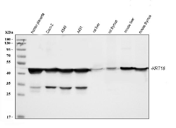
Figure 1. Western blot analysis of Cytokeratin 18 using anti-Cytokeratin 18 antibody (A01357-1).
Electrophoresis was performed on a 5-20% SDS-PAGE gel at 70V (Stacking gel) / 90V (Resolving gel) for 2-3 hours. The sample well of each lane was loaded with 30 ug of sample under reducing conditions.
Lane 1: human placenta tissue lysates,
Lane 2: human CACO-2 whole cell lysates,
Lane 3: human A549 whole cell lysates,
Lane 4: human A431 whole cell lysates,
Lane 5: rat liver tissue lysates,
Lane 6: rat thymus tissue lysates,
Lane 7: mouse liver tissue lysates,
Lane 8: mouse thymus tissue lysates.
After electrophoresis, proteins were transferred to a nitrocellulose membrane at 150 mA for 50-90 minutes. Blocked the membrane with 5% non-fat milk/TBS for 1.5 hour at RT. The membrane was incubated with rabbit anti-Cytokeratin 18 antigen affinity purified polyclonal antibody (Catalog # A01357-1) at 0.5 μg/mL overnight at 4°C, then washed with TBS-0.1%Tween 3 times with 5 minutes each and probed with a goat anti-rabbit IgG-HRP secondary antibody at a dilution of 1:5000 for 1.5 hour at RT. The signal is developed using an Enhanced Chemiluminescent detection (ECL) kit (Catalog # EK1002) with Tanon 5200 system. A specific band was detected for Cytokeratin 18 at approximately 48 kDa. The expected band size for Cytokeratin 18 is at 48 kDa.

Click image to see more details
Figure 2. IHC analysis of Cytokeratin 18 using anti-Cytokeratin 18 antibody (A01357-1).
Cytokeratin 18 was detected in paraffin-embedded section of mouse intestine tissues. Heat mediated antigen retrieval was performed in citrate buffer (pH6, epitope retrieval solution) for 20 mins. The tissue section was blocked with 10% goat serum. The tissue section was then incubated with 1μg/ml rabbit anti-Cytokeratin 18 Antibody (A01357-1) overnight at 4°C. Biotinylated goat anti-rabbit IgG was used as secondary antibody and incubated for 30 minutes at 37°C. The tissue section was developed using Strepavidin-Biotin-Complex (SABC)(Catalog # SA1022) with DAB as the chromogen.

Click image to see more details
Figure 3. IHC analysis of Cytokeratin 18 using anti-Cytokeratin 18 antibody (A01357-1).
Cytokeratin 18 was detected in paraffin-embedded section of mouse kidney tissues. Heat mediated antigen retrieval was performed in citrate buffer (pH6, epitope retrieval solution) for 20 mins. The tissue section was blocked with 10% goat serum. The tissue section was then incubated with 1μg/ml rabbit anti-Cytokeratin 18 Antibody (A01357-1) overnight at 4°C. Biotinylated goat anti-rabbit IgG was used as secondary antibody and incubated for 30 minutes at 37°C. The tissue section was developed using Strepavidin-Biotin-Complex (SABC)(Catalog # SA1022) with DAB as the chromogen.

Click image to see more details
Figure 4. IHC analysis of Cytokeratin 18 using anti-Cytokeratin 18 antibody (A01357-1).
Cytokeratin 18 was detected in paraffin-embedded section of rat intestine tissues. Heat mediated antigen retrieval was performed in citrate buffer (pH6, epitope retrieval solution) for 20 mins. The tissue section was blocked with 10% goat serum. The tissue section was then incubated with 1μg/ml rabbit anti-Cytokeratin 18 Antibody (A01357-1) overnight at 4°C. Biotinylated goat anti-rabbit IgG was used as secondary antibody and incubated for 30 minutes at 37°C. The tissue section was developed using Strepavidin-Biotin-Complex (SABC)(Catalog # SA1022) with DAB as the chromogen.

Click image to see more details
Figure 5. IHC analysis of Cytokeratin 18 using anti-Cytokeratin 18 antibody (A01357-1).
Cytokeratin 18 was detected in paraffin-embedded section of rat kidney tissues. Heat mediated antigen retrieval was performed in citrate buffer (pH6, epitope retrieval solution) for 20 mins. The tissue section was blocked with 10% goat serum. The tissue section was then incubated with 1μg/ml rabbit anti-Cytokeratin 18 Antibody (A01357-1) overnight at 4°C. Biotinylated goat anti-rabbit IgG was used as secondary antibody and incubated for 30 minutes at 37°C. The tissue section was developed using Strepavidin-Biotin-Complex (SABC)(Catalog # SA1022) with DAB as the chromogen.

Click image to see more details
Figure 6. IHC analysis of Cytokeratin 18 using anti-Cytokeratin 18 antibody (A01357-1).
Cytokeratin 18 was detected in paraffin-embedded section of human intestinal cancer tissues. Heat mediated antigen retrieval was performed in citrate buffer (pH6, epitope retrieval solution) for 20 mins. The tissue section was blocked with 10% goat serum. The tissue section was then incubated with 1μg/ml rabbit anti-Cytokeratin 18 Antibody (A01357-1) overnight at 4°C. Biotinylated goat anti-rabbit IgG was used as secondary antibody and incubated for 30 minutes at 37°C. The tissue section was developed using Strepavidin-Biotin-Complex (SABC)(Catalog # SA1022) with DAB as the chromogen.

Click image to see more details
Figure 7. IHC analysis of Cytokeratin 18 using anti-Cytokeratin 18 antibody (A01357-1).
Cytokeratin 18 was detected in paraffin-embedded section of human placenta tissues. Heat mediated antigen retrieval was performed in citrate buffer (pH6, epitope retrieval solution) for 20 mins. The tissue section was blocked with 10% goat serum. The tissue section was then incubated with 1μg/ml rabbit anti-Cytokeratin 18 Antibody (A01357-1) overnight at 4°C. Biotinylated goat anti-rabbit IgG was used as secondary antibody and incubated for 30 minutes at 37°C. The tissue section was developed using Strepavidin-Biotin-Complex (SABC)(Catalog # SA1022) with DAB as the chromogen.

Click image to see more details
Figure 8. IF analysis of Cytokeratin 18 using anti-Cytokeratin 18 antibody (A01357-1)
Cytokeratin 18 was detected in paraffin-embedded section of human intestinal cancer tissues. Heat mediated antigen retrieval was performed in citrate buffer (pH6, epitope retrieval solution ) for 20 mins. The tissue section was blocked with 10% goat serum. The tissue section was then incubated with 1μg/mL rabbit anti-Cytokeratin 18 Antibody (A01357-1) overnight at 4°C. Biotin conjugated goat anti-rabbit IgG (BA1003) was used as secondary antibody and incubated for 30 minutes at 37°C. The tissue section was developed using Cy3 Conjugated Avidin (BA1037). The section was counterstained with DAPI. Visualize using a fluorescence microscope and filter sets appropriate for the label used.

Click image to see more details
Figure 9. IF analysis of Cytokeratin 18 using anti-Cytokeratin 18 antibody (A01357-1)
Cytokeratin 18 was detected in paraffin-embedded section of human lung cancer tissues. Heat mediated antigen retrieval was performed in citrate buffer (pH6, epitope retrieval solution ) for 20 mins. The tissue section was blocked with 10% goat serum. The tissue section was then incubated with 1μg/mL rabbit anti-Cytokeratin 18 Antibody (A01357-1) overnight at 4°C. Biotin conjugated goat anti-rabbit IgG (BA1003) was used as secondary antibody and incubated for 30 minutes at 37°C. The tissue section was developed using Cy3 Conjugated Avidin (BA1037). The section was counterstained with DAPI. Visualize using a fluorescence microscope and filter sets appropriate for the label used.

Click image to see more details
Figure 10. Flow Cytometry analysis of A431 cells using anti-Cytokeratin 18 antibody (A01357-1).
Overlay histogram showing A431 cells stained with A01357-1 (Blue line).The cells were blocked with 10% normal goat serum. And then incubated with rabbit anti-Cytokeratin 18 Antibody (A01357-1,1μg/1x106 cells) for 30 min at 20°C. DyLight®488 conjugated goat anti-rabbit IgG (BA1127, 5-10μg/1x106 cells) was used as secondary antibody for 30 minutes at 20°C. Isotype control antibody (Green line) was rabbit IgG (1μg/1x106) used under the same conditions. Unlabelled sample (Red line) was also used as a control.

Click image to see more details
Figure 11. Flow Cytometry analysis of A549 cells using anti-Cytokeratin 18 antibody (A01357-1).
Overlay histogram showing A549 cells stained with A01357-1 (Blue line).The cells were blocked with 10% normal goat serum. And then incubated with rabbit anti-Cytokeratin 18 Antibody (A01357-1,1μg/1x106 cells) for 30 min at 20°C. DyLight®488 conjugated goat anti-rabbit IgG (BA1127, 5-10μg/1x106 cells) was used as secondary antibody for 30 minutes at 20°C. Isotype control antibody (Green line) was rabbit IgG (1μg/1x106) used under the same conditions. Unlabelled sample (Red line) was also used as a control.
Protein Target Info & Infographic
Gene/Protein Information For KRT18 (Source: Uniprot.org, NCBI)
Gene Name
KRT18
Full Name
Keratin, type I cytoskeletal 18
Weight
48.058kDa
Superfamily
intermediate filament family
Alternative Names
Cell proliferation-inducing gene 46 protein; cell proliferation-inducing protein 46; CK-18; CYK18; Cytokeratin 18; cytokeratin-18; K18; Keratin 18; keratin, type I cytoskeletal 18; keratin-18; KRT18 KRT18 CK-18, CYK18, K18 keratin 18 keratin, type I cytoskeletal 18|cell proliferation-inducing gene 46 protein|cytokeratin 18|keratin 18, type I
*If product is indicated to react with multiple species, protein info is based on the gene entry specified above in "Species".For more info on KRT18, check out the KRT18 Infographic

We have 30,000+ of these available, one for each gene! Check them out.
In this infographic, you will see the following information for KRT18: database IDs, superfamily, protein function, synonyms, molecular weight, chromosomal locations, tissues of expression, subcellular locations, post-translational modifications, and related diseases, research areas & pathways. If you want to see more information included, or would like to contribute to it and be acknowledged, please contact [email protected].
Specific Publications For Anti-Cytokeratin 18/KRT18 Antibody Picoband™ (A01357-1)
Hello CJ!
A01357-1 has been cited in 15 publications:
*The publications in this section are manually curated by our staff scientists. They may differ from Bioz's machine gathered results. Both are accurate. If you find a publication citing this product but is missing from this list, please let us know we will issue you a thank-you coupon.
Expression of hepatitis B virus 1.3-fold genome plasmid in an SV40 T-antigen-immortalized mouse hepatic cell line
Sodium alginate-bioglass-encapsulated hAECs restore ovarian function in premature ovarian failure by stimulating angiogenic factor secretion
Transplantation of human amniotic epithelial cells promotes morphological and functional regeneration in a rat uterine scar model
The influence of different microenvironments on melanoma invasiveness and microcirculation patterns: an animal experiment study in the mouse model
The effects of human platelet lysate on dental pulp stem cells derived from impacted human third molars
Species: Human
The critical role of CD133 + CD44 +/high tumor cells in hematogenous metastasis of liver cancers
Species: Human
Shi R,Liu L,Wang F,He Y,Niu Y,Wang C,Zhang X,Zhang X,Zhang H,Chen M,Wang Y.Downregulation of cytokeratin 18 induces cellular partial EMT and stemness through increasing EpCAM expression in breast cancer.Cell Signal.2020 Dec;76:109810.doi:10.1016/j.cellsig
Species: Human
A01357-1 usage in article: APP:IF, SAMPLE:BREAST CANCER CELLS AND BREAST TISSUE, DILUTION:1:100
Kong Y, Zhao C, Huang Y, Liu Y, Liu S, Guo Y, Li M, Xu T, Zhao B, Wang J. Angiopoietin-like protein 4 promotes very-low-density lipoprotein assembly and secretion in bovine hepatocytes in vitro. IUBMB Life. 2020 Nov 17. doi: 10.1002/iub.2403. Epub ahead o
Species: Calf
A01357-1 usage in article: APP:WB, SAMPLE:HEPATOCYTES, DILUTION:NA
Small intestinal intraepithelial lymphocytes expressing CD8 and T cell receptor %u03B3%u03B4 are involved in bacterial clearance during Salmonella enterica serovar Typhimurium %u2026
miR-182 aids in receptive endometrium development in dairy goats by down-regulating PTN expression
Recommended Resources
Here are featured tools and databases that you might find useful.
- Boster's Pathways Library
- Protein Databases
- Bioscience Research Protocol Resources
- Data Processing & Analysis Software
- Photo Editing Software
- Scientific Literature Resources
- Research Paper Management Tools
- Molecular Biology Software
- Primer Design Tools
- Bioinformatics Tools
- Phylogenetic Tree Analysis
Customer Reviews
Have you used Anti-Cytokeratin 18/KRT18 Antibody Picoband™?
Submit a review and receive an Amazon gift card.
- $30 for a review with an image
Be the first to review Anti-Cytokeratin 18/KRT18 Antibody Picoband™
*The first user to submit a review for a product is eligible for Boster's Innovating Scientists Reward, which gives product credits. This is in addition to the gift card reward.
Customer Q&As
Have a question?
Find answers in Q&As, reviews.
Can't find your answer?
Submit your question
16 Customer Q&As for Anti-Cytokeratin 18/KRT18 Antibody Picoband™
Question
I was wanting to use your anti-Cytokeratin 18/KRT18 antibody for IHC-P for rat liver on frozen tissues, but I want to know if it has been validated for this particular application. Has this antibody been validated and is this antibody a good choice for rat liver identification?
Verified Customer
Verified customer
Asked: 2020-04-28
Answer
You can see on the product datasheet, A01357-1 anti-Cytokeratin 18/KRT18 antibody has been tested for IF, IHC-P, WB on human, mouse, rat tissues. We have an innovator award program that if you test this antibody and show it works in rat liver in IHC-frozen, you can get your next antibody for free.
Boster Scientific Support
Answered: 2020-04-28
Question
We need to test anti-Cytokeratin 18/KRT18 antibody A01357-1 on rat liver for research purposes, then I may be interested in using anti-Cytokeratin 18/KRT18 antibody A01357-1 for diagnostic purposes as well. Is the antibody suitable for diagnostic purposes?
Verified Customer
Verified customer
Asked: 2020-04-28
Answer
The products we sell, including anti-Cytokeratin 18/KRT18 antibody A01357-1, are only intended for research use. They would not be suitable for use in diagnostic work. If you have the means to develop a product into diagnostic use, and are interested in collaborating with us and develop our product into an IVD product, please contact us for more discussions.
Boster Scientific Support
Answered: 2020-04-28
Question
We have tried in the past anti-Cytokeratin 18/KRT18 antibody for IHC-P on pancreas last year. I am using mouse, and We intend to use the antibody for IF next. We are interested in examining pancreas as well as vulva in our next experiment. Could you please give me some suggestion on which antibody would work the best for IF?
Verified Customer
Verified customer
Asked: 2020-04-20
Answer
I viewed the website and datasheets of our anti-Cytokeratin 18/KRT18 antibody and it appears that A01357-1 has been tested on mouse in both IHC-P and IF. Thus A01357-1 should work for your application. Our Boster satisfaction guarantee will cover this product for IF in mouse even if the specific tissue type has not been validated. We do have a comprehensive range of products for IF detection and you can check out our website bosterbio.com to find out more information about them.
Boster Scientific Support
Answered: 2020-04-20
Question
We appreciate helping with my inquiry over the phone. Here are the WB image, lot number and protocol we used for liver using anti-Cytokeratin 18/KRT18 antibody A01357-1. Let me know if you need anything else.
Verified Customer
Verified customer
Asked: 2020-03-30
Answer
I appreciate the data. You have provided everything we needed. Our lab team are working to resolve your inquiry as quickly as possible, and we appreciate your patience and understanding! Please let me know if there is anything you need in the meantime.
Boster Scientific Support
Answered: 2020-03-30
Question
We have observed staining in mouse placenta. Are there any suggestions? Is anti-Cytokeratin 18/KRT18 antibody supposed to stain placenta positively?
Verified Customer
Verified customer
Asked: 2020-03-11
Answer
According to literature placenta does express KRT18. According to Uniprot.org, KRT18 is expressed in adenohypophysis, placenta, cervix, colon, pancreas, placenta uterus, vulva, liver, colon carcinoma, cervix carcinoma, cervix carcinoma erythroleukemia, among other tissues. Regarding which tissues have KRT18 expression, here are a few articles citing expression in various tissues:
Cervix carcinoma, Pubmed ID: 17081983, 17924679, 18669648, 18691976, 20068231
Cervix carcinoma, and Erythroleukemia, Pubmed ID: 23186163
Cervix, Colon, Pancreas, Placenta, and Uterus, Pubmed ID: 15489334
Colon carcinoma, Pubmed ID: 9150948, 24129315
Liver, Pubmed ID: 2422083, 24275569
Placenta, Pubmed ID: 2434380
Vulva, Pubmed ID: 2434381
Boster Scientific Support
Answered: 2020-03-11
Question
Can you help my question with product A01357-1, anti-Cytokeratin 18/KRT18 antibody. I was wondering if it would be possible to conjugate this antibody with biotin. I would need it to be without BSA or sodium azide. I am planning on using a buffer exchange of sodium azide with PBS only. Would there be problems for me to conjugate the antibody and store it in -20 degrees in small aliquots?
Verified Customer
Verified customer
Asked: 2020-02-27
Answer
It is not recommended storing this antibody with PBS buffer only in -20 degrees. If you want to store it in -20 degrees it is best to add some cryoprotectant like glycerol. If you want carrier free A01357-1 anti-Cytokeratin 18/KRT18 antibody, we can provide it to you in a special formula with trehalose and/or glycerol. These molecules will not interfere with conjugation chemistry and provide a good level of protection for the antibody from degradation. Please be sure to specify this in your purchase order.
Boster Scientific Support
Answered: 2020-02-27
Question
We are currently using anti-Cytokeratin 18/KRT18 antibody A01357-1 for rat tissue, and we are content with the IHC-P results. The species of reactivity given in the datasheet says human, mouse, rat. Is it true that the antibody can work on goat tissues as well?
Verified Customer
Verified customer
Asked: 2019-12-25
Answer
The anti-Cytokeratin 18/KRT18 antibody (A01357-1) has not been tested for cross reactivity specifically with goat tissues, though there is a good chance of cross reactivity. We have an innovator award program that if you test this antibody and show it works in goat you can get your next antibody for free. Please contact me if I can help you with anything.
Boster Scientific Support
Answered: 2019-12-25
Question
Will anti-Cytokeratin 18/KRT18 antibody A01357-1 work for IHC-P with liver?
Verified Customer
Verified customer
Asked: 2019-12-04
Answer
According to the expression profile of liver, KRT18 is highly expressed in liver. So, it is likely that anti-Cytokeratin 18/KRT18 antibody A01357-1 will work for IHC-P with liver.
Boster Scientific Support
Answered: 2019-12-04
Question
I see that the anti-Cytokeratin 18/KRT18 antibody A01357-1 works with IHC-P, what is the protocol used to produce the result images on the product page?
Verified Customer
Verified customer
Asked: 2019-11-11
Answer
You can find protocols for IHC-P on the "support/technical resources" section of our navigation menu. If you have any further questions, please send an email to [email protected]
Boster Scientific Support
Answered: 2019-11-11
Question
Please see the WB image, lot number and protocol we used for liver using anti-Cytokeratin 18/KRT18 antibody A01357-1. Please let me know if you require anything else.
Verified Customer
Verified customer
Asked: 2019-09-20
Answer
Thank you very much for the data. Our lab team are working to resolve this as quickly as possible, and we appreciate your patience and understanding! You have provided everything we needed. Please let me know if there is anything you need in the meantime.
Boster Scientific Support
Answered: 2019-09-20
Question
Does A01357-1 anti-Cytokeratin 18/KRT18 antibody work on parafin embedded sections? If so, which fixation method do you recommend we use (PFA, paraformaldehyde, other)?
Verified Customer
Verified customer
Asked: 2019-06-14
Answer
It shows on the product datasheet, A01357-1 anti-Cytokeratin 18/KRT18 antibody as been tested on IHC-P. It is best to use PFA for fixation because it has better tissue penetration ability. PFA needs to be prepared fresh before use. Long term stored PFA turns into formalin, as the PFA molecules congregate and become formalin.
Boster Scientific Support
Answered: 2019-06-14
Question
Our team were satisfied with the WB result of your anti-Cytokeratin 18/KRT18 antibody. However we have seen positive staining in vulva cytoplasm using this antibody. Is that expected? Could you tell me where is KRT18 supposed to be expressed?
Verified Customer
Verified customer
Asked: 2018-11-16
Answer
Based on literature, vulva does express KRT18. Generally KRT18 expresses in cytoplasm, perinuclear region. nucleus,. Regarding which tissues have KRT18 expression, here are a few articles citing expression in various tissues:
Cervix carcinoma, Pubmed ID: 17081983, 17924679, 18669648, 18691976, 20068231
Cervix carcinoma, and Erythroleukemia, Pubmed ID: 23186163
Cervix, Colon, Pancreas, Placenta, and Uterus, Pubmed ID: 15489334
Colon carcinoma, Pubmed ID: 9150948, 24129315
Liver, Pubmed ID: 2422083, 24275569
Placenta, Pubmed ID: 2434380
Vulva, Pubmed ID: 2434381
Boster Scientific Support
Answered: 2018-11-16
Question
Is this A01357-1 anti-Cytokeratin 18/KRT18 antibody reactive to the isotypes of KRT18?
Verified Customer
Verified customer
Asked: 2018-03-30
Answer
The immunogen of A01357-1 anti-Cytokeratin 18/KRT18 antibody is E.coli-derived human Cytokeratin 18 recombinant protein (Position: E204-H430). Human Cytokeratin 18 shares 87.7% and 85.9% amino acid (aa) sequence identity with mouse and rat Cytokeratin 18, respectively. Could you tell me which isotype you are interested in so I can help see if the immunogen is part of this isotype?
Boster Scientific Support
Answered: 2018-03-30
Question
Is a blocking peptide available for product anti-Cytokeratin 18/KRT18 antibody (A01357-1)?
Verified Customer
Verified customer
Asked: 2017-08-04
Answer
We do provide the blocking peptide for product anti-Cytokeratin 18/KRT18 antibody (A01357-1). If you would like to place an order for it please contact [email protected] and make a special request.
Boster Scientific Support
Answered: 2017-08-04
Question
Is there a BSA free version of anti-Cytokeratin 18/KRT18 antibody A01357-1 available?
R. Anderson
Verified customer
Asked: 2013-04-05
Answer
Thanks for your recent telephone inquiry. I can confirm that some lots of this anti-Cytokeratin 18/KRT18 antibody A01357-1 are BSA free. For now, these lots are available and we can make a BSA free formula for you free of charge. It will take 3 extra days to prepare. If you require this antibody BSA free again in future, please do not hesitate to contact me and I will be pleased to check which lots we have in stock that are BSA free.
Boster Scientific Support
Answered: 2013-04-05
Question
I am interested in using your anti-Cytokeratin 18/KRT18 antibody for cell cycle studies. Has this antibody been tested with western blotting on hepg2 whole cell lysates? We would like to see some validation images before ordering.
G. Gonzalez
Verified customer
Asked: 2013-01-22
Answer
Thanks for your inquiry. This A01357-1 anti-Cytokeratin 18/KRT18 antibody is validated on rat kidney, mouse liver, hela whole cell lysates, hepg2 whole cell lysates, a431 cells, a549 cells. It is guaranteed to work for IF, IHC-P, WB in human, mouse, rat. Our Boster guarantee will cover your intended experiment even if the sample type has not been be directly tested.
Boster Scientific Support
Answered: 2013-01-22
