Click image to see more details
Product Info Summary
| SKU: | M02101-2 |
|---|---|
| Size: | 100 μg/vial |
| Reactive Species: | Human |
| Host: | Mouse |
| Application: | IF, IHC, WB |
Customers Who Bought This Also Bought


Product info
Product Name
Anti-Cytokeratin 19 KRT19 Antibody Picoband™ (monoclonal, 3D4)
View all Cytokeratin 19 Antibodies
SKU/Catalog Number
M02101-2
Size
100 μg/vial
Form
Lyophilized
Description
Boster Bio Anti-Cytokeratin 19 KRT19 Antibody Picoband™ (monoclonal, 3D4) catalog # M02101-2. Tested in IF, IHC, WB applications. This antibody reacts with Human.
Storage & Handling
Store at -20˚C for one year from date of receipt. After reconstitution, at 4˚C for one month. It can also be aliquotted and stored frozen at -20˚C for six months. Avoid repeated freeze-thaw cycles.
Cite This Product
Anti-Cytokeratin 19 KRT19 Antibody Picoband™ (monoclonal, 3D4) (Boster Biological Technology, Pleasanton CA, USA, Catalog # M02101-2)
Host
Mouse
Contents
Each vial contains 4mg Trehalose, 0.9mg NaCl, 0.2mg Na2HPO4, 0.05mg NaN3.
Clonality
Monoclonal
Clone Number
3D4
Isotype
Mouse IgG1
Immunogen
A synthetic peptide corresponding to a sequence at the C-terminus of human Cytokeratin 19, different from the related mouse and rat sequences by nine amino acids.
*Blocking peptide can be purchased. Costs vary based on immunogen length. Contact us for pricing.
Cross-reactivity
No cross-reactivity with other proteins.
Reactive Species
M02101-2 is reactive to KRT19 in Human
Applications
M02101-2 is guaranteed for IF, IHC, WB Boster Guarantee
Observed Molecular Weight
44 kDa
Calculated molecular weight
44.106kDa
Background of Cytokeratin 19
Keratin, type I cytoskeletal 19 is a protein that in humans is encoded by the KRT19 gene. The protein encoded by this gene is a member of the keratin family. It is specifically expressed in the periderm, the transiently superficial layer that envelops the developing epidermis. The type I cytokeratins are clustered in a region of chromosome 17q12-q21. Due to its high sensitivity, KRT19 is the most used marker for the RT-PCR-mediated detection of tumor cells disseminated in lymph nodes, peripheral blood, and bone marrow of breast cancer patients. Keratin 19 is often used together with keratin 8 and keratin 18 to differentiate cells of epithelial origin from hematopoietic cells in tests that enumerate circulating tumor cells in blood.
Antibody Validation
Boster validates all antibodies on WB, IHC, ICC, Immunofluorescence, and ELISA with known positive control and negative samples to ensure specificity and high affinity, including thorough antibody incubations.
Innovating Scientists Reward
If you are the first to review this product, or if you have results for a special sample, species or application this product is not validated in, share your results with us and receive product credits you can use towards any Boster products! Applicable to all scientists worldwide.
Submit A Review
Assay dilution & Images
Reconsitution
Add 0.2ml of distilled water will yield a concentration of 500ug/ml.
Assay Dilutions Recommendation
The recommendations below provide a starting point for assay optimization. The actual working concentration varies and should be decided by the user.
Western blot, 0.1-0.5μg/ml
Immunohistochemistry (Paraffin-embedded Section), 0.5-1μg/ml
Immunofluorescence, 2μg/ml
Validation Images & Assay Conditions
Click image to see more details
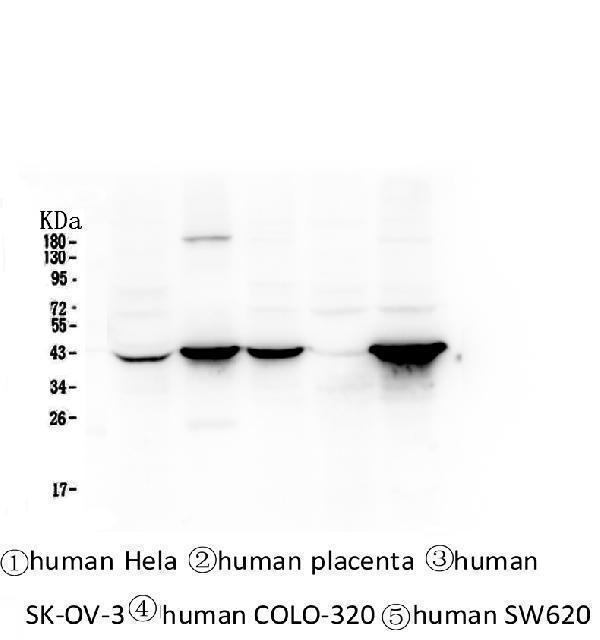
Figure 1. Western blot analysis of Cytokeratin 19 using anti-Cytokeratin 19 antibody (M02101-2).
Electrophoresis was performed on a 5-20% SDS-PAGE gel at 70V (Stacking gel) / 90V (Resolving gel) for 2-3 hours. The sample well of each lane was loaded with 50ug of sample under reducing conditions.
Lane 1: human Hela whole cell lysates,
Lane 2: human placenta tissue lysates,
Lane 3: human SK-OV-3 whole cell lysates,
Lane 4: human COLO-320 whole cell lysates.
After Electrophoresis, proteins were transferred to a Nitrocellulose membrane at 150mA for 50-90 minutes. Blocked the membrane with 5% Non-fat Milk/ TBS for 1.5 hour at RT. The membrane was incubated with mouse anti-Cytokeratin 19 antigen affinity purified monoclonal antibody (Catalog # M02101-2) at 0.5 μg/mL overnight at 4°C, then washed with TBS-0.1%Tween 3 times with 5 minutes each and probed with a goat anti-mouse IgG-HRP secondary antibody at a dilution of 1:10000 for 1.5 hour at RT. The signal is developed using an Enhanced Chemiluminescent detection (ECL) kit (Catalog # EK1001) with Tanon 5200 system.

Click image to see more details
Figure 2. IHC analysis of Cytokeratin 19 using anti-Cytokeratin 19 antibody (M02101-2).
Cytokeratin 19 was detected in paraffin-embedded section of human intestinal cancer tissue. Heat mediated antigen retrieval was performed in citrate buffer (pH6, epitope retrieval solution) for 20 mins. The tissue section was blocked with 10% goat serum. The tissue section was then incubated with 2μg/ml mouse anti-Cytokeratin 19 Antibody (M02101-2) overnight at 4°C. Biotinylated goat anti-mouse IgG was used as secondary antibody and incubated for 30 minutes at 37°C. The tissue section was developed using Strepavidin-Biotin-Complex (SABC)(Catalog # SA1021) with DAB as the chromogen.

Click image to see more details
Figure 3. IF analysis of Cytokeratin 19 using anti-Cytokeratin 19 antibody (M02101-2).
Cytokeratin 19 was detected in paraffin-embedded section of human intestinal cancer tissues. Heat mediated antigen retrieval was performed in citrate buffer (pH6, epitope retrieval solution ) for 20 mins. The tissue section was blocked with 10% goat serum. The tissue section was then incubated with 1μg/mL mouse anti-Cytokeratin 19 Antibody (M02101-2) overnight at 4°C. DyLight®550 Conjugated Goat Anti-Mouse IgG was used as secondary antibody at 1:200 dilution and incubated for 30 minutes at 37°C. Visualize using a fluorescence microscope and filter sets appropriate for the label used.
Protein Target Info & Infographic
Gene/Protein Information For KRT19 (Source: Uniprot.org, NCBI)
Gene Name
KRT19
Full Name
Keratin, type I cytoskeletal 19
Weight
44.106kDa
Superfamily
intermediate filament family
Alternative Names
40-kDa keratin intermediate filament; CK19; CK-19; Cytokeratin 19; EndoC; K19; K1CS; keratin 19; keratin, type I cytoskeletal 19; keratin, type I, 40-kd; keratin-19; Krt19; MGC15366 KRT19 CK19, K19, K1CS keratin 19 keratin, type I cytoskeletal 19|40-kDa keratin intermediate filament|CK-19|cytokeratin 19|keratin 19, type I|keratin, type I, 40-kd
*If product is indicated to react with multiple species, protein info is based on the gene entry specified above in "Species".For more info on KRT19, check out the KRT19 Infographic

We have 30,000+ of these available, one for each gene! Check them out.
In this infographic, you will see the following information for KRT19: database IDs, superfamily, protein function, synonyms, molecular weight, chromosomal locations, tissues of expression, subcellular locations, post-translational modifications, and related diseases, research areas & pathways. If you want to see more information included, or would like to contribute to it and be acknowledged, please contact [email protected].
Specific Publications For Anti-Cytokeratin 19 KRT19 Antibody Picoband™ (monoclonal, 3D4) (M02101-2)
Hello CJ!
M02101-2 has been cited in 5 publications:
*The publications in this section are manually curated by our staff scientists. They may differ from Bioz's machine gathered results. Both are accurate. If you find a publication citing this product but is missing from this list, please let us know we will issue you a thank-you coupon.
Selective tropism of liver stem cells to hepatocellular carcinoma in vivo.
Wang W, Said A, Wang B, Qu G, Xu Q, Liu B, Shen Z. PLoS One. 2018 Mar 1;13(3):e0193876. doi: 10.1371/journal.pone.0193876. eCollection 2018. Establishment and evaluation of the goose embryo epithelial (GEE) cell line as a new model for propagation...
Quan, J., Du, Q., Hou, Y., Wang, Z., & Zhang, J. (2017). Utilization of E-cadherin by monocytes from tumour cells plays key roles in the progression of bone invasion by oral squamous cell carcinoma. Oncology Reports, 38(2), 850-858. Advance online...
Lu J, Gu Yp, Xu X, Liu Ml, Xie P, Song Hp. World J Gastroenterol. 2005 Jun 14;11(22):3426-30. Adult Islets Cultured In Collagen Gel Transdifferentiate Into Duct-Like Cells.
Li M, Zhang B, Zhang Z, Liu X, Qi X, Zhao J, Jiang Y, Zhai H, Ji Y, Luo D. Biomed Res Int. 2014;2014:981261. Doi: 10.1155/2014/981261. Epub 2014 May 22. Stem Cell-Like Circulating Tumor Cells Indicate Poor Prognosis In Gastric Cancer.
Recommended Resources
Here are featured tools and databases that you might find useful.
- Boster's Pathways Library
- Protein Databases
- Bioscience Research Protocol Resources
- Data Processing & Analysis Software
- Photo Editing Software
- Scientific Literature Resources
- Research Paper Management Tools
- Molecular Biology Software
- Primer Design Tools
- Bioinformatics Tools
- Phylogenetic Tree Analysis
Customer Reviews
Have you used Anti-Cytokeratin 19 KRT19 Antibody Picoband™ (monoclonal, 3D4)?
Submit a review and receive an Amazon gift card.
- $30 for a review with an image
Be the first to review Anti-Cytokeratin 19 KRT19 Antibody Picoband™ (monoclonal, 3D4)
*The first user to submit a review for a product is eligible for Boster's Innovating Scientists Reward, which gives product credits. This is in addition to the gift card reward.
Customer Q&As
Have a question?
Find answers in Q&As, reviews.
Can't find your answer?
Submit your question
4 Customer Q&As for Anti-Cytokeratin 19 KRT19 Antibody Picoband™ (monoclonal, 3D4)
Question
We ordered your anti-Cytokeratin 19 antibody (monoclonal, 3D4) for IF on colon carcinoma in a previous experiment. I am using human, and We intend to use the antibody for IHC next. My question regards examining colon carcinoma as well as peripheral blood leukocyte in our next experiment. Could you please give me some suggestion on which antibody would work the best for IHC?
Verified Customer
Verified customer
Asked: 2020-05-04
Answer
I looked at the website and datasheets of our anti-Cytokeratin 19 antibody (monoclonal, 3D4) and it appears that M02101-2 has been validated on human in both IF and IHC. Thus M02101-2 should work for your application. Our Boster satisfaction guarantee will cover this product for IHC in human even if the specific tissue type has not been validated. We do have a comprehensive range of products for IHC detection and you can check out our website bosterbio.com to find out more information about them.
Boster Scientific Support
Answered: 2020-05-04
Question
We have observed staining in human mammary gland. Any tips? Is anti-Cytokeratin 19 antibody (monoclonal, 3D4) supposed to stain mammary gland positively?
Verified Customer
Verified customer
Asked: 2019-12-12
Answer
Based on literature mammary gland does express KRT19. Based on Uniprot.org, KRT19 is expressed in lower esophagus mucosa, placenta, mammary gland, pancreas placenta, peripheral blood leukocyte, keratinocyte, lymph node, cervix carcinoma erythroleukemia, colon carcinoma, among other tissues. Regarding which tissues have KRT19 expression, here are a few articles citing expression in various tissues:
Cervix carcinoma, and Erythroleukemia, Pubmed ID: 23186163
Colon carcinoma, Pubmed ID: 24129315
Keratinocyte, Pubmed ID: 1286667
Lymph node, Pubmed ID: 10212274
Mammary gland, Pancreas, and Placenta, Pubmed ID: 15489334
Peripheral blood leukocyte, Pubmed ID: 11682035
Placenta, Pubmed ID: 2447559, 2469734, 14702039
Boster Scientific Support
Answered: 2019-12-12
Question
Our lab want to know about using your anti-Cytokeratin 19 antibody (monoclonal, 3D4) for viral process studies. Has this antibody been tested with western blotting on intestinal cancer tissue? We would like to see some validation images before ordering.
Verified Customer
Verified customer
Asked: 2019-08-15
Answer
Thank you for your inquiry. This M02101-2 anti-Cytokeratin 19 antibody (monoclonal, 3D4) is validated on human placenta tissue, hela whole cell lysates, intestinal cancer tissue. It is guaranteed to work for IF, IHC, WB in human. Our Boster guarantee will cover your intended experiment even if the sample type has not been be directly tested.
Boster Scientific Support
Answered: 2019-08-15
Question
We are currently using anti-Cytokeratin 19 antibody (monoclonal, 3D4) M02101-2 for human tissue, and we are well pleased with the IF results. The species of reactivity given in the datasheet says human. Is it likely that the antibody can work on dog tissues as well?
J. Kulkarni
Verified customer
Asked: 2013-02-22
Answer
The anti-Cytokeratin 19 antibody (monoclonal, 3D4) (M02101-2) has not been validated for cross reactivity specifically with dog tissues, but there is a good chance of cross reactivity. We have an innovator award program that if you test this antibody and show it works in dog you can get your next antibody for free. Please contact me if I can help you with anything.
Boster Scientific Support
Answered: 2013-02-22
