Click image to see more details
Product Info Summary
| SKU: | PA2185 |
|---|---|
| Size: | 100 μg/vial |
| Reactive Species: | Human, Mouse, Rat |
| Host: | Rabbit |
| Application: | IHC, WB |
Customers Who Bought This Also Bought

Product info
Product Name
Anti-EAAT1/SLC1A3 Antibody Picoband®
SKU/Catalog Number
PA2185
BA0892 is an alternative SKU for this antibody, used in previous lots.
Size
100 μg/vial
Form
Lyophilized
Description
Boster Bio Anti-EAAT1/SLC1A3 Antibody catalog # PA2185. Tested in IHC, WB applications. This antibody reacts with Human, Mouse, Rat. The brand Picoband indicates this is a premium antibody that guarantees superior quality, high affinity, and strong signals with minimal background in Western blot applications. Only our best-performing antibodies are designated as Picoband, ensuring unmatched performance.
Storage & Handling
Store at -20˚C for one year from date of receipt. After reconstitution, at 4˚C for one month. It can also be aliquotted and stored frozen at -20˚C for six months. Avoid repeated freeze-thaw cycles.
Cite This Product
Anti-EAAT1/SLC1A3 Antibody Picoband® (Boster Biological Technology, Pleasanton CA, USA, Catalog # PA2185)
Host
Rabbit
Contents
Each vial contains antibody formulated with stabilizing components, 0.9mg NaCl, 0.2mg Na2HPO4, 0.05mg Thimerosal, 0.05mg NaN3.
*This antibody is supplied in a stabilized formulation.
Compatibility with conjugation reactions depends on the chemistry of the conjugation method used.
For conjugation methods that are not compatible with the stabilizing components present in this formulation, a carrier-free antibody format is required.
Clonality
Polyclonal
Isotype
Rabbit IgG
Immunogen
A synthetic peptide corresponding to a sequence at the C-terminus of human EAAT1, different from the related rat and mouse sequences by three amino acids.
Cross-reactivity
No cross-reactivity with other proteins
Reactive Species
PA2185 is reactive to SLC1A3 in Human, Mouse, Rat
Observed Molecular Weight
60 kDa
Calculated molecular weight
59.6 kDa
Background of SLC1A3
Solute carrier family 1 (glial high-affinity glutamate transporter), member 3, also known as SLC1A3, EAAT1 or GLAST, is a protein that in humans is encoded by the SLC1A3 gene. This gene is a member of high affinity glutamate transporter family. SLC1A3 is mapped to chromosome 5p13.2 by fluorescence in situ hybridization (FISH). This gene transports L-glutamate and also L- and D-aspartate. It is essential for terminating the postsynaptic action of glutamate by rapidly removing released glutamate from the synaptic cleft. This gene acts as a symport by cotransporting sodium.
Antibody Validation
Boster validates all antibodies on WB, IHC, ICC, Immunofluorescence, and ELISA with known positive control and negative samples to ensure specificity and high affinity, including thorough antibody incubations.
Application & Images
Applications
PA2185 is guaranteed for IHC, WB Boster Guarantee
Assay Dilutions Recommendation
The recommendations below provide a starting point for assay optimization. The actual working concentration varies and should be decided by the user.
Western blot, 0.1-0.5μg/ml, Human, Mouse, Rat
Immunohistochemistry (Paraffin-embedded Section), 0.5-1μg/ml, Human, Mouse, Rat
Immunohistochemistry (Frozen Section), 0.5-1μg/ml, Rat, Human
Positive Control
WB: human SH-SY5Y whole cell, rat brain tissu, mouse brain tissue
IHC: human brain tissue, ,mouse brain tissue, rat brain tissu
Validation Images & Assay Conditions
Click image to see more details
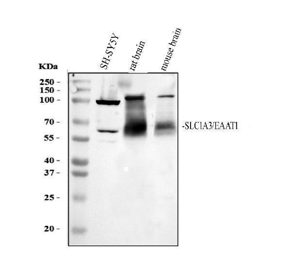
Western blot analysis of GLAST/SLC1A3 using anti-GLAST/SLC1A3 antibody (PA2185).
Electrophoresis was performed on a 10% SDS-PAGE gel at 80V (Stacking gel) / 120V (Resolving gel) for 2 hours. The sample well of each lane was loaded with 30 ug of sample under reducing conditions.
Lane 1: human SH-SY5Y whole cell lysates,
Lane 2: rat kidney tissue lysates,
Lane 3: mouse kidney tissue lysates.
After electrophoresis, proteins were transferred to a nitrocellulose membrane at 150 mA for 50-90 minutes. Blocked the membrane with 5% non-fat milk/TBS for 1.5 hour at RT. The membrane was incubated with rabbit anti-GLAST/SLC1A3 antigen affinity purified polyclonal antibody (PA2185) at 0.5 μg/mL overnight at 4°C, then washed with TBS-0.1%Tween 3 times with 5 minutes each and probed with a goat anti-rabbit IgG-HRP secondary antibody (Catalog # BA1054) at a dilution of 1:5000 for 1.5 hour at RT. The signal is developed using an ECL Plus Western Blotting Substrate (Catalog # AR1196-200) with Tanon 5200 system. A specific band was detected for GLAST/SLC1A3 at approximately 60 kDa. The expected band size for GLAST/SLC1A3 is at 60 kDa.

Click image to see more details
IHC analysis of GLAST/SLC1A3 using anti-GLAST/SLC1A3 antibody (PA2185).
GLAST/SLC1A3 was detected in a paraffin-embedded section of human brain tissue. Heat mediated antigen retrieval was performed in EDTA buffer (pH 8.0, epitope retrieval solution). The tissue section was blocked with 10% goat serum. The tissue section was then incubated with 2 μg/ml rabbit anti-GLAST/SLC1A3 Antibody (PA2185) overnight at 4°C. Peroxidase Conjugated Goat Anti-rabbit IgG was used as secondary antibody and incubated for 30 minutes at 37°C. The tissue section was developed using HRP Conjugated Rabbit IgG Super Vision Assay Kit (Catalog # SV0002) with DAB as the chromogen.

Click image to see more details
IHC analysis of GLAST/SLC1A3 using anti-GLAST/SLC1A3 antibody (PA2185).
GLAST/SLC1A3 was detected in a paraffin-embedded section of mouse brain tissue. Heat mediated antigen retrieval was performed in EDTA buffer (pH 8.0, epitope retrieval solution). The tissue section was blocked with 10% goat serum. The tissue section was then incubated with 2 μg/ml rabbit anti-GLAST/SLC1A3 Antibody (PA2185) overnight at 4°C. Peroxidase Conjugated Goat Anti-rabbit IgG was used as secondary antibody and incubated for 30 minutes at 37°C. The tissue section was developed using HRP Conjugated Rabbit IgG Super Vision Assay Kit (Catalog # SV0002) with DAB as the chromogen.

Click image to see more details
IHC analysis of GLAST/SLC1A3 using anti-GLAST/SLC1A3 antibody (PA2185).
GLAST/SLC1A3 was detected in a paraffin-embedded section of rat brain tissue. Heat mediated antigen retrieval was performed in EDTA buffer (pH 8.0, epitope retrieval solution). The tissue section was blocked with 10% goat serum. The tissue section was then incubated with 2 μg/ml rabbit anti-GLAST/SLC1A3 Antibody (PA2185) overnight at 4°C. Peroxidase Conjugated Goat Anti-rabbit IgG was used as secondary antibody and incubated for 30 minutes at 37°C. The tissue section was developed using HRP Conjugated Rabbit IgG Super Vision Assay Kit (Catalog # SV0002) with DAB as the chromogen.
Specific Publications For Anti-EAAT1/SLC1A3 Antibody Picoband® (PA2185)
Loading publications
Recommended Resources
Here are featured tools and databases that you might find useful.
- Boster's Pathways Library
- Protein Databases
- Bioscience Research Protocol Resources
- Data Processing & Analysis Software
- Photo Editing Software
- Scientific Literature Resources
- Research Paper Management Tools
- Molecular Biology Software
- Primer Design Tools
- Bioinformatics Tools
- Phylogenetic Tree Analysis
Customer Reviews
Have you used Anti-EAAT1/SLC1A3 Antibody Picoband®?
Share your experimental results or join a short interview to earn up to $1,000 in product credits or other rewards.
0 Reviews For Anti-EAAT1/SLC1A3 Antibody Picoband®
Customer Q&As
Have a question?
Find answers in Q&As, reviews.
Can't find your answer?
Submit your question
