Click image to see more details
-

-
-
-
-
+4
Product Info Summary
| SKU: | M04372 |
|---|---|
| Size: | 100 μg/vial |
| Reactive Species: | Human |
| Host: | Mouse |
| Application: | Flow Cytometry, IF, IHC, ICC, WB |
Customers Who Bought This Also Bought

Product info
Product Name
Anti-FACL4/ACSL4 Antibody Picoband™ (monoclonal, 4I7)
SKU/Catalog Number
M04372
Size
100 μg/vial
Form
Lyophilized
Description
Boster Bio Anti-FACL4/ACSL4 Antibody Picoband™ (monoclonal, 4I7) catalog # M04372. Tested in Flow Cytometry, IF, IHC, ICC, WB applications. This antibody reacts with Human.
Storage & Handling
At -20°C for one year from date of receipt. After reconstitution, at 4°C for one month. It can also be aliquotted and stored frozen at -20°C for six months. Avoid repeated freezing and thawing.
Cite This Product
Anti-FACL4/ACSL4 Antibody Picoband™ (monoclonal, 4I7) (Boster Biological Technology, Pleasanton CA, USA, Catalog # M04372)
Host
Mouse
Contents
Each vial contains 4 mg Trehalose, 0.9 mg NaCl and 0.2 mg Na2HPO4.
Clonality
Monoclonal
Clone Number
4I7
Isotype
Mouse IgG1
Immunogen
A synthetic peptide corresponding to a sequence at the C-terminus of human FACL4/ACSL4.
*Blocking peptide can be purchased. Costs vary based on immunogen length. Contact us for pricing.
Cross-reactivity
No cross-reactivity with other proteins.
Reactive Species
M04372 is reactive to ACSL4 in Human
Applications
M04372 is guaranteed for Flow Cytometry, IF, IHC, ICC, WB Boster Guarantee
Observed Molecular Weight
79 kDa
Calculated molecular weight
79.188kDa
Background of FACL4
Long-chain-fatty-acid—CoA ligase 4 is an enzyme that in humans is encoded by the ACSL4 gene. It is mapped to Xq23. The protein encoded by this gene is an isozyme of the long-chain fatty-acid-coenzyme A ligase family. Although differing in substrate specificity, subcellular localization, and tissue distribution, all isozymes of this family convert free long-chain fatty acids into fatty acyl-CoA esters, and thereby play a key role in lipid biosynthesis and fatty acid degradation. This isozyme preferentially utilizes arachidonate as substrate. The absence of this enzyme may contribute to the cognitive disability or Alport syndrome. Alternative splicing of this gene generates multiple transcript variants.
Antibody Validation
Boster validates all antibodies on WB, IHC, ICC, Immunofluorescence, and ELISA with known positive control and negative samples to ensure specificity and high affinity, including thorough antibody incubations.
Innovating Scientists Reward
If you are the first to review this product, or if you have results for a special sample, species or application this product is not validated in, share your results with us and receive product credits you can use towards any Boster products! Applicable to all scientists worldwide.
Submit A Review
Assay dilution & Images
Reconsitution
Adding 0.2 ml of distilled water will yield a concentration of 500 μg/ml.
Assay Dilutions Recommendation
The recommendations below provide a starting point for assay optimization. The actual working concentration varies and should be decided by the user.
Western blot, 0.25-0.5 μg/ml, Human
Immunohistochemistry(Paraffin-embedded Section), 2-5 μg/ml, Human
Immunocytochemistry/Immunofluorescence, 5 μg/ml, Human
Flow Cytometry, 1-3 μg/1x106 cells, Human
Validation Images & Assay Conditions
Click image to see more details
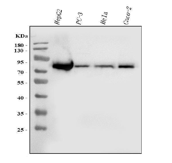
Figure 1. Western blot analysis of FACL4/ACSL4 using anti-FACL4/ACSL4 antibody (M04372).
Electrophoresis was performed on a 5-20% SDS-PAGE gel at 70V (Stacking gel) / 90V (Resolving gel) for 2-3 hours. The sample well of each lane was loaded with 30 ug of sample under reducing conditions.
Lane 1: human HepG2 whole cell lysates,
Lane 2: human PC-3 whole cell lysates,
Lane 3: human Hela whole cell lysates,
Lane 4: human Caco-2 whole cell lysates.
After electrophoresis, proteins were transferred to a nitrocellulose membrane at 150 mA for 50-90 minutes. Blocked the membrane with 5% non-fat milk/TBS for 1.5 hour at RT. The membrane was incubated with mouse anti-FACL4/ACSL4 antigen affinity purified monoclonal antibody (Catalog # M04372) at 0.5 μg/mL overnight at 4°C, then washed with TBS-0.1%Tween 3 times with 5 minutes each and probed with a goat anti-mouse IgG-HRP secondary antibody at a dilution of 1:10000 for 1.5 hour at RT. The signal is developed using an Enhanced Chemiluminescent detection (ECL) kit (Catalog # EK1001) with Tanon 5200 system. A specific band was detected for FACL4/ACSL4 at approximately 79 kDa. The expected band size for FACL4/ACSL4 is at 68 kDa.

Click image to see more details
Figure 2. IHC analysis of FACL4/ACSL4 using anti-FACL4/ACSL4 antibody (M04372).
FACL4/ACSL4 was detected in a paraffin-embedded section of human bladder epithelial carcinoma tissue. Heat mediated antigen retrieval was performed in EDTA buffer (pH 8.0, epitope retrieval solution). The tissue section was blocked with 10% goat serum. The tissue section was then incubated with 2 μg/ml mouse anti-FACL4/ACSL4 Antibody (M04372) overnight at 4°C. Biotinylated goat anti-mouse IgG was used as secondary antibody and incubated for 30 minutes at 37°C. The tissue section was developed using Strepavidin-Biotin-Complex (SABC) (Catalog # SA1021) with DAB as the chromogen.

Click image to see more details
Figure 3. IHC analysis of FACL4/ACSL4 using anti-FACL4/ACSL4 antibody (M04372).
FACL4/ACSL4 was detected in a paraffin-embedded section of human lung cancer tissue. Heat mediated antigen retrieval was performed in EDTA buffer (pH 8.0, epitope retrieval solution). The tissue section was blocked with 10% goat serum. The tissue section was then incubated with 2 μg/ml mouse anti-FACL4/ACSL4 Antibody (M04372) overnight at 4°C. Biotinylated goat anti-mouse IgG was used as secondary antibody and incubated for 30 minutes at 37°C. The tissue section was developed using Strepavidin-Biotin-Complex (SABC) (Catalog # SA1021) with DAB as the chromogen.

Click image to see more details
Figure 4. IHC analysis of FACL4/ACSL4 using anti-FACL4/ACSL4 antibody (M04372).
FACL4/ACSL4 was detected in a paraffin-embedded section of human metaplasia of squamous cells of the renal pelvis tissue. Heat mediated antigen retrieval was performed in EDTA buffer (pH 8.0, epitope retrieval solution). The tissue section was blocked with 10% goat serum. The tissue section was then incubated with 2 μg/ml mouse anti-FACL4/ACSL4 Antibody (M04372) overnight at 4°C. Biotinylated goat anti-mouse IgG was used as secondary antibody and incubated for 30 minutes at 37°C. The tissue section was developed using Strepavidin-Biotin-Complex (SABC) (Catalog # SA1021) with DAB as the chromogen.

Click image to see more details
Figure 5. IHC analysis of FACL4/ACSL4 using anti-FACL4/ACSL4 antibody (M04372).
FACL4/ACSL4 was detected in a paraffin-embedded section of human ovarian cancer tissue. Heat mediated antigen retrieval was performed in EDTA buffer (pH 8.0, epitope retrieval solution). The tissue section was blocked with 10% goat serum. The tissue section was then incubated with 2 μg/ml mouse anti-FACL4/ACSL4 Antibody (M04372) overnight at 4°C. Biotinylated goat anti-mouse IgG was used as secondary antibody and incubated for 30 minutes at 37°C. The tissue section was developed using Strepavidin-Biotin-Complex (SABC) (Catalog # SA1021) with DAB as the chromogen.

Click image to see more details
Figure 6. IHC analysis of FACL4/ACSL4 using anti-FACL4/ACSL4 antibody (M04372).
FACL4/ACSL4 was detected in a paraffin-embedded section of human rectal moderately differentiated adenocarcinoma tissue. Heat mediated antigen retrieval was performed in EDTA buffer (pH 8.0, epitope retrieval solution). The tissue section was blocked with 10% goat serum. The tissue section was then incubated with 2 μg/ml mouse anti-FACL4/ACSL4 Antibody (M04372) overnight at 4°C. Biotinylated goat anti-mouse IgG was used as secondary antibody and incubated for 30 minutes at 37°C. The tissue section was developed using Strepavidin-Biotin-Complex (SABC) (Catalog # SA1021) with DAB as the chromogen.

Click image to see more details
Figure 7. IF analysis of FACL4/ACSL4 using anti-FACL4/ACSL4 antibody (M04372).
FACL4/ACSL4 was detected in an immunocytochemical section of SiHa cells. Enzyme antigen retrieval was performed using IHC enzyme antigen retrieval reagent (AR0022) for 15 mins. The cells were blocked with 10% goat serum. And then incubated with 5 μg/mL mouse anti-FACL4/ACSL4 Antibody (M04372) overnight at 4°C. DyLight®488 Conjugated Goat Anti-Mouse IgG (BA1126) was used as secondary antibody at 1:100 dilution and incubated for 30 minutes at 37°C. The section was counterstained with DAPI. Visualize using a fluorescence microscope and filter sets appropriate for the label used.

Click image to see more details
Figure 8. Flow Cytometry analysis of HepG2 cells using anti-FACL4/ACSL4 antibody (M04372).
Overlay histogram showing HepG2 cells stained with M04372 (Blue line). The cells were blocked with 10% normal goat serum. And then incubated with mouse anti-FACL4/ACSL4 Antibody (M04372, 1 μg/1x106 cells) for 30 min at 20°C. DyLight®488 conjugated goat anti-mouse IgG (BA1126, 5-10 μg/1x106 cells) was used as secondary antibody for 30 minutes at 20°C. Isotype control antibody (Green line) was mouse IgG (1 μg/1x106) used under the same conditions. Unlabelled sample (Red line) was also used as a control.
Protein Target Info & Infographic
Gene/Protein Information For ACSL4 (Source: Uniprot.org, NCBI)
Gene Name
ACSL4
Full Name
Long-chain-fatty-acid--CoA ligase 4
Weight
79.188kDa
Superfamily
ATP-dependent AMP-binding enzyme family
Alternative Names
ACS4mental retardation, X-linked 68; acyl-CoA synthetase 4; acyl-CoA synthetase long-chain family member 4; EC 6.2.1.3; FACL4long-chain 4; LACS 4; LACS4MRX68; lignoceroyl-CoA synthase; Long-chain acyl-CoA synthetase 4; long-chain fatty-acid-Coenzyme A ligase 4; long-chain-fatty-acid--CoA ligase 4; mental retardation, X-linked 63; MRX63 ACSL4 ACS4, FACL4, LACS4, MRX63, MRX68 acyl-CoA synthetase long chain family member 4 long-chain-fatty-acid--CoA ligase 4|acyl-CoA synthetase 4|arachidonate--CoA ligase|fatty-acid-Coenzyme A ligase, long-chain 4|lignoceroyl-CoA synthase|long-chain acyl-CoA synthetase 4|long-chain fatty-acid-Coenzyme A ligase 4
*If product is indicated to react with multiple species, protein info is based on the gene entry specified above in "Species".For more info on ACSL4, check out the ACSL4 Infographic

We have 30,000+ of these available, one for each gene! Check them out.
In this infographic, you will see the following information for ACSL4: database IDs, superfamily, protein function, synonyms, molecular weight, chromosomal locations, tissues of expression, subcellular locations, post-translational modifications, and related diseases, research areas & pathways. If you want to see more information included, or would like to contribute to it and be acknowledged, please contact [email protected].
Specific Publications For Anti-FACL4/ACSL4 Antibody Picoband™ (monoclonal, 4I7) (M04372)
Hello CJ!
No publications found for M04372
*Do you have publications using this product? Share with us and receive a reward. Ask us for more details.
Recommended Resources
Here are featured tools and databases that you might find useful.
- Boster's Pathways Library
- Protein Databases
- Bioscience Research Protocol Resources
- Data Processing & Analysis Software
- Photo Editing Software
- Scientific Literature Resources
- Research Paper Management Tools
- Molecular Biology Software
- Primer Design Tools
- Bioinformatics Tools
- Phylogenetic Tree Analysis
Customer Reviews
Have you used Anti-FACL4/ACSL4 Antibody Picoband™ (monoclonal, 4I7)?
Submit a review and receive an Amazon gift card.
- $30 for a review with an image
Be the first to review Anti-FACL4/ACSL4 Antibody Picoband™ (monoclonal, 4I7)
*The first user to submit a review for a product is eligible for Boster's Innovating Scientists Reward, which gives product credits. This is in addition to the gift card reward.
Customer Q&As
Have a question?
Find answers in Q&As, reviews.
Can't find your answer?
Submit your question
