Click image to see more details
Product Info Summary
| SKU: | PA1239 |
|---|---|
| Size: | 100 μg/vial |
| Reactive Species: | Human, Mouse, Rat |
| Host: | Rabbit |
| Application: | IF, IHC, WB |
Customers Who Bought This Also Bought

Product info
Product Name
Anti-GFAP Antibody
SKU/Catalog Number
PA1239
Size
100 μg/vial
Form
Lyophilized
Description
Boster Bio Anti-GFAP Antibody catalog # PA1239. Tested in IF, IHC, WB applications. This antibody reacts with Human, Mouse, Rat.
Storage & Handling
Store at -20˚C for one year from date of receipt. After reconstitution, at 4˚C for one month. It can also be aliquotted and stored frozen at -20˚C for six months. Avoid repeated freeze-thaw cycles.
Cite This Product
Anti-GFAP Antibody (Boster Biological Technology, Pleasanton CA, USA, Catalog # PA1239)
Host
Rabbit
Contents
Each vial contains 5mg BSA, 0.9mg NaCl, 0.2mg Na2HPO4, 0.05mg Thimerosal, 0.05mg NaN3.
Clonality
Polyclonal
Isotype
Rabbit IgG
Immunogen
A synthetic peptide corresponding to a sequence at the C-terminus of human GFAP, identical to the related rat sequence, and different from the related mouse sequence by two amino acids.
*Blocking peptide can be purchased. Costs vary based on immunogen length. Contact us for pricing.
Cross-reactivity
No cross-reactivity with other proteins
Reactive Species
PA1239 is reactive to GFAP in Human, Mouse, Rat
Applications
PA1239 is guaranteed for IF, IHC, WB Boster Guarantee
Observed Molecular Weight
49 kDa
Calculated molecular weight
49.88kDa
Background of GFAP
Glial fibrillary acidic protein (GFAP) is an intermediate-filament (IF) protein that is highly specific for cells of astroglial lineage, although its tissue-specific role is speculative. GFAP has been located in rat kidney glomeruli and peritubular fibroblasts, leydig cells of testis, skin keratinocytes, osteocytes of bones, chondrocytes of epiglottis, bronchus, and stellate-shaped cells of the pancreas and liver. Its expression is essential for normal white matter architecture and blood-brain barrier integrity, and its absence leads to late-onset CNS dysmyelination. GFAP has also been shown to play a role in mitosis by adjusting the filament network present in the cell. During mitosis, there is an increase in the amount of phosphorylated GFAP, and a movement of this modified protein to the cleavage furrow.
Antibody Validation
Boster validates all antibodies on WB, IHC, ICC, Immunofluorescence, and ELISA with known positive control and negative samples to ensure specificity and high affinity, including thorough antibody incubations.
Innovating Scientists Reward
If you are the first to review this product, or if you have results for a special sample, species or application this product is not validated in, share your results with us and receive product credits you can use towards any Boster products! Applicable to all scientists worldwide.
Submit A Review
Assay dilution & Images
Reconsitution
Add 0.2ml of distilled water will yield a concentration of 500ug/ml.
Assay Dilutions Recommendation
The recommendations below provide a starting point for assay optimization. The actual working concentration varies and should be decided by the user.
Immunohistochemistry (Paraffin-embedded Section), 0.5-1μg/ml, Rat, Mouse, By Heat
Western blot, 0.1-0.5μg/ml, Human, Mouse, Rat
Immunofluorescence, 2μg/ml, Mouse
Validation Images & Assay Conditions
Click image to see more details
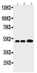
Figure 1. Western blot analysis of GFAP using anti-GFAP antibody (PA1239).
Electrophoresis was performed on a 5-20% SDS-PAGE gel at 70V (Stacking gel) / 90V (Resolving gel) for 2-3 hours. The sample well of each lane was loaded with 50ug of sample under reducing conditions.
Lane 1: Rat Brain Tissue Lysate,
Lane 2: Mouse Brain Tissue Lysate,
Lane 3: U87 Whole Cell Lysate.
After Electrophoresis, proteins were transferred to a Nitrocellulose membrane at 150mA for 50-90 minutes. Blocked the membrane with 5% Non-fat Milk/ TBS for 1.5 hour at RT. The membrane was incubated with rabbit anti-GFAP antigen affinity purified polyclonal antibody (Catalog # PA1239) at 0.5 μg/mL overnight at 4°C, then washed with TBS-0.1%Tween 3 times with 5 minutes each and probed with a goat anti-rabbit IgG-HRP secondary antibody at a dilution of 1:10000 for 1.5 hour at RT. The signal is developed using an Enhanced Chemiluminescent detection (ECL) kit (Catalog # EK1002) with Tanon 5200 system. A specific band was detected for GFAP at approximately 49 KD. The expected band size for GFAP is at 49 KD.

Click image to see more details
Figure 2. IHC analysis of GFAP using anti-GFAP antibody (PA1239).
GFAP was detected in paraffin-embedded section of rat brain tissues. Heat mediated antigen retrieval was performed in citrate buffer (pH6, epitope retrieval solution) for 20 mins. The tissue section was blocked with 10% goat serum. The tissue section was then incubated with 1μg/ml rabbit anti-GFAP Antibody (PA1239) overnight at 4°C. Biotinylated goat anti-rabbit IgG was used as secondary antibody and incubated for 30 minutes at 37°C. The tissue section was developed using Strepavidin-Biotin-Complex (SABC)(Catalog # SA1022) with DAB as the chromogen.

Click image to see more details
Figure 3. IF analysis of GFAP using anti-GFAP antibody (PA1239).
GFAP was detected in paraffin-embedded section of mouse brain tissues. Heat mediated antigen retrieval was performed in citrate buffer (pH6, epitope retrieval solution ) for 20 mins. The tissue section was blocked with 10% goat serum. The tissue section was then incubated with 1μg/mL rabbit anti-GFAP Antibody (PA1239) overnight at 4°C. Cy3 Conjugated Goat Anti-Rabbit IgG (BA1032) was used as secondary antibody at 1:100 dilution and incubated for 30 minutes at 37°C. The section was counterstained with DAPI. Visualize using a fluorescence microscope and filter sets appropriate for the label used.
Protein Target Info & Infographic
Gene/Protein Information For GFAP (Source: Uniprot.org, NCBI)
Gene Name
GFAP
Full Name
Glial fibrillary acidic protein
Weight
49.88kDa
Superfamily
intermediate filament family
Alternative Names
FLJ45472; GFAP astrocytes; GFAP immunohistochemistry; GFAP mouse; GFAP rabbit; GFAP stain; GFAP; glial fibrillary acidic protein GFAP ALXDRD glial fibrillary acidic protein glial fibrillary acidic protein
*If product is indicated to react with multiple species, protein info is based on the gene entry specified above in "Species".For more info on GFAP, check out the GFAP Infographic

We have 30,000+ of these available, one for each gene! Check them out.
In this infographic, you will see the following information for GFAP: database IDs, superfamily, protein function, synonyms, molecular weight, chromosomal locations, tissues of expression, subcellular locations, post-translational modifications, and related diseases, research areas & pathways. If you want to see more information included, or would like to contribute to it and be acknowledged, please contact [email protected].
Specific Publications For Anti-GFAP Antibody (PA1239)
Hello CJ!
PA1239 has been cited in 111 publications:
*The publications in this section are manually curated by our staff scientists. They may differ from Bioz's machine gathered results. Both are accurate. If you find a publication citing this product but is missing from this list, please let us know we will issue you a thank-you coupon.
OLIG2 Drives Abnormal Neurodevelopmental Phenotypes in Human iPSC-Based Organoid and Chimeric Mouse Models of Down Syndrome
Species: Mouse
Xiang Z,Jiang X,Ji R,Yuan H.Enhanced expression of P2X4 purinoceptors in pyramidal neurons of the rat hippocampal CA1 region may be involved ischemia-reperfusion injury.Purinergic Signal.2021 May 9.doi:10.1007/s11302-021-09780-z.Epub ahead of print.PMID:33966147.
Species: Rat
PA1239 usage in article: APP:IF, SAMPLE:BRAIN TISSUE, DILUTION:1:400
Xiang S,Zhao D,Hao H,Wang XU,Li L,Yang T.α-Helical protein absorption at post-traumatic epileptic foci monitored by Fourier transform infrared mapping.J Biosci.2020;45:55.PMID:32345781.
Species: Rat
PA1239 usage in article: APP:IHC, SAMPLE:FRONTAL BRAIN LOBE, DILUTION:1:100
Zhang Y,Zhang M,Zhu W,Pan X,Wang Q,Gao X,Wang C,Zhang X,Liu Y,Li S,Sun H.Role of Elevated Thrombospondin-1 in Kainic Acid-Induced Status Epilepticus.Neurosci Bull.2020 Mar;36(3):263-276.doi:10.1007/s12264-019-00437-x.Epub 2019 Oct 29.PMID:31664678;PMCID:P
Species: Rat
PA1239 usage in article: APP:WB, SAMPLE:HIPPOCAMPUS AND CORTEX, DILUTION:1:1000
Chen X,Zhang L,Hua F,Zhuang Y,Liu H,Wang S.EphA4 Obstructs Spinal Cord Neuron Regeneration by Promoting Excessive Activation of Astrocytes.Cell Mol Neurobiol.2021 Feb 17.doi:10.1007/s10571-021-01046-x.Epub ahead of print.PMID:33595805.
Species: Mouse
PA1239 usage in article: APP:IF, SAMPLE:SPINAL CORD TISSUE, DILUTION:1:500
Zhang LY,Jin QQ,Hölscher C,Li L.Glucagon-like peptide-1/glucose-dependent insulinotropic polypeptide dual receptor agonist DA-CH5 is superior to exendin-4 in protecting neurons in the 6-hydroxydopamine rat Parkinson model. Neural Regen Res 2021;16:1660-70
Species: Human,Rat
PA1239 usage in article: APP:IF, SAMPLE:SN TISSUE, DILUTION:1:100
Wu,H.,Jiang,X.,Li,Y.,Zhou,Y.,Zhang,T.,Zhi,P.,Gao,J.,Engineering Stem Cell Derived Biomimetic Vesicles for Versatility and Effective Targeted Delivery.Adv. Funct.Mater.2020, 30, 2006169.https://doi.org/10.1002/adfm.202006169
Species: Rat,Mouse
PA1239 usage in article: APP:IF, SAMPLE:BRAIN TISSUE, DILUTION:1:200
Jiangong Wang,Bin Liu,Yong Xu et al. Inhibition of Histamine H3 receptor Attenuates Neuroinflammation and Cognitive Impairments in Alzheimer’s Disease via activating CREB Pathway,16 December 2020, PREPRINT (Version 1) available at Research Square [https:/
Species: Mouse
PA1239 usage in article: APP:IHC, SAMPLE:BRAIN TISSUE, DILUTION:1:200
Ahmed S. Ahmed,JAK-1/STAT-3 pathway mediated role in aging cerebellar cortex degenerative changes of albino wistar rats, Translational Research in Anatomy, 2020,100089,ISSN 2214-854X, https://doi.org/10. 1016/j.tria.2020.100089.
Species: Rat
PA1239 usage in article: APP:IHC, SAMPLE:CEREBELLAR CORTEX, DILUTION:1:20
Zhou Feng,Shengyan Liu,Qianwei Chen,Qiang Tan,Jishu Xian,Hua Feng,Zhi Chen,Gang Li,uPA alleviates kaolin-induced hydrocephalus by promoting the release and activation of hepatocyte growth factor in rats, Neuroscience Letters,Volume 731,2020,135011,ISSN 03
Species: Rat
PA1239 usage in article: APP:IF, SAMPLE:BRAIN TISSUE,
Recommended Resources
Here are featured tools and databases that you might find useful.
- Boster's Pathways Library
- Protein Databases
- Bioscience Research Protocol Resources
- Data Processing & Analysis Software
- Photo Editing Software
- Scientific Literature Resources
- Research Paper Management Tools
- Molecular Biology Software
- Primer Design Tools
- Bioinformatics Tools
- Phylogenetic Tree Analysis
Customer Reviews
Have you used Anti-GFAP Antibody?
Submit a review and receive an Amazon gift card.
- $30 for a review with an image
1 Reviews For Anti-GFAP Antibody
0
This Antibody Works Perfectly!--Maria Teresa Dell'Anno, NEUROSCIENCE, YALE UNIVERSITY, POSTDOC
Excellent

Source: Biocompare.com
| Applications | Immunohistochemistry |
|---|---|
| Sample | Spinal cord |
| Detection | Confocal microscope |
"I used it for IHC on frozen sections at a dilution of 1:500. It did not need several trials to optimize the protocol. No bad things overall. I will purchase it again."
Maria Teresa Dell'Anno
Verified customer
Submitted 2016-08-24
Customer Q&As
Have a question?
Find answers in Q&As, reviews.
Can't find your answer?
Submit your question
16 Customer Q&As for Anti-GFAP Antibody
Question
Is a blocking peptide available for product anti-GFAP antibody (PA1239)?
Verified Customer
Verified customer
Asked: 2020-05-06
Answer
We do provide the blocking peptide for product anti-GFAP antibody (PA1239). If you would like to place an order for it please contact [email protected] and make a special request.
Boster Scientific Support
Answered: 2020-05-06
Question
Will PA1239 anti-GFAP antibody work on parafin embedded sections? If so, which fixation method do you recommend we use (PFA, paraformaldehyde, other)?
Verified Customer
Verified customer
Asked: 2020-04-22
Answer
You can see on the product datasheet, PA1239 anti-GFAP antibody as been validated on IHC. It is best to use PFA for fixation because it has better tissue penetration ability. PFA needs to be prepared fresh before use. Long term stored PFA turns into formalin, as the PFA molecules congregate and become formalin.
Boster Scientific Support
Answered: 2020-04-22
Question
We have been able to see staining in rat fetal brain cortex. Any tips? Is anti-GFAP antibody supposed to stain fetal brain cortex positively?
Verified Customer
Verified customer
Asked: 2020-02-18
Answer
From what I have seen in literature fetal brain cortex does express GFAP. From what I have seen in Uniprot.org, GFAP is expressed in dorsal motor nucleus of vagus nerve, brain thalamus, brain, kidney, fetal brain cortex, fetal brain, blood, among other tissues. Regarding which tissues have GFAP expression, here are a few articles citing expression in various tissues:
Blood, Pubmed ID: 12837269
Brain, Pubmed ID: 15489334
Brain, and Thalamus, Pubmed ID: 14702039
Fetal brain, Pubmed ID: 12058025
Fetal brain cortex, Pubmed ID: 2780570
Kidney, Pubmed ID: 17974005
Boster Scientific Support
Answered: 2020-02-18
Question
I have attached the WB image, lot number and protocol we used for fetal brain using anti-GFAP antibody PA1239. Please let me know if you require anything else.
Verified Customer
Verified customer
Asked: 2019-12-23
Answer
Thank you very much for the data. Our lab team are working to resolve this as quickly as possible, and we appreciate your patience and understanding! You have provided everything we needed. Please let me know if there is anything you need in the meantime.
Boster Scientific Support
Answered: 2019-12-23
Question
Thank you for helping with my inquiry over the phone. Here are the WB image, lot number and protocol we used for fetal brain using anti-GFAP antibody PA1239. Let me know if you need anything else.
L. Rodriguez
Verified customer
Asked: 2019-12-16
Answer
We appreciate the data. You have provided everything we needed. Our lab team are working to resolve your inquiry as quickly as possible, and we appreciate your patience and understanding! Please let me know if there is anything you need in the meantime.
Boster Scientific Support
Answered: 2019-12-16
Question
Can you help my question with product PA1239, anti-GFAP antibody. I was wondering if it would be possible to conjugate this antibody with biotin. I would need it to be without BSA or sodium azide. I am planning on using a buffer exchange of sodium azide with PBS only. Would there be problems for me to conjugate the antibody and store it in -20 degrees in small aliquots?
Verified Customer
Verified customer
Asked: 2019-08-29
Answer
It is not recommended storing this antibody with PBS buffer only in -20 degrees. If you want to store it in -20 degrees it is best to add some cryoprotectant like glycerol. If you want carrier free PA1239 anti-GFAP antibody, we can provide it to you in a special formula with trehalose and/or glycerol. These molecules will not interfere with conjugation chemistry and provide a good level of protection for the antibody from degradation. Please be sure to specify this in your purchase order.
Boster Scientific Support
Answered: 2019-08-29
Question
I was wanting to use your anti-GFAP antibody for IHC for rat fetal brain on frozen tissues, but I want to know if it has been validated for this particular application. Has this antibody been validated and is this antibody a good choice for rat fetal brain identification?
Verified Customer
Verified customer
Asked: 2019-07-22
Answer
You can see on the product datasheet, PA1239 anti-GFAP antibody has been tested for IF, IHC, WB on human, mouse, rat tissues. We have an innovator award program that if you test this antibody and show it works in rat fetal brain in IHC-frozen, you can get your next antibody for free.
Boster Scientific Support
Answered: 2019-07-22
Question
Is this PA1239 anti-GFAP antibody reactive to the isotypes of GFAP?
Z. Carter
Verified customer
Asked: 2019-07-15
Answer
The immunogen of PA1239 anti-GFAP antibody is A synthetic peptide corresponding to a sequence at the C-terminus of human GFAP(417-432aa DGEVIKESKQEHKDVM), identical to the related rat sequence, and different from the related mouse sequence by two amino acids. Could you tell me which isotype you are interested in so I can help see if the immunogen is part of this isotype?
Boster Scientific Support
Answered: 2019-07-15
Question
I see that the anti-GFAP antibody PA1239 works with IHC, what is the protocol used to produce the result images on the product page?
Verified Customer
Verified customer
Asked: 2019-06-12
Answer
You can find protocols for IHC on the "support/technical resources" section of our navigation menu. If you have any further questions, please send an email to [email protected]
Boster Scientific Support
Answered: 2019-06-12
Question
Is there a BSA free version of anti-GFAP antibody PA1239 available?
Verified Customer
Verified customer
Asked: 2019-05-15
Answer
We appreciate your recent telephone inquiry. I can confirm that some lots of this anti-GFAP antibody PA1239 are BSA free. For now, these lots are available and we can make a BSA free formula for you free of charge. It will take 3 extra days to prepare. If you require this antibody BSA free again in future, please do not hesitate to contact me and I will be pleased to check which lots we have in stock that are BSA free.
Boster Scientific Support
Answered: 2019-05-15
Question
I was wanting to use using your anti-GFAP antibody for regulation of chaperone-mediated autophagy studies. Has this antibody been tested with western blotting on u87 whole cell lysate? We would like to see some validation images before ordering.
Verified Customer
Verified customer
Asked: 2019-03-20
Answer
We appreciate your inquiry. This PA1239 anti-GFAP antibody is validated on rat brain tissue, tissue lysate, mouse brain, u87 whole cell lysate. It is guaranteed to work for IF, IHC, WB in human, mouse, rat. Our Boster guarantee will cover your intended experiment even if the sample type has not been be directly tested.
Boster Scientific Support
Answered: 2019-03-20
Question
Will anti-GFAP antibody PA1239 work for IHC with fetal brain?
Verified Customer
Verified customer
Asked: 2018-12-20
Answer
According to the expression profile of fetal brain, GFAP is highly expressed in fetal brain. So, it is likely that anti-GFAP antibody PA1239 will work for IHC with fetal brain.
Boster Scientific Support
Answered: 2018-12-20
Question
We are currently using anti-GFAP antibody PA1239 for rat tissue, and we are happy with the IHC results. The species of reactivity given in the datasheet says human, mouse, rat. Is it possible that the antibody can work on zebrafish tissues as well?
Verified Customer
Verified customer
Asked: 2018-07-11
Answer
The anti-GFAP antibody (PA1239) has not been tested for cross reactivity specifically with zebrafish tissues, though there is a good chance of cross reactivity. We have an innovator award program that if you test this antibody and show it works in zebrafish you can get your next antibody for free. Please contact me if I can help you with anything.
Boster Scientific Support
Answered: 2018-07-11
Question
I am looking for to test anti-GFAP antibody PA1239 on rat fetal brain for research purposes, then I may be interested in using anti-GFAP antibody PA1239 for diagnostic purposes as well. Is the antibody suitable for diagnostic purposes?
Verified Customer
Verified customer
Asked: 2017-09-19
Answer
The products we sell, including anti-GFAP antibody PA1239, are only intended for research use. They would not be suitable for use in diagnostic work. If you have the means to develop a product into diagnostic use, and are interested in collaborating with us and develop our product into an IVD product, please contact us for more discussions.
Boster Scientific Support
Answered: 2017-09-19
Question
We were content with the WB result of your anti-GFAP antibody. However we have seen positive staining in brain thalamus cytoplasm. using this antibody. Is that expected? Could you tell me where is GFAP supposed to be expressed?
Verified Customer
Verified customer
Asked: 2017-07-19
Answer
From literature, brain thalamus does express GFAP. Generally GFAP expresses in cytoplasm. Regarding which tissues have GFAP expression, here are a few articles citing expression in various tissues:
Blood, Pubmed ID: 12837269
Brain, Pubmed ID: 15489334
Brain, and Thalamus, Pubmed ID: 14702039
Fetal brain, Pubmed ID: 12058025
Fetal brain cortex, Pubmed ID: 2780570
Kidney, Pubmed ID: 17974005
Boster Scientific Support
Answered: 2017-07-19
Question
We purchased anti-GFAP antibody for IF on fetal brain in the past. I am using rat, and We are going to use the antibody for IHC next. I would like examining fetal brain as well as dorsal motor nucleus of vagus nerve in our next experiment. Could you please give me some suggestion on which antibody would work the best for IHC?
H. Dhar
Verified customer
Asked: 2016-04-15
Answer
I viewed the website and datasheets of our anti-GFAP antibody and it seems that PA1239 has been tested on rat in both IF and IHC. Thus PA1239 should work for your application. Our Boster satisfaction guarantee will cover this product for IHC in rat even if the specific tissue type has not been validated. We do have a comprehensive range of products for IHC detection and you can check out our website bosterbio.com to find out more information about them.
Boster Scientific Support
Answered: 2016-04-15
