Click image to see more details
Product Info Summary
| SKU: | A00514-3 |
|---|---|
| Size: | 100 μg/vial |
| Reactive Species: | Human, Mouse, Rat |
| Host: | Rabbit |
| Application: | ELISA, Flow Cytometry, IF, IHC, ICC, WB |
Customers Who Bought This Also Bought


Product info
Product Name
Anti-KEAP1 Antibody Picoband™
SKU/Catalog Number
A00514-3
Size
100 μg/vial
Form
Lyophilized
Description
Boster Bio Anti-KEAP1 Antibody Picoband™ catalog # A00514-3. Tested in ELISA, Flow Cytometry, IF, IHC, ICC, WB applications. This antibody reacts with Human, Mouse, Rat.
Storage & Handling
At -20°C for one year from date of receipt. After reconstitution, at 4°C for one month. It can also be aliquotted and stored frozen at -20°C for six months. Avoid repeated freezing and thawing.
Cite This Product
Anti-KEAP1 Antibody Picoband™ (Boster Biological Technology, Pleasanton CA, USA, Catalog # A00514-3)
Host
Rabbit
Contents
Each vial contains 4 mg Trehalose, 0.9 mg NaCl, 0.2 mg Na2HPO4.
Clonality
Polyclonal
Isotype
Rabbit IgG
Immunogen
E.coli-derived human KEAP1 recombinant protein (Position: K84-K312).
*Blocking peptide can be purchased. Costs vary based on immunogen length. Contact us for pricing.
Cross-reactivity
No cross-reactivity with other proteins.
Reactive Species
A00514-3 is reactive to KEAP1 in Human, Mouse, Rat
Applications
A00514-3 is guaranteed for ELISA, Flow Cytometry, IF, IHC, ICC, WB Boster Guarantee
Observed Molecular Weight
66-72 kDa
Calculated molecular weight
69.666kDa
Background of KEAP1
KEAP1 (KELCH-LIKE ECH-ASSOCIATED PROTEIN 1), is a protein that in humans is encoded by the Keap1 gene. The KIAA0132 gene is mapped on 19p13.2. Keap1 contains a central BTB/POZ domain and a C-terminal double glycine repeat (DGR), or Kelch, module. Keap1 has been shown to interact with Nrf2, a master regulator of the antioxidant response, which is important for the amelioration of oxidative stress. In the presence of the electrophilic agent diethylmalate, Nrf2 activity is released from Keap1 and Nrf2 translocate to the nucleus. Under quiescent conditions, Nrf2 is anchored in the cytoplasm through binding to Keap1, which, in turn, facilitates the ubiquitination and subsequent proteolysis of Nrf2. Because Nrf2 activation leads to a coordinated antioxidant and anti-inflammatory response, and Keap1 represses Nrf2 activation, Keap1 has become a very attractive drug target.
Antibody Validation
Boster validates all antibodies on WB, IHC, ICC, Immunofluorescence, and ELISA with known positive control and negative samples to ensure specificity and high affinity, including thorough antibody incubations.
Innovating Scientists Reward
If you are the first to review this product, or if you have results for a special sample, species or application this product is not validated in, share your results with us and receive product credits you can use towards any Boster products! Applicable to all scientists worldwide.
Submit A Review
Assay dilution & Images
Reconsitution
Adding 0.2 ml of distilled water will yield a concentration of 500 μg/ml.
Assay Dilutions Recommendation
The recommendations below provide a starting point for assay optimization. The actual working concentration varies and should be decided by the user.
Western blot, 0.25-0.5 μg/ml, Human, Mouse, Rat
Immunohistochemistry(Paraffin-embedded Section), 2-5 μg/ml, Human, Rat
Immunocytochemistry/Immunofluorescence, 5 μg/ml, Human
Flow Cytometry, 1-3 μg/1x106 cells, Human
Direct ELISA, 0.1-0.5 μg/ml, Human
Validation Images & Assay Conditions
Click image to see more details
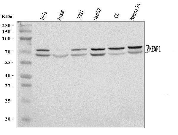
Figure 1. Western blot analysis of KEAP1 using anti-KEAP1 antibody (A00514-3).
Electrophoresis was performed on a 5-20% SDS-PAGE gel at 70V (Stacking gel) / 90V (Resolving gel) for 2-3 hours. The sample well of each lane was loaded with 30 ug of sample under reducing conditions.
Lane 1: human Hela whole cell lysates,
Lane 2: human Jurkat whole cell lysates,
Lane 3: human 293T whole cell lysates,
Lane 4: human HepG2 whole cell lysates,
Lane 5: rat C6 whole cell lysates,
Lane 6: mouse Neuro-2a whole cell lysates.
After electrophoresis, proteins were transferred to a nitrocellulose membrane at 150 mA for 50-90 minutes. Blocked the membrane with 5% non-fat milk/TBS for 1.5 hour at RT. The membrane was incubated with rabbit anti-KEAP1 antigen affinity purified polyclonal antibody (Catalog # A00514-3) at 0.5 μg/mL overnight at 4°C, then washed with TBS-0.1%Tween 3 times with 5 minutes each and probed with a goat anti-rabbit IgG-HRP secondary antibody at a dilution of 1:5000 for 1.5 hour at RT. The signal is developed using an Enhanced Chemiluminescent detection (ECL) kit (Catalog # EK1002) with Tanon 5200 system. A specific band was detected for KEAP1 at approximately 66-72 kDa. The expected band size for KEAP1 is at 70 kDa.

Click image to see more details
Figure 2. IF analysis of KEAP1 using anti-KEAP1 antibody (A00514-3).
KEAP1 was detected in an immunocytochemical section of A431 cells. Enzyme antigen retrieval was performed using IHC enzyme antigen retrieval reagent (AR0022) for 15 mins. The cells were blocked with 10% goat serum. And then incubated with 5 μg/mL rabbit anti-KEAP1 Antibody (A00514-3) overnight at 4°C. DyLight®488 Conjugated Goat Anti-Rabbit IgG (BA1127) was used as secondary antibody at 1:100 dilution and incubated for 30 minutes at 37°C. The section was counterstained with DAPI. Visualize using a fluorescence microscope and filter sets appropriate for the label used.

Click image to see more details
Figure 3. Flow Cytometry analysis of U251 cells using anti-KEAP1 antibody (A00514-3).
Overlay histogram showing U251 cells stained with A00514-3 (Blue line). The cells were blocked with 10% normal goat serum. And then incubated with rabbit anti-KEAP1 Antibody (A00514-3, 1 μg/1x106 cells) for 30 min at 20°C. DyLight®488 conjugated goat anti-rabbit IgG (BA1127, 5-10 μg/1x106 cells) was used as secondary antibody for 30 minutes at 20°C. Isotype control antibody (Green line) was rabbit IgG (1 μg/1x106) used under the same conditions. Unlabelled sample (Red line) was also used as a control.
Protein Target Info & Infographic
Gene/Protein Information For KEAP1 (Source: Uniprot.org, NCBI)
Gene Name
KEAP1
Full Name
Kelch-like ECH-associated protein 1
Weight
69.666kDa
Superfamily
KEAP1 family
Alternative Names
INrf2; INrf2MGC9454; Keap1; kelch-like ECH-associated protein 1; Kelch-like protein 19; KIAA0132MGC4407; KLHL19Cytosolic inhibitor of Nrf2; MGC10630; MGC1114; MGC20887 KEAP1 INrf2, KLHL19 kelch like ECH associated protein 1 kelch-like ECH-associated protein 1|KEAP1 delta C|cytosolic inhibitor of Nrf2|kelch-like family member 19|kelch-like protein 19
*If product is indicated to react with multiple species, protein info is based on the gene entry specified above in "Species".For more info on KEAP1, check out the KEAP1 Infographic

We have 30,000+ of these available, one for each gene! Check them out.
In this infographic, you will see the following information for KEAP1: database IDs, superfamily, protein function, synonyms, molecular weight, chromosomal locations, tissues of expression, subcellular locations, post-translational modifications, and related diseases, research areas & pathways. If you want to see more information included, or would like to contribute to it and be acknowledged, please contact [email protected].
Specific Publications For Anti-KEAP1 Antibody Picoband™ (A00514-3)
Hello CJ!
A00514-3 has been cited in 3 publications:
*The publications in this section are manually curated by our staff scientists. They may differ from Bioz's machine gathered results. Both are accurate. If you find a publication citing this product but is missing from this list, please let us know we will issue you a thank-you coupon.
Individual and combined antioxidant effects of ginsenoside F2 and cyanidin-3-O-glucoside in human embryonic kidney 293 cells
Polysaccharide from Ostrea rivularis attenuates reproductive oxidative stress damage via activating Keap1-Nrf2/ARE pathway
Andrographolide protects chondrocytes from oxidative stress injury by activation of the Keap1–Nrf2–Are signaling pathway
Recommended Resources
Here are featured tools and databases that you might find useful.
- Boster's Pathways Library
- Protein Databases
- Bioscience Research Protocol Resources
- Data Processing & Analysis Software
- Photo Editing Software
- Scientific Literature Resources
- Research Paper Management Tools
- Molecular Biology Software
- Primer Design Tools
- Bioinformatics Tools
- Phylogenetic Tree Analysis
Customer Reviews
Have you used Anti-KEAP1 Antibody Picoband™?
Submit a review and receive an Amazon gift card.
- $30 for a review with an image
Be the first to review Anti-KEAP1 Antibody Picoband™
*The first user to submit a review for a product is eligible for Boster's Innovating Scientists Reward, which gives product credits. This is in addition to the gift card reward.
Customer Q&As
Have a question?
Find answers in Q&As, reviews.
Can't find your answer?
Submit your question
