Click image to see more details
-

-
-
-
-
+4
Product Info Summary
| SKU: | A03522 |
|---|---|
| Size: | 100 μg/vial |
| Reactive Species: | Human, Mouse, Rat |
| Host: | Rabbit |
| Application: | IF, IHC, WB |
Customers Who Bought This Also Bought


Product info
Product Name
Anti-Laminin/Lamc1/Lamc2/Lamc3 Antibody Picoband™
View all Laminin gamma 1 Antibodies
SKU/Catalog Number
A03522
Size
100 μg/vial
Form
Lyophilized
Description
Boster Bio Anti-Laminin/Lamc1/Lamc2/Lamc3 Antibody Picoband™ catalog # A03522. Tested in IF, IHC, WB applications. This antibody reacts with Human, Mouse, Rat.
Storage & Handling
Store at -20˚C for one year from date of receipt. After reconstitution, at 4˚C for one month. It can also be aliquotted and stored frozen at -20˚C for six months. Avoid repeated freeze-thaw cycles.
Cite This Product
Anti-Laminin/Lamc1/Lamc2/Lamc3 Antibody Picoband™ (Boster Biological Technology, Pleasanton CA, USA, Catalog # A03522)
Host
Rabbit
Contents
Each vial contains 4mg Trehalose, 0.9mg NaCl, 0.2mg Na2HPO4, 0.05mg NaN3.
Clonality
Polyclonal
Isotype
Rabbit IgG
Immunogen
Peptide mixture of laminin gamma1,2,3 (NKLNEIEGSLNKAKDEMKAS; DLEERVRRQRNHLHLLETSI; LQLDSHGALHHKLRQLEEES). Laminin gamma has only three subtypes of antibody to gamma1-3 reactive with all isoforms of laminin.
*Blocking peptide can be purchased. Costs vary based on immunogen length. Contact us for pricing.
Cross-reactivity
No cross-reactivity with other proteins.
Reactive Species
A03522 is reactive to LAMC1 in Human, Mouse, Rat
Applications
A03522 is guaranteed for IF, IHC, WB Boster Guarantee
Observed Molecular Weight
150, 220-250 kDa
Calculated molecular weight
177.603kDa
Background of Laminin gamma 1
Tumor necrosis factor ligand superfamily member 14 is a protein that in humans is encoded by the TNFSF14 gene. TNFSF14 has also been designated as CD258, as well as LIGHT. It was mapped on chromosome 19p13.3. The protein encoded by this gene is a member of the tumor necrosis factor(TNF) ligand family. This protein may function as a costimulatory factor for the activation of lymphoid cells and as a deterrent to infection by herpesvirus. It has been shown to stimulate the proliferation of T cells, and trigger apoptosis of various tumor cells. This protein is also reported to prevent tumor necrosis factor alpha mediated apoptosis in primary hepatocyte. Two alternatively spliced transcript variant encoding distinct isoforms have been reported.
Antibody Validation
Boster validates all antibodies on WB, IHC, ICC, Immunofluorescence, and ELISA with known positive control and negative samples to ensure specificity and high affinity, including thorough antibody incubations.
Innovating Scientists Reward
If you are the first to review this product, or if you have results for a special sample, species or application this product is not validated in, share your results with us and receive product credits you can use towards any Boster products! Applicable to all scientists worldwide.
Submit A Review
Assay dilution & Images
Reconsitution
Add 0.2ml of distilled water will yield a concentration of 500ug/ml.
Assay Dilutions Recommendation
The recommendations below provide a starting point for assay optimization. The actual working concentration varies and should be decided by the user.
Western blot, 0.1-0.5μg/ml, Human, Mouse, Rat,
Immunohistochemistry (Paraffin-embedded Section), 0.5-1μg/ml, Mouse, Rat, By Heat
Immunofluorescence, 2μg/ml, Mouse, Rat
Validation Images & Assay Conditions
Click image to see more details
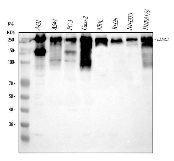
Figure 1. Western blot analysis of Laminin using anti-Laminin antibody (A03522).
Electrophoresis was performed on a 5-20% SDS-PAGE gel at 70V (Stacking gel) / 90V (Resolving gel) for 2-3 hours. The sample well of each lane was loaded with 30 ug of sample under reducing conditions.
Lane 1: human A431 whole cell lysates,
Lane 2: human A549 whole cell lysates,
Lane 3: human PC-3 whole cell lysates,
Lane 4: human CACO-2 whole cell lysates,
Lane 5: rat NRK whole cell lysates,
Lane 6: rat RH35 whole cell lysates,
Lane 7: mouse NIH/3T3 whole cell lysates,
Lane 8: mouse HEPA1-6 whole cell lysates.
After electrophoresis, proteins were transferred to a nitrocellulose membrane at 150 mA for 50-90 minutes. Blocked the membrane with 5% non-fat milk/TBS for 1.5 hour at RT. The membrane was incubated with rabbit anti-Laminin antigen affinity purified polyclonal antibody (Catalog # A03522) at 0.5 μg/mL overnight at 4°C, then washed with TBS-0.1%Tween 3 times with 5 minutes each and probed with a goat anti-rabbit IgG-HRP secondary antibody at a dilution of 1:5000 for 1.5 hour at RT. The signal is developed using an Enhanced Chemiluminescent detection (ECL) kit (Catalog # EK1002) with Tanon 5200 system. A specific band was detected for Laminin at approximately 150, 220-250 kDa. The expected band size for Laminin is at 177 kDa.

Click image to see more details
Figure 2. IHC analysis of Laminin using anti-Laminin antibody (A03522).
Laminin was detected in paraffin-embedded section of mouse heart tissue . Heat mediated antigen retrieval was performed in citrate buffer (pH6, epitope retrieval solution) for 20 mins. The tissue section was blocked with 10% goat serum. The tissue section was then incubated with 1μg/ml rabbit anti-Laminin Antibody (A03522) overnight at 4°C. Biotinylated goat anti-rabbit IgG was used as secondary antibody and incubated for 30 minutes at 37°C. The tissue section was developed using Strepavidin-Biotin-Complex (SABC)(Catalog # SA1022) with DAB as the chromogen.

Click image to see more details
Figure 3. IHC analysis of Laminin using anti-Laminin antibody (A03522).
Laminin was detected in paraffin-embedded section of mouse kidney tissue . Heat mediated antigen retrieval was performed in citrate buffer (pH6, epitope retrieval solution) for 20 mins. The tissue section was blocked with 10% goat serum. The tissue section was then incubated with 1μg/ml rabbit anti-Laminin Antibody (A03522) overnight at 4°C. Biotinylated goat anti-rabbit IgG was used as secondary antibody and incubated for 30 minutes at 37°C. The tissue section was developed using Strepavidin-Biotin-Complex (SABC)(Catalog # SA1022) with DAB as the chromogen.

Click image to see more details
Figure 4. IHC analysis of Laminin using anti-Laminin antibody (A03522).
Laminin was detected in paraffin-embedded section of rat cardiac muscle tissue. Heat mediated antigen retrieval was performed in citrate buffer (pH6, epitope retrieval solution) for 20 mins. The tissue section was blocked with 10% goat serum. The tissue section was then incubated with 1μg/ml rabbit anti-Laminin Antibody (A03522) overnight at 4°C. Biotinylated goat anti-rabbit IgG was used as secondary antibody and incubated for 30 minutes at 37°C. The tissue section was developed using Strepavidin-Biotin-Complex (SABC)(Catalog # SA1022) with DAB as the chromogen.

Click image to see more details
Figure 5. IHC analysis of Laminin using anti-Laminin antibody (A03522).
Laminin was detected in paraffin-embedded section of rat kidney tissue . Heat mediated antigen retrieval was performed in citrate buffer (pH6, epitope retrieval solution) for 20 mins. The tissue section was blocked with 10% goat serum. The tissue section was then incubated with 1μg/ml rabbit anti-Laminin Antibody (A03522) overnight at 4°C. Biotinylated goat anti-rabbit IgG was used as secondary antibody and incubated for 30 minutes at 37°C. The tissue section was developed using Strepavidin-Biotin-Complex (SABC)(Catalog # SA1022) with DAB as the chromogen.

Click image to see more details
Figure 6. IF analysis of Laminin using anti-Laminin antibody (A03522).
Laminin was detected in a paraffin-embedded section of mouse skeletal muscle tissue. Heat mediated antigen retrieval was performed in EDTA buffer (pH 8.0, epitope retrieval solution). The tissue section was blocked with 10% goat serum. The tissue section was then incubated with 5 μg/mL rabbit anti-Laminin Antibody (A03522) overnight at 4°C. DyLight®488 Conjugated Goat Anti-Rabbit IgG (BA1127) was used as secondary antibody at 1:500 dilution and incubated for 30 minutes at 37°C. The section was counterstained with DAPI. Visualize using a fluorescence microscope and filter sets appropriate for the label used.

Click image to see more details
Figure 7. IF analysis of Laminin using anti-Laminin antibody (A03522).
Laminin was detected in a paraffin-embedded section of mouse skeletal muscle tissue. Heat mediated antigen retrieval was performed in EDTA buffer (pH 8.0, epitope retrieval solution). The tissue section was blocked with 10% goat serum. The tissue section was then incubated with 5 μg/mL rabbit anti-Laminin Antibody (A03522) overnight at 4°C. DyLight®488 Conjugated Goat Anti-Rabbit IgG (BA1127) was used as secondary antibody at 1:500 dilution and incubated for 30 minutes at 37°C. The section was counterstained with DAPI. Visualize using a fluorescence microscope and filter sets appropriate for the label used.

Click image to see more details
Figure 8. IF analysis of Laminin using anti-Laminin antibody (A03522).
Laminin was detected in a paraffin-embedded section of rat skeletal muscle tissue. Heat mediated antigen retrieval was performed in EDTA buffer (pH 8.0, epitope retrieval solution). The tissue section was blocked with 10% goat serum. The tissue section was then incubated with 5 μg/mL rabbit anti-Laminin Antibody (A03522) overnight at 4°C. DyLight®488 Conjugated Goat Anti-Rabbit IgG (BA1127) was used as secondary antibody at 1:500 dilution and incubated for 30 minutes at 37°C. The section was counterstained with DAPI. Visualize using a fluorescence microscope and filter sets appropriate for the label used.
Protein Target Info & Infographic
Gene/Protein Information For LAMC1 (Source: Uniprot.org, NCBI)
Gene Name
LAMC1
Full Name
Laminin subunit gamma-1
Weight
177.603kDa
Alternative Names
LAMB2Laminin-7 subunit gamma; LAMC1; Laminin B2 chain; Laminin B2; Laminin gamma 1; laminin subunit gamma-1; laminin, gamma 1 (formerly LAMB2); Laminin-1 subunit gamma; Laminin-10 subunit gamma; Laminin-11 subunit gamma; Laminin-2 subunit gamma; Laminin-3 subunit gamma; Laminin-4 subunit gamma; Laminin-6 subunit gamma; Laminin-8 subunit gamma; Laminin-9 subunit gamma; MGC87297; S-LAM gamma; S-laminin subunit gamma LAMC1 LAMB2 laminin subunit gamma 1 laminin subunit gamma-1|S-LAM gamma|S-laminin subunit gamma|laminin B2 chain|laminin, gamma 1 (formerly LAMB2)|laminin-10 subunit gamma|laminin-11 subunit gamma|laminin-2 subunit gamma|laminin-3 subunit gamma|laminin-4 subunit gamma|laminin-6 subunit gamma|laminin-7 subunit gamma|laminin-8 subunit gamma|laminin-9 subunit gamma
*If product is indicated to react with multiple species, protein info is based on the gene entry specified above in "Species".For more info on LAMC1, check out the LAMC1 Infographic

We have 30,000+ of these available, one for each gene! Check them out.
In this infographic, you will see the following information for LAMC1: database IDs, superfamily, protein function, synonyms, molecular weight, chromosomal locations, tissues of expression, subcellular locations, post-translational modifications, and related diseases, research areas & pathways. If you want to see more information included, or would like to contribute to it and be acknowledged, please contact [email protected].
Specific Publications For Anti-Laminin/Lamc1/Lamc2/Lamc3 Antibody Picoband™ (A03522)
Hello CJ!
A03522 has been cited in 27 publications:
*The publications in this section are manually curated by our staff scientists. They may differ from Bioz's machine gathered results. Both are accurate. If you find a publication citing this product but is missing from this list, please let us know we will issue you a thank-you coupon.
Effects of transforming growth factor-β2 on myocilin expression and secretion in human primary cultured trabecular meshwork cells
The role of ursodeoxycholic acid on cholestatic hepatic fibrosis in infant rats
Effects of in vitro cultivated Calculus Bovis compound on pulmonary lesions in rabbits with schistosomiasis
Electroacupuncture Promotes the Differentiation of Transplanted Bone Marrow Mesenchymal Stem Cells Overexpressing TrkC into Neuron-Like Cells in Transected Spinal Cord of Rats:
Bone Marrow Mesenchymal Stem Cells and Electroacupuncture Downregulate the Inhibitor Molecules and Promote the Axonal Regeneration in the Transected Spinal Cord of Rats:
Observation of vascularization and protein after implanting porous silk fibroin films in rat
Effect of Chinese traditional compound, Gan‐fu‐kang, on CCl4‐induced liver fibrosis in rats and its probable molecular mechanisms
miR-194-Loaded Gelatin Nanospheres Target MEF2C to Suppress Muscle Atrophy in a Mechanical Unloading Model
Apoptosis and death receptor signaling in diaphragm of burnt rats
Implantation of adult bone marrow-derived mesenchymal stem cells transfected with the neurotrophin-3 gene and pretreated with retinoic acid in completely transected spinal cord
Recommended Resources
Here are featured tools and databases that you might find useful.
- Boster's Pathways Library
- Protein Databases
- Bioscience Research Protocol Resources
- Data Processing & Analysis Software
- Photo Editing Software
- Scientific Literature Resources
- Research Paper Management Tools
- Molecular Biology Software
- Primer Design Tools
- Bioinformatics Tools
- Phylogenetic Tree Analysis
Customer Reviews
Have you used Anti-Laminin/Lamc1/Lamc2/Lamc3 Antibody Picoband™?
Submit a review and receive an Amazon gift card.
- $30 for a review with an image
Be the first to review Anti-Laminin/Lamc1/Lamc2/Lamc3 Antibody Picoband™
*The first user to submit a review for a product is eligible for Boster's Innovating Scientists Reward, which gives product credits. This is in addition to the gift card reward.
Customer Q&As
Have a question?
Find answers in Q&As, reviews.
Can't find your answer?
Submit your question
