Click image to see more details
Product Info Summary
| SKU: | PA1969 |
|---|---|
| Size: | 100 μg/vial |
| Reactive Species: | Human, Mouse, Rat |
| Host: | Rabbit |
| Application: | IF, IHC, ICC, WB |
Customers Who Bought This Also Bought

Product info
Product Name
Anti-NRG1 Antibody Picoband®
SKU/Catalog Number
PA1969
BA3235 is an alternative SKU for this antibody, used in previous lots.
Size
100 μg/vial
Form
Lyophilized
Description
Boster Bio Anti-NRG1 Antibody catalog # PA1969. Tested in IF, IHC, ICC, WB applications. This antibody reacts with Human, Mouse, Rat. The brand Picoband indicates this is a premium antibody that guarantees superior quality, high affinity, and strong signals with minimal background in Western blot applications. Only our best-performing antibodies are designated as Picoband, ensuring unmatched performance.
Storage & Handling
Store at -20˚C for one year from date of receipt. After reconstitution, at 4˚C for one month. It can also be aliquotted and stored frozen at -20˚C for six months. Avoid repeated freeze-thaw cycles.
Cite This Product
Anti-NRG1 Antibody Picoband® (Boster Biological Technology, Pleasanton CA, USA, Catalog # PA1969)
Host
Rabbit
Contents
Each vial contains 4mg Trehalose, 0.9mg NaCl and 0.2mg Na2HPO4.
Clonality
Polyclonal
Isotype
Rabbit IgG
Immunogen
A synthetic peptide corresponding to a sequence at the C-terminus of human NRG1, different from the related rat and mouse sequences by one amino acids.
Cross-reactivity
No cross-reactivity with other proteins
Reactive Species
PA1969 is reactive to NRG1 in Human, Mouse, Rat
Observed Molecular Weight
40, 65, 100 kDa
Calculated molecular weight
70.4 kDa
Background of NRG1
NRG1 (Neuregulin 1), also known as ARIA, NDF or HRGA, is a protein that in humans is encoded by the NRG1 gene. NRG1 is one of four proteins in the neuregulin family that act on the EGFR family of receptors. By in situ hybridization of a tritium-labeled probe to human metaphase spreads, Orr-Urtreger et al. (1993) localized the NDF gene to 8p21-p12. In mouse embryos 14.5 days postcoitum, Orr-Urtreger et al. (1993) found that NDF expression is confined predominantly to the central and peripheral nervous systems, including the neuroepithelium that lines the lateral ventricles of the brain, the ventral horn of the spinal cord, and the intestinal as well as dorsal root ganglia.
Antibody Validation
Boster validates all antibodies on WB, IHC, ICC, Immunofluorescence, and ELISA with known positive control and negative samples to ensure specificity and high affinity, including thorough antibody incubations.
Application & Images
Applications
PA1969 is guaranteed for IF, IHC, ICC, WB Boster Guarantee
Recommend Dilution
| Application | Dilution | Species |
|---|---|---|
| Western blot | 0.1-0.5μg/ml | Human, Mouse, Rat |
| Immunohistochemistry (Paraffin-embedded Section) | 2-5μg/ml | Human |
| Immunocytochemistry/Immunofluorescence | 2-5μg/ml | Human |
Tested application
Suggested blocking solution with 5% non-fat milk or BSA; (*)Recommended protein loading: 20-40 µg per lane
Use TE buffer pH 9.0 for antigen retrieval; (*) citrate buffer pH 6.0 is an alternative.
Validation Images & Assay Conditions
Click image to see more details
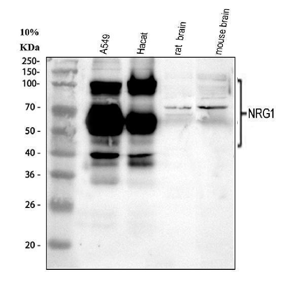
Western blot analysis of NRG1 using anti-NRG1 antibody (PA1969).
Electrophoresis was performed on a 5-20% SDS-PAGE gel at 70V (Stacking gel) / 90V (Resolving gel) for 2-3 hours. The sample well of each lane was loaded with 30 ug of sample under reducing conditions.
Lane 1: huamn A549 whole cell lysates,
Lane 2: human Hacat whole cell lysates,
Lane 3: rat brain tissue lysates,
Lane 4: mouse brain tissue lysates.
After electrophoresis, proteins were transferred to a nitrocellulose membrane at 150 mA for 50-90 minutes. Blocked the membrane with 5% non-fat milk/TBS for 1.5 hour at RT. The membrane was incubated with rabbit anti-NRG1 antigen affinity purified polyclonal antibody (Catalog # PA1969) at 0.5 μg/mL overnight at 4°C, then washed with TBS-0.1%Tween 3 times with 5 minutes each and probed with a goat anti-rabbit IgG-HRP secondary antibody at a dilution of 1:5000 for 1.5 hour at RT. The signal is developed using an Enhanced Chemiluminescent detection (ECL) kit (Catalog # EK1002) with Tanon 5200 system. A specific band was detected for NRG1 at approximately 40, 65 and 100 kDa. The expected band size for NRG1 is at 25, 40, 65 and 70 kDa.

Click image to see more details
IHC analysis of NRG1 using anti-NRG1 antibody (PA1969).
NRG1 was detected in a paraffin-embedded section of human tonsil tissue. Heat mediated antigen retrieval was performed in EDTA buffer (pH 8.0, epitope retrieval solution). The tissue section was blocked with 10% goat serum. The tissue section was then incubated with 2 μg/ml rabbit anti-NRG1 Antibody (PA1969) overnight at 4°C. Peroxidase Conjugated Goat Anti-rabbit IgG was used as secondary antibody and incubated for 30 minutes at 37°C. The tissue section was developed using HRP Conjugated Rabbit IgG Super Vision Assay Kit (Catalog # SV0002) with DAB as the chromogen.

Click image to see more details
IF analysis of NRG1 using anti-NRG1 antibody (PA1969) and anti-Beta Tubulin antibody (M01857-3).
NRG1 was detected in immunocytochemical section of U2OS cell. Enzyme antigen retrieval was performed using IHC enzyme antigen retrieval reagent (AR0022) for 15 mins. The cells were blocked with 10% goat serum. And then incubated with 5 μg/mL rabbit anti-NRG1 Antibody (PA1969) and mouse anti-Beta Tubulin antibody (M01857-3) overnight at 4°C. DyLight®488 Conjugated Goat Anti-Rabbit IgG (BA1127) and DyLight®550 Conjugated Goat Anti-Mouse IgG (BA1133) were used as secondary antibody at 1:500 dilution and incubated for 30 minutes at 37°C. Visualize using a fluorescence microscope and filter sets appropriate for the label used.
Specific Publications For Anti-NRG1 Antibody Picoband® (PA1969)
Loading publications
Recommended Resources
Here are featured tools and databases that you might find useful.
- Boster's Pathways Library
- Protein Databases
- Bioscience Research Protocol Resources
- Data Processing & Analysis Software
- Photo Editing Software
- Scientific Literature Resources
- Research Paper Management Tools
- Molecular Biology Software
- Primer Design Tools
- Bioinformatics Tools
- Phylogenetic Tree Analysis
Customer Reviews
Have you used Anti-NRG1 Antibody Picoband®?
Share your experimental results or join a short interview to earn up to $1,000 in product credits or other rewards.
0 Reviews For Anti-NRG1 Antibody Picoband®
Customer Q&As
Have a question?
Find answers in Q&As, reviews.
Can't find your answer?
Submit your question
1 Customer Q&As for Anti-NRG1 Antibody Picoband®
Question
Which isoforms of the protein are detected by this antibody? Keywords: specificity, immunogen information, reactivity, isoform detection
Verified Customer
Verified customer
Asked: 2019-07-29
Answer
The antibody should detect isoforms 1,2, 6, 7, and 11. It is not specific to beta 1.
Boster Scientific Support
Answered: 2019-07-29
