
Click image to see more details
Product Info Summary
| SKU: | P00104 |
|---|---|
| Size: | 100 μl |
| Reactive Species: | Human, Mouse, Rat |
| Host: | Rabbit |
| Application: | IP, WB |
Customers Who Bought This Also Bought

Product info
Product Name
Anti-Phospho-Erk1 (T202/Y204) + Erk2 (T185/Y187) MAPK3 Rabbit Monoclonal Antibody
SKU/Catalog Number
P00104
Size
100 μl
Form
Liquid
Description
Boster Bio Anti-Phospho-Erk1 (T202/Y204) + Erk2 (T185/Y187) MAPK3 Rabbit Monoclonal Antibody catalog # P00104. Tested in WB, IP applications. This antibody reacts with Human, Mouse, Rat.
Storage & Handling
Store at -20°C for one year. For short term storage and frequent use, store at 4°C for up to one month. Avoid repeated freeze-thaw cycles.
Cite This Product
Anti-Phospho-Erk1 (T202/Y204) + Erk2 (T185/Y187) MAPK3 Rabbit Monoclonal Antibody (Boster Biological Technology, Pleasanton CA, USA, Catalog # P00104)
Host
Rabbit
Contents
Rabbit IgG in phosphate buffered saline, pH 7.4, 150mM NaCl, 0.02% sodium azide and 50% glycerol, 0.4-0.5mg/ml BSA.
Clonality
Monoclonal
Clone Number
BIH-13
Isotype
Rabbit IgG
Immunogen
A synthesized peptide derived from human Phospho-ERK1 (T202/Y204) + ERK2 (T185/Y187)
*Blocking peptide can be purchased. Costs vary based on immunogen length. Contact us for pricing.
Reactive Species
P00104 is reactive to MAPK3 in Human, Mouse, Rat
Applications
P00104 is guaranteed for IP, WB Boster Guarantee
Observed Molecular Weight
50-70 kDa
Calculated molecular weight
43.136kDa
Antibody Validation
Boster validates all antibodies on WB, IHC, ICC, Immunofluorescence, and ELISA with known positive control and negative samples to ensure specificity and high affinity, including thorough antibody incubations.
Innovating Scientists Reward
If you are the first to review this product, or if you have results for a special sample, species or application this product is not validated in, share your results with us and receive product credits you can use towards any Boster products! Applicable to all scientists worldwide.
Submit A Review
Assay dilution & Images
Assay Dilutions Recommendation
The recommendations below provide a starting point for assay optimization. The actual working concentration varies and should be decided by the user.
WB 1:500-1:2000
IP 1:50
Validation Images & Assay Conditions
Click image to see more details
All lanes use the Antibody at 1:1K dilution for 1 hour at room temperature.

Click image to see more details
All lanes use the Antibody at 1:2K dilution for 1 hour at room temperature.

Click image to see more details
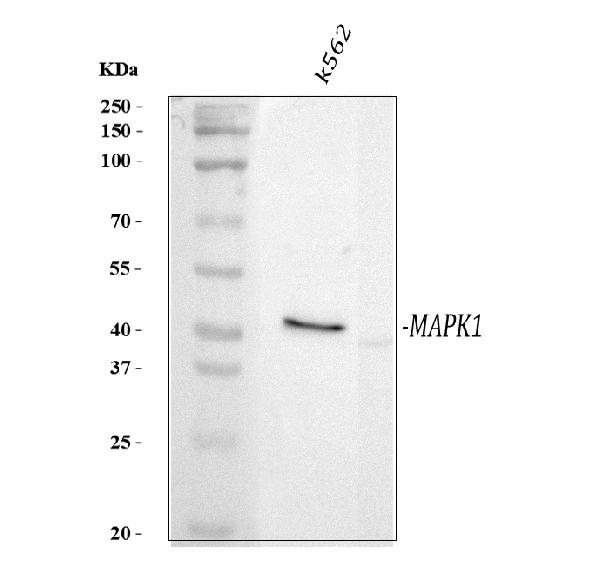
Western blot analysis of Phospho-Erk1 (T202/Y204) + Erk2 (T185/Y187) expression in A431 cell lysate treated with EGF.
Protein Target Info & Infographic
Gene/Protein Information For MAPK3 (Source: Uniprot.org, NCBI)
Gene Name
MAPK3
Full Name
Mitogen-activated protein kinase 3
Weight
43.136kDa
Superfamily
protein kinase superfamily
Alternative Names
EC 2.7.11; ERK1; ERK-1; ERK1p44-MAPK; ERT2; Extracellular signal-regulated kinase 1; extracellular signal-related kinase 1; HS44KDAP; HUMKER1A; Insulin-stimulated MAP2 kinase; MAP kinase 1; MAP kinase 3; MAP kinase isoform p44; MAPK 1; MAPK 3; MAPK3; MGC20180; Microtubule-associated protein 2 kinase; Mitogen-activated protein kinase 1; mitogen-activated protein kinase 3; P44ERK1; p44-ERK1; p44mapk; PRKM3; PRKM3EC 2.7.11.24 MAPK3 ERK-1, ERK1, ERT2, HS44KDAP, HUMKER1A, P44ERK1, P44MAPK, PRKM3, p44-ERK1, p44-MAPK mitogen-activated protein kinase 3 mitogen-activated protein kinase 3|MAPK 1|extracellular signal-regulated kinase 1|extracellular signal-related kinase 1|insulin-stimulated MAP2 kinase|microtubule-associated protein 2 kinase
*If product is indicated to react with multiple species, protein info is based on the gene entry specified above in "Species".For more info on MAPK3, check out the MAPK3 Infographic

We have 30,000+ of these available, one for each gene! Check them out.
In this infographic, you will see the following information for MAPK3: database IDs, superfamily, protein function, synonyms, molecular weight, chromosomal locations, tissues of expression, subcellular locations, post-translational modifications, and related diseases, research areas & pathways. If you want to see more information included, or would like to contribute to it and be acknowledged, please contact [email protected].
Specific Publications For Anti-Phospho-Erk1 (T202/Y204) + Erk2 (T185/Y187) MAPK3 Rabbit Monoclonal Antibody (P00104)
Hello CJ!
P00104 has been cited in 10 publications:
*The publications in this section are manually curated by our staff scientists. They may differ from Bioz's machine gathered results. Both are accurate. If you find a publication citing this product but is missing from this list, please let us know we will issue you a thank-you coupon.
Overexpression of sprouty 1 protein in human oral squamous cell carcinogenesis
Wang L,Xiong Q,Li P,Chen G,Tariq N,Wu C.The negative charge of the 343 site is essential for maintaining physiological functions of CXCR4.BMC Mol Cell Biol.2021 Jan 23;22(1):8.doi:10.1186/s12860-021-00347-9.PMID:33485325;PMCID:PMC7825245.
Species: Human
Ma G,Kimatu BM,Yang W,Pei F,Zhao L,Du H,Su A,Hu Q,Xiao H.Preparation of newly identified polysaccharide from Pleurotus eryngii and its anti-inflammation activities potential.J Food Sci.2020 Sep;85(9):2822-2831. doi:10.1111/1750-3841.15375.Epub 2020 Aug 14
Species: Mouse
P00104 usage in article: APP:WB, SAMPLE:RAW264.7 CELL, DILUTION:NA
Wang C,Wang J,Liu X,Han Z,Aimin Jiang,Wei Z,Yang Z.Cl-amidine attenuates lipopolysaccharide-induced mouse mastitis by inhibiting NF-κB, MAPK, NLRP3 signaling pathway and neutrophils extracellular traps release.Microb Pathog.2020 Sep 24;149:104530.doi:10.1
Species: Mouse
P00104 usage in article: APP:WB, SAMPLE:MAMMARY TISSUE, DILUTION:1:1000
Inhibition of extracellular signal-regulated kinases ameliorates hypertension-induced renal vascular remodeling in rat models
Synthesis and biological evaluation of novel N, N%u2032-disubstituted urea and thiourea derivatives as potential anti-melanoma agents
Disulfiram inhibits TGF-?-induced epithelial-mesenchymal transition and stem-like features in breast cancer via ERK/NF-?B/Snail pathway
Fructus phyllanthi tannin fraction induces apoptosis and inhibits migration and invasion of human lung squamous carcinoma cells in vitro via MAPK/MMP pathways
Ma Hr, Wang J, Chen Yf, Chen H, Wang Ws, Aisa Ha. Int J Mol Med. 2014 Jun;33(6):1627-34. Doi: 10.3892/Ijmm.2014.1722. Epub 2014 Apr 3. Icariin And Icaritin Stimulate The Proliferation Of Skbr3 Cells Through The Gper1-Mediated Modulation Of The Egf...
Hu Cp, Feng Jt, Tang Yl, Zhu Jq, Lin Mj, Yu Me. Mediators Inflamm. 2006;2006(5):84829. Lif Upregulates Expression Of Nk-1R In Nhbe Cells.
Recommended Resources
Here are featured tools and databases that you might find useful.
- Boster's Pathways Library
- Protein Databases
- Bioscience Research Protocol Resources
- Data Processing & Analysis Software
- Photo Editing Software
- Scientific Literature Resources
- Research Paper Management Tools
- Molecular Biology Software
- Primer Design Tools
- Bioinformatics Tools
- Phylogenetic Tree Analysis
Customer Reviews
Have you used Anti-Phospho-Erk1 (T202/Y204) + Erk2 (T185/Y187) MAPK3 Rabbit Monoclonal Antibody?
Submit a review and receive an Amazon gift card.
- $30 for a review with an image
Be the first to review Anti-Phospho-Erk1 (T202/Y204) + Erk2 (T185/Y187) MAPK3 Rabbit Monoclonal Antibody
*The first user to submit a review for a product is eligible for Boster's Innovating Scientists Reward, which gives product credits. This is in addition to the gift card reward.
Customer Q&As
Have a question?
Find answers in Q&As, reviews.
Can't find your answer?
Submit your question
6 Customer Q&As for Anti-Phospho-Erk1 (T202/Y204) + Erk2 (T185/Y187) MAPK3 Rabbit Monoclonal Antibody
Question
We have been able to see staining in rat leukemic t-cell. Any tips? Is anti-Phospho-Erk1 (T202/Y204) + Erk2 (T185/Y187) Rabbit Monoclonal antibody supposed to stain leukemic t-cell positively?
A. Johnson
Verified customer
Asked: 2020-01-13
Answer
Based on literature leukemic t-cell does express MAPK3. Based on Uniprot.org, MAPK3 is expressed in right frontal lobe, hepatoma, lymph, cervix carcinoma, leukemic t-cell, cervix carcinoma erythroleukemia, among other tissues. Regarding which tissues have MAPK3 expression, here are a few articles citing expression in various tissues:
Cervix carcinoma, Pubmed ID: 17081983, 18669648, 18691976, 20068231
Cervix carcinoma, and Erythroleukemia, Pubmed ID: 23186163
Hepatoma, Pubmed ID: 1540184, 7687743
Leukemic T-cell, Pubmed ID: 19690332
Lymph, Pubmed ID: 15489334
Boster Scientific Support
Answered: 2020-01-13
Question
We are interested in using your anti-Phospho-Erk1 (T202/Y204) + Erk2 (T185/Y187) Rabbit Monoclonal antibody for thyroid gland development studies. Has this antibody been tested with western blotting on a431 cell lysate? We would like to see some validation images before ordering.
Verified Customer
Verified customer
Asked: 2019-06-20
Answer
We appreciate your inquiry. This P00104 anti-Phospho-Erk1 (T202/Y204) + Erk2 (T185/Y187) Rabbit Monoclonal antibody is validated on a431 cell lysate. It is guaranteed to work for IP, WB in human, mouse, rat. Our Boster guarantee will cover your intended experiment even if the sample type has not been be directly tested.
Boster Scientific Support
Answered: 2019-06-20
Question
Can P00104 detect a protein with phosphorylation at Y204 only, or T202 and Y204?
Verified customer
Asked: 2019-03-28
Answer
The sequence for the Anti-Phospho-Erk1 (T202/Y204) + Erk2 (T185/Y187) MAPK3 Rabbit Monoclonal Antibody (P00104) is 192DPEHDHTGFL-pT-E-pY-VATRWYR211. According to this sequence, P00104 detects a protein with phosphorylation at T202 and Y204.
Boster Scientific Support
Answered: 2019-04-04
Question
We are currently using anti-Phospho-Erk1 (T202/Y204) + Erk2 (T185/Y187) Rabbit Monoclonal antibody P00104 for human tissue, and we are happy with the WB results. The species of reactivity given in the datasheet says human, mouse, rat. Is it true that the antibody can work on horse tissues as well?
Verified Customer
Verified customer
Asked: 2018-09-05
Answer
The anti-Phospho-Erk1 (T202/Y204) + Erk2 (T185/Y187) Rabbit Monoclonal antibody (P00104) has not been validated for cross reactivity specifically with horse tissues, though there is a good chance of cross reactivity. We have an innovator award program that if you test this antibody and show it works in horse you can get your next antibody for free. Please contact me if I can help you with anything.
Boster Scientific Support
Answered: 2018-09-05
Question
Our team were happy with the WB result of your anti-Phospho-Erk1 (T202/Y204) + Erk2 (T185/Y187) Rabbit Monoclonal antibody. However we have observed positive staining in cervix carcinoma erythroleukemia cytoplasm. nucleus. membrane using this antibody. Is that expected? Could you tell me where is MAPK3 supposed to be expressed?
Verified Customer
Verified customer
Asked: 2018-08-27
Answer
From what I have seen in literature, cervix carcinoma erythroleukemia does express MAPK3. Generally MAPK3 expresses in cytoplasm. nucleus. membrane, caveola. Regarding which tissues have MAPK3 expression, here are a few articles citing expression in various tissues:
Cervix carcinoma, Pubmed ID: 17081983, 18669648, 18691976, 20068231
Cervix carcinoma, and Erythroleukemia, Pubmed ID: 23186163
Hepatoma, Pubmed ID: 1540184, 7687743
Leukemic T-cell, Pubmed ID: 19690332
Lymph, Pubmed ID: 15489334
Boster Scientific Support
Answered: 2018-08-27
Question
We ordered your anti-Phospho-Erk1 (T202/Y204) + Erk2 (T185/Y187) Rabbit Monoclonal antibody for WB on lymph a few months ago. I am using mouse, and I plan to use the antibody for IP next. We need examining lymph as well as leukemic t-cell in our next experiment. Do you have any suggestion on which antibody would work the best for IP?
F. Singh
Verified customer
Asked: 2015-09-07
Answer
I looked at the website and datasheets of our anti-Phospho-Erk1 (T202/Y204) + Erk2 (T185/Y187) Rabbit Monoclonal antibody and it appears that P00104 has been validated on mouse in both WB and IP. Thus P00104 should work for your application. Our Boster satisfaction guarantee will cover this product for IP in mouse even if the specific tissue type has not been validated. We do have a comprehensive range of products for IP detection and you can check out our website bosterbio.com to find out more information about them.
Boster Scientific Support
Answered: 2015-09-07
