Click image to see more details
-

-
-
-
-
+8
Product Info Summary
| SKU: | PB9359 |
|---|---|
| Size: | 100 μg/vial |
| Reactive Species: | Human, Mouse, Rat |
| Host: | Rabbit |
| Application: | IF, IHC, WB |
Customers Who Bought This Also Bought

Product info
Product Name
Anti-Vimentin Antibody Picoband™
SKU/Catalog Number
PB9359
Size
100 μg/vial
Form
Lyophilized
Description
Boster Bio Anti-Vimentin Antibody Picoband™ catalog # PB9359. Tested in IF, IHC, WB applications. This antibody reacts with Human, Mouse, Rat.
Storage & Handling
Store at -20˚C for one year from date of receipt. After reconstitution, at 4˚C for one month. It can also be aliquotted and stored frozen at -20˚C for six months. Avoid repeated freeze-thaw cycles.
Cite This Product
Anti-Vimentin Antibody Picoband™ (Boster Biological Technology, Pleasanton CA, USA, Catalog # PB9359)
Host
Rabbit
Contents
Each vial contains 4 mg Trehalose, 0.9 mg NaCl, 0.2mg Na2HPO4, 0.05 mg NaN3.
Clonality
Polyclonal
Isotype
Rabbit IgG
Immunogen
A synthetic peptide corresponding to a sequence at the C-terminus of human Vimentin, identical to the related mouse and rat sequences.
*Blocking peptide can be purchased. Costs vary based on immunogen length. Contact us for pricing.
Cross-reactivity
No cross-reactivity with other proteins.
Reactive Species
PB9359 is reactive to VIM in Human, Mouse, Rat
Applications
PB9359 is guaranteed for IF, IHC, WB Boster Guarantee
Observed Molecular Weight
54 kDa
Calculated molecular weight
53.652kDa
Background of Vimentin
VIM (vimentin) is also known as HEL113 or CTRCT30. This gene encodes a member of the intermediate filament family. Intermediate filamentents, along with microtubules and actin microfilaments, make up the cytoskeleton. The protein encoded by this gene is responsible for maintaining cell shape, integrity of the cytoplasm, and stabilizing cytoskeletal interactions. It is also involved in the immune response, and controls the transport of low-density lipoprotein (LDL)-derived cholesterol from a lysosome to the site of esterification. It functions as an organizer of a number of critical proteins involved in attachment, migration, and cell signaling. Mutations in this gene causes a dominant, pulverulent cataract.
Antibody Validation
Boster validates all antibodies on WB, IHC, ICC, Immunofluorescence, and ELISA with known positive control and negative samples to ensure specificity and high affinity, including thorough antibody incubations.
Innovating Scientists Reward
If you are the first to review this product, or if you have results for a special sample, species or application this product is not validated in, share your results with us and receive product credits you can use towards any Boster products! Applicable to all scientists worldwide.
Submit A Review
Assay dilution & Images
Reconsitution
Add 0.2ml of distilled water will yield a concentration of 500ug/ml.
Assay Dilutions Recommendation
The recommendations below provide a starting point for assay optimization. The actual working concentration varies and should be decided by the user.
Western blot, 0.1-0.5μg/ml, Human, Mouse, Rat
Immunohistochemistry (Paraffin-embedded Section), 0.5-1μg/ml, Human, Mouse, Rat, By Heat
Immunofluorescence, 5 μg/ml, Mouse, Rat
Validation Images & Assay Conditions
Click image to see more details
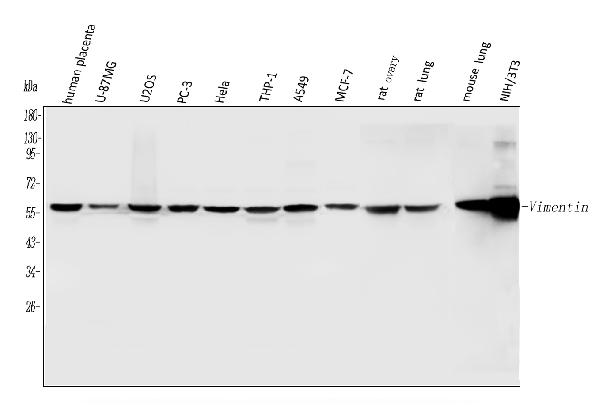
Figure 1. Western blot analysis of Vimentin using anti-Vimentin antibody (PB9359).
Electrophoresis was performed on a 5-20% SDS-PAGE gel at 70V (Stacking gel) / 90V (Resolving gel) for 2-3 hours. The sample well of each lane was loaded with 30 ug of sample under reducing conditions.
Lane 1: human placenta tissue lysates,
Lane 2: human U-87MG whole cell lysates,
Lane 3: human U20S whole cell lysates,
Lane 4: human PC-3 whole cell lysates,
Lane 5: human Hela whole cell lysates,
Lane 6: human THP-1 whole cell lysates,
Lane 7: human A549 whole cell lysates,
Lane 8: human MCF-7 whole cell lysates,
Lane 9: rat ovary tissue lysates,
Lane 10: rat lung tissue lysates,
Lane 11: mouse lung tissue lysates,
Lane 12: mouse NIH/3T3 whole cell lysates.
After electrophoresis, proteins were transferred to a nitrocellulose membrane at 150 mA for 50-90 minutes. Blocked the membrane with 5% non-fat milk/TBS for 1.5 hour at RT. The membrane was incubated with rabbit anti-Vimentin antigen affinity purified polyclonal antibody (Catalog # PB9359) at 0.5 μg/mL overnight at 4°C, then washed with TBS-0.1%Tween 3 times with 5 minutes each and probed with a goat anti-rabbit IgG-HRP secondary antibody at a dilution of 1:5000 for 1.5 hour at RT. The signal is developed using an Enhanced Chemiluminescent detection (ECL) kit (Catalog # EK1002) with Tanon 5200 system. A specific band was detected for Vimentin at approximately 54 kDa. The expected band size for Vimentin is at 54 kDa.

Click image to see more details
Figure 2. IHC analysis of Vimentin using anti-Vimentin antibody (PB9359).
Vimentin was detected in a paraffin-embedded section of mouse cardiac muscle tissue. Heat mediated antigen retrieval was performed in EDTA buffer (pH 8.0, epitope retrieval solution). The tissue section was blocked with 10% goat serum. The tissue section was then incubated with 2 μg/ml rabbit anti-Vimentin Antibody (PB9359) overnight at 4°C. Biotinylated goat anti-rabbit IgG was used as secondary antibody and incubated for 30 minutes at 37°C. The tissue section was developed using Strepavidin-Biotin-Complex (SABC) (Catalog # SA1022) with DAB as the chromogen.

Click image to see more details
Figure 3. IHC analysis of Vimentin using anti-Vimentin antibody (PB9359).
Vimentin was detected in a paraffin-embedded section of rat cardiac muscle tissue. Heat mediated antigen retrieval was performed in EDTA buffer (pH 8.0, epitope retrieval solution). The tissue section was blocked with 10% goat serum. The tissue section was then incubated with 2 μg/ml rabbit anti-Vimentin Antibody (PB9359) overnight at 4°C. Biotinylated goat anti-rabbit IgG was used as secondary antibody and incubated for 30 minutes at 37°C. The tissue section was developed using Strepavidin-Biotin-Complex (SABC) (Catalog # SA1022) with DAB as the chromogen.

Click image to see more details
Figure 4. IHC analysis of Vimentin using anti-Vimentin antibody (PB9359).
Vimentin was detected in a paraffin-embedded section of human mammary cancer tissue. Heat mediated antigen retrieval was performed in EDTA buffer (pH 8.0, epitope retrieval solution). The tissue section was blocked with 10% goat serum. The tissue section was then incubated with 2 μg/ml rabbit anti-Vimentin Antibody (PB9359) overnight at 4°C. Biotinylated goat anti-rabbit IgG was used as secondary antibody and incubated for 30 minutes at 37°C. The tissue section was developed using Strepavidin-Biotin-Complex (SABC) (Catalog # SA1022) with DAB as the chromogen.

Click image to see more details
Figure 5. IHC analysis of Vimentin using anti-Vimentin antibody (PB9359).
Vimentin was detected in a paraffin-embedded section of human placenta tissue. Heat mediated antigen retrieval was performed in EDTA buffer (pH 8.0, epitope retrieval solution). The tissue section was blocked with 10% goat serum. The tissue section was then incubated with 2 μg/ml rabbit anti-Vimentin Antibody (PB9359) overnight at 4°C. Peroxidase Conjugated Goat Anti-rabbit IgG was used as secondary antibody and incubated for 30 minutes at 37°C. The tissue section was developed using HRP Conjugated Rabbit IgG Super Vision Assay Kit (Catalog # SV0002) with DAB as the chromogen.

Click image to see more details
Figure 6. IHC analysis of Vimentin using anti-Vimentin antibody (PB9359).
Vimentin was detected in a paraffin-embedded section of human lung adenocarcinoma tissue. Heat mediated antigen retrieval was performed in EDTA buffer (pH 8.0, epitope retrieval solution). The tissue section was blocked with 10% goat serum. The tissue section was then incubated with 2 μg/ml rabbit anti-Vimentin Antibody (PB9359) overnight at 4°C. Peroxidase Conjugated Goat Anti-rabbit IgG was used as secondary antibody and incubated for 30 minutes at 37°C. The tissue section was developed using HRP Conjugated Rabbit IgG Super Vision Assay Kit (Catalog # SV0002) with DAB as the chromogen.

Click image to see more details
Figure 7. IHC analysis of Vimentin using anti-Vimentin antibody (PB9359).
Vimentin was detected in a paraffin-embedded section of human colorectal adenocarcinoma tissue. Heat mediated antigen retrieval was performed in EDTA buffer (pH 8.0, epitope retrieval solution). The tissue section was blocked with 10% goat serum. The tissue section was then incubated with 2 μg/ml rabbit anti-Vimentin Antibody (PB9359) overnight at 4°C. Peroxidase Conjugated Goat Anti-rabbit IgG was used as secondary antibody and incubated for 30 minutes at 37°C. The tissue section was developed using HRP Conjugated Rabbit IgG Super Vision Assay Kit (Catalog # SV0002) with DAB as the chromogen.

Click image to see more details
Figure 8. IHC analysis of Vimentin using anti-Vimentin antibody (PB9359).
Vimentin was detected in a paraffin-embedded section of human invasive urothelial carcinoma tissue. Heat mediated antigen retrieval was performed in EDTA buffer (pH 8.0, epitope retrieval solution). The tissue section was blocked with 10% goat serum. The tissue section was then incubated with 2 μg/ml rabbit anti-Vimentin Antibody (PB9359) overnight at 4°C. Peroxidase Conjugated Goat Anti-rabbit IgG was used as secondary antibody and incubated for 30 minutes at 37°C. The tissue section was developed using HRP Conjugated Rabbit IgG Super Vision Assay Kit (Catalog # SV0002) with DAB as the chromogen.

Click image to see more details
Figure 9. IHC analysis of Vimentin using anti-Vimentin antibody (PB9359).
Vimentin was detected in a paraffin-embedded section of human liver cancer tissue. Heat mediated antigen retrieval was performed in EDTA buffer (pH 8.0, epitope retrieval solution). The tissue section was blocked with 10% goat serum. The tissue section was then incubated with 2 μg/ml rabbit anti-Vimentin Antibody (PB9359) overnight at 4°C. Peroxidase Conjugated Goat Anti-rabbit IgG was used as secondary antibody and incubated for 30 minutes at 37°C. The tissue section was developed using HRP Conjugated Rabbit IgG Super Vision Assay Kit (Catalog # SV0002) with DAB as the chromogen.

Click image to see more details
Figure 10. IHC analysis of Vimentin using anti-Vimentin antibody (PB9359).
Vimentin was detected in a paraffin-embedded section of human tonsil tissue. Heat mediated antigen retrieval was performed in EDTA buffer (pH 8.0, epitope retrieval solution). The tissue section was blocked with 10% goat serum. The tissue section was then incubated with 2 μg/ml rabbit anti-Vimentin Antibody (PB9359) overnight at 4°C. Peroxidase Conjugated Goat Anti-rabbit IgG was used as secondary antibody and incubated for 30 minutes at 37°C. The tissue section was developed using HRP Conjugated Rabbit IgG Super Vision Assay Kit (Catalog # SV0002) with DAB as the chromogen.

Click image to see more details
Figure 11. IF analysis of Vimentin using anti-Vimentin antibody (PB9359).
Vimentin was detected in a paraffin-embedded section of mouse brain tissue. Heat mediated antigen retrieval was performed in EDTA buffer (pH 8.0, epitope retrieval solution). The tissue section was blocked with 10% goat serum. The tissue section was then incubated with 5 μg/mL rabbit anti-Vimentin Antibody (PB9359) overnight at 4°C. DyLight488 Conjugated Goat Anti-Rabbit IgG (BA1127) was used as secondary antibody at 1:500 dilution and incubated for 30 minutes at 37°C. The section was counterstained with DAPI. Visualize using a fluorescence microscope and filter sets appropriate for the label used.

Click image to see more details
Figure 12. IF analysis of Vimentin using anti-Vimentin antibody (PB9359).
Vimentin was detected in a paraffin-embedded section of rat brain tissue. Heat mediated antigen retrieval was performed in EDTA buffer (pH 8.0, epitope retrieval solution). The tissue section was blocked with 10% goat serum. The tissue section was then incubated with 5 μg/mL rabbit anti-Vimentin Antibody (PB9359) overnight at 4°C. DyLight488 Conjugated Goat Anti-Rabbit IgG (BA1127) was used as secondary antibody at 1:500 dilution and incubated for 30 minutes at 37°C. The section was counterstained with DAPI. Visualize using a fluorescence microscope and filter sets appropriate for the label used.
Protein Target Info & Infographic
Gene/Protein Information For VIM (Source: Uniprot.org, NCBI)
Gene Name
VIM
Full Name
Vimentin
Weight
53.652kDa
Superfamily
intermediate filament family
Alternative Names
FLJ36605; VIM; Vimentin VIM vimentin vimentin|epididymis secretory sperm binding protein
*If product is indicated to react with multiple species, protein info is based on the gene entry specified above in "Species".For more info on VIM, check out the VIM Infographic

We have 30,000+ of these available, one for each gene! Check them out.
In this infographic, you will see the following information for VIM: database IDs, superfamily, protein function, synonyms, molecular weight, chromosomal locations, tissues of expression, subcellular locations, post-translational modifications, and related diseases, research areas & pathways. If you want to see more information included, or would like to contribute to it and be acknowledged, please contact [email protected].
Specific Publications For Anti-Vimentin Antibody Picoband™ (PB9359)
Hello CJ!
PB9359 has been cited in 120 publications:
*The publications in this section are manually curated by our staff scientists. They may differ from Bioz's machine gathered results. Both are accurate. If you find a publication citing this product but is missing from this list, please let us know we will issue you a thank-you coupon.
LINC00184 involved in the regulatory network of ANGPT2 via ceRNA mediated miR-145 inhibition in gastric cancer
Mechanism of transmembrane and coiled‑coil domain 1 in the regulation of proliferation and migration of A549 cells
Histone deacetylase HDAC4 promotes the proliferation and invasion of glioma cells
Microrchidia family CW‑type zinc finger 2 promotes the proliferation, invasion, migration and epithelial‑mesenchymal transition of glioma by regulating PTEN/PI3K/AKT signaling via binding to N‑myc downstream regulated gene 1 promoter
Effect of Porphyromonas gingivalis lipopolysaccharide on calcification of human umbilical artery smooth muscle cells co-cultured with human periodontal ligament cells
Development and Radiation Response Assessment in A Novel Syngeneic Mouse Model of Tongue Cancer: 2D Culture, 3D Organoids and Orthotopic Allografts
Baicalein inhibits inflammatory response and promotes osteogenic activity in periodontal ligament cells challenged with lipopolysaccharides
Identification of Metastasis-Related Proteins and Their Clinical Relevance to Triple-Negative Human Breast Cancer
Elevated Circular RNA PVT1 Promotes Eutopic Endometrial Cell Proliferation and Invasion of Adenomyosis via miR-145/Talin1 Axis
The Therapeutic Effects of Exosomes Derived from Human Umbilical Cord Mesenchymal Stem Cells on Scleroderma
Recommended Resources
Here are featured tools and databases that you might find useful.
- Boster's Pathways Library
- Protein Databases
- Bioscience Research Protocol Resources
- Data Processing & Analysis Software
- Photo Editing Software
- Scientific Literature Resources
- Research Paper Management Tools
- Molecular Biology Software
- Primer Design Tools
- Bioinformatics Tools
- Phylogenetic Tree Analysis
Customer Reviews
Have you used Anti-Vimentin Antibody Picoband™?
Submit a review and receive an Amazon gift card.
- $30 for a review with an image
Be the first to review Anti-Vimentin Antibody Picoband™
*The first user to submit a review for a product is eligible for Boster's Innovating Scientists Reward, which gives product credits. This is in addition to the gift card reward.
Customer Q&As
Have a question?
Find answers in Q&As, reviews.
Can't find your answer?
Submit your question
5 Customer Q&As for Anti-Vimentin Antibody Picoband™
Question
We purchased anti-Vimentin antibody for IHC on leukemic t-cell in a previous project. I am using human, and We intend to use the antibody for WB next. We are interested in examining leukemic t-cell as well as cervix in our next experiment. Could you please give me some suggestion on which antibody would work the best for WB?
Verified Customer
Verified customer
Asked: 2020-04-03
Answer
I looked at the website and datasheets of our anti-Vimentin antibody and I see that PB9359 has been validated on human in both IHC and WB. Thus PB9359 should work for your application. Our Boster satisfaction guarantee will cover this product for WB in human even if the specific tissue type has not been validated. We do have a comprehensive range of products for WB detection and you can check out our website bosterbio.com to find out more information about them.
Boster Scientific Support
Answered: 2020-04-03
Question
My team were well pleased with the WB result of your anti-Vimentin antibody. However we have seen positive staining in lymphoblast cytoplasm using this antibody. Is that expected? Could you tell me where is VIM supposed to be expressed?
Verified Customer
Verified customer
Asked: 2020-02-11
Answer
Based on literature, lymphoblast does express VIM. Generally VIM expresses in cytoplasm. Regarding which tissues have VIM expression, here are a few articles citing expression in various tissues:
Cervix carcinoma, Pubmed ID: 16964243, 17081983, 17924679, 18220336, 18669648, 18691976, 20068231
Cervix carcinoma, and Erythroleukemia, Pubmed ID: 23186163
Embryo, Placenta, and Stomach, Pubmed ID: 14702039
Fibroblast, Pubmed ID: 3371665
Leukemic T-cell, Pubmed ID: 19690332
Liver, Pubmed ID: 24275569
Lymphoblast, Pubmed ID: 14654843
Lymphoma, Pubmed ID: 14996095
Mammary carcinoma, Pubmed ID: 9150946
Osteosarcoma, Pubmed ID: 2323579
Boster Scientific Support
Answered: 2020-02-11
Question
We have observed staining in human cervix carcinoma. Are there any suggestions? Is anti-Vimentin antibody supposed to stain cervix carcinoma positively?
Verified Customer
Verified customer
Asked: 2019-12-20
Answer
From literature cervix carcinoma does express VIM. From Uniprot.org, VIM is expressed in dorsal root ganglion, lymphoma, testis, embryo, placenta stomach, adipose tissue coronary artery, cervix, placenta testis, mammary carcinoma, t-cell, hepatoma, brain, cajal-retzius cell fetal brain cortex, fibroblast, osteosarcoma, lymphoblast, cervix carcinoma, leukemic t-cell, cervix carcinoma erythroleukemia, liver, among other tissues. Regarding which tissues have VIM expression, here are a few articles citing expression in various tissues:
Cervix carcinoma, Pubmed ID: 16964243, 17081983, 17924679, 18220336, 18669648, 18691976, 20068231
Cervix carcinoma, and Erythroleukemia, Pubmed ID: 23186163
Embryo, Placenta, and Stomach, Pubmed ID: 14702039
Fibroblast, Pubmed ID: 3371665
Leukemic T-cell, Pubmed ID: 19690332
Liver, Pubmed ID: 24275569
Lymphoblast, Pubmed ID: 14654843
Lymphoma, Pubmed ID: 14996095
Mammary carcinoma, Pubmed ID: 9150946
Osteosarcoma, Pubmed ID: 2323579
Boster Scientific Support
Answered: 2019-12-20
Question
We are currently using anti-Vimentin antibody PB9359 for mouse tissue, and we are happy with the IHC results. The species of reactivity given in the datasheet says human, mouse, rat. Is it possible that the antibody can work on monkey tissues as well?
A. Roberts
Verified customer
Asked: 2017-10-12
Answer
The anti-Vimentin antibody (PB9359) has not been tested for cross reactivity specifically with monkey tissues, though there is a good chance of cross reactivity. We have an innovator award program that if you test this antibody and show it works in monkey you can get your next antibody for free. Please contact me if I can help you with anything.
Boster Scientific Support
Answered: 2017-10-12
Question
I am interested in using your anti-Vimentin antibody for cellular response to interferon-gamma studies. Has this antibody been tested with western blotting on rat kidney tissue? We would like to see some validation images before ordering.
G. Rodriguez
Verified customer
Asked: 2015-02-16
Answer
We appreciate your inquiry. This PB9359 anti-Vimentin antibody is validated on ht1080 whole cell lysate, nih whole cell lysate, jurkat whole cell lysate, hut whole cell lysate, hela whole cell lysate, human placenta tissue, tissue lysate, rat kidney tissue, testis tissue, mouse kidney tissue, cardiac muscle tissue, mammary cancer tissue. It is guaranteed to work for IHC, WB in human, mouse, rat. Our Boster guarantee will cover your intended experiment even if the sample type has not been be directly tested.
Boster Scientific Support
Answered: 2015-02-16
