AIFM1 / Apoptosis-inducing factor 1, mitochondrial · IHC design guide

Design Immunohistochemistry for AIFM1

This guide helps plan AIFM1 paraffin IHC around granular cytoplasmic tissue staining (HPA tissue IHC). It covers controls and scoring of cytoplasmic and nuclear signal, since apoptosis can move AIFM1 into nuclei (UniProt).

Evidence assembled Oct 2026 · For research use; verify linked source records and product datasheet before use
Immunohistochemistry protocol sheet for AIFM1 (IHC for AIFM1): expected localisation Tissue: granular cytoplasm (HPA tissue IHC); mitochondrial IMS expected (UniProt), antibody PB9366, validated IHC image, and IHC protocol steps
Printable AIFM1 IHC protocol sheet — expected localisation Tissue: granular cytoplasm (HPA tissue IHC); mitochondrial IMS expected (UniProt), antibody PB9366, controls and protocol steps. Open the full AIFM1 IHC guide →

AIFM1 Immunohistochemistry Experimental Design Guide

Expected localisation, validated protocols, controls and antibodies — the at-a-glance facts below, then the full design guide.

Must know before staining
Expected localisation Tissue: granular cytoplasm (HPA tissue IHC); mitochondrial IMS expected (UniProt)
Staining pattern Generally granular cytoplasmic staining (HPA tissue IHC)
Antigen retrieval Citrate pH 6 HIER, heat-mediated (datasheet PB9366)
Positive control ⓘ Adrenal gland+4 more · see all
Negative control ⓘ Adipose tissue+4 more · see all
Important caveats
Reasons your staining may differ from the expected pattern.
Fixation Keep fixation conditions consistent across sections. (standard IHC practice; not target-specific)
Caveat Apoptosis can shift staining toward nuclei (UniProt)
Regulation Apoptotic stimuli redistribute AIFM1 (UniProt)
Isoform / epitope Six isoforms; mature chain starts at residue 102—check epitope position (UniProt)
Section 1

Recommended AIFM1 IHC & IF Protocols

Compare the catalog antibody's IHC-P protocol (datasheet: PB9366) with the published human HNSC tissue protocol (PMC12055095).

Recommended immunohistochemistry (IHC-P) protocol parameters
SampleParaffin-embedded Rat Cardiac Muscle tissue; fixative not specified (datasheet PB9366)
FixationImage fixative and duration unreported (datasheet PB9366); verify before use.
Sectioning4–5 µm sections on charged slides (standard)
DeparaffinisationXylene, graded ethanol series to water (standard)
Antigen retrievalHeat retrieval: Citrate pH 6, 20 min (datasheet PB9366)
Peroxidase block3% H2O2, 10 min, room temperature (standard)
Blocking10% goat serum (datasheet PB9366)
Primary antibodyRabbit anti-AIFM1, 0.5-1μg/ml (datasheet PB9366)
Primary incubationOvernight at 4 °C (datasheet PB9366)
DetectionStreptavidin-biotin complex (SABC), DAB chromogen (datasheet PB9366)
CounterstainHematoxylin, blue, dehydrate and mount (standard)
Expected resultAIFM1-positive staining in glandular cells of adrenal gland (HPA tissue IHC: High). HPA tissue profile: General granular cytoplasmic expression. No signal in the no-primary control.
💡Decision noteStart with heat-mediated citrate pH 6 retrieval (datasheet: PB9366). The published protocol specifies citrate for 15 minutes without reporting pH or heating method (PMC12055095).
Section 2

What Is the Expected AIFM1 Staining Pattern?

AIFM1 is chiefly mitochondrial, with an inner-membrane-anchored form in the intermembrane space (UniProt O95831 localization and processing). In paraffin-section IHC, expect granular cytoplasmic staining in selected cells, including pancreatic exocrine glandular cells and adrenal glandular cells with high HPA staining (HPA tissue IHC). HPA rates its tissue IHC evidence Enhanced, reflecting agreement between staining and RNA expression (HPA tissue IHC).

What am I looking at on my slide?
Granular cytoplasmic staining in pancreatic exocrine glandular cells or adrenal glandular cells, with clearly defined cell outlines.This matches HPA's general granular cytoplasmic profile and its High calls for those cells (HPA tissue IHC). Granularity is compatible with mitochondrial localization (UniProt O95831 localization); chromogenic IHC alone does not prove mitochondrial colocalization.
Predominantly nuclear staining in otherwise unremarkable cells, without a convincing granular cytoplasmic pattern.Treat an isolated nuclear pattern cautiously: HPA describes general granular cytoplasmic tissue staining (HPA tissue IHC). AIFM1 can move from mitochondria to nuclei during apoptosis (UniProt O95831 localization and function), so nuclear staining needs biological context and independent confirmation before interpretation.
Strong staining in adipocytes or bone-marrow hematopoietic cells while a matched known-positive cell population stains weakly.HPA reports those cell populations as Not detected (HPA tissue IHC). Review morphology and controls for cross-reactivity or endogenous detection activity (general IHC practice). A tissue-wide label must not replace a cell-specific reading.
Broad, diffuse color covers cells and surrounding tissue, obscuring granular cytoplasmic detail.That appearance cannot establish the reported AIFM1 pattern (HPA tissue IHC). Check the no-primary control, blocking, washing and chromogen development for background (general IHC practice), then score only signal that can be assigned to identifiable cells.
No convincing signal in pancreatic exocrine glandular cells on an otherwise interpretable section.These cells are High in HPA tissue IHC (HPA tissue IHC), so an absent result warrants a run-level check. Confirm tissue identity and preservation, then review the antibody dilution, retrieval, detection reagents and positive control (general IHC practice); absence alone does not show loss of AIFM1.
💡Expected AIFM1 appearanceCall a positive result when identifiable glandular cells show granular cytoplasmic staining, especially in HPA High populations such as pancreatic exocrine glandular cells (HPA tissue IHC); diffuse color, staining restricted to an unexpected cell population, or nuclear-only staining without corroboration is not sufficient to call the expected pattern.
How each factor affects the staining
Cell population and tissue choiceHPA calls pancreatic exocrine and adrenal glandular cells High, colon glandular cells Low, and adipocytes Not detected (HPA tissue IHC). Compare named cell populations under the microscope; a negative call in one population does not characterize every cell in its tissue.
Mitochondrial processing and redistributionAIFM1 forms an inner-membrane-anchored mature form and can yield a soluble form that moves to cytoplasm and nucleus during apoptosis (UniProt O95831 processing and localization). Interpret a changed compartment alongside morphology and an independent marker; location alone cannot identify the processed form.
Antibody evidenceHPA lists HPA030611 and CAB003764 as IHC Enhanced (HPA antibodies). That supports the reported tissue pattern; it does not establish that every staining event is specific. Include appropriate positive and no-primary controls when assessing a new IHC run (general IHC practice).
Isoform and epitope limitsUniProt lists 6 AIFM1 isoforms and a processed chain spanning residues 102–613 (UniProt O95831 isoforms and processing). The supplied antibody evidence does not locate either antibody's epitope (HPA antibodies); do not infer which isoforms or processed forms a stain detects.
IF/ICC Q&A: What pattern should I expect?Mainly mitochondrial signal is the HPA ICC-IF finding; connecting-piece localization is additional (HPA subcellular ICC-IF). That supports a localization comparison, not an IF protocol or a claim that paraffin-section chromogenic granules resolve individual mitochondria.
Why is my staining missing, weak or wrong?
SituationLikely causeNext action
Known-positive glandular cells appear unstained.A failed IHC run or unsuitable section remains possible; pancreatic exocrine glandular cells are High in HPA tissue IHC (HPA tissue IHC).Check tissue identity, section quality and the run's positive control; then review retrieval, catalog-antibody dilution and detection steps (general IHC practice). Do not assign an AIFM1-specific fixation effect from these data.
Color is widespread and granular detail is lost.Nonspecific reagent binding, inadequate washing or excess chromogen development can produce background (general IHC practice).Inspect the no-primary control and staining distribution. Adjust blocking, washes or detection conditions as indicated by that control (general IHC practice), then reassess the HPA granular cytoplasmic pattern (HPA tissue IHC).
An HPA Not detected cell population stains strongly.For adipocytes or bone-marrow hematopoietic cells, this conflicts with the reported HPA cell-level call (HPA tissue IHC); cross-reactivity or endogenous detection activity is possible (general IHC practice).Confirm cell identity, compare a positive population and inspect no-primary and detection controls before scoring the unexpected signal (general IHC practice).
Nuclear staining dominates the section.AIFM1 nuclear translocation is linked to apoptosis (UniProt O95831 localization and function), but nuclear color alone does not establish that event.Check cell morphology and controls, then seek independent evidence of apoptosis or localization before reporting redistribution (general IHC practice). Compare with HPA's granular cytoplasmic baseline (HPA tissue IHC).
A Low population stains less than a High population.This can fit HPA's cell-level calls: colon glandular cells are Low and pancreatic exocrine glandular cells are High (HPA tissue IHC).Score the named cell types separately and use the same scoring criteria across sections (general IHC practice). Avoid treating a weak Low population as a failed run when its positive control performs.
Granular cytoplasmic staining is present, but mitochondrial identity is uncertain.HPA tissue IHC reports granularity, while mitochondrial localization comes from UniProt and HPA ICC-IF (HPA tissue IHC; UniProt O95831 localization; HPA subcellular ICC-IF).Report the visible chromogenic pattern as granular cytoplasmic. If mitochondrial assignment matters, assess colocalization in a separately validated localization experiment (general IHC/IF practice).

Sample controls for AIFM1 IHC & IF

🧪Run kidney first and look for staining in Bowman's capsule cells (HPA: High in Bowman's capsule). Use adipose tissue as the negative comparator, focusing on adipocytes (HPA: Not detected in adipocytes); on the kidney slide, treat cells without visible staining as background comparators, without assuming any kidney cell type is AIFM1-negative (UniProt O95831: expressed in all tested tissues).
Positive control tissue: Adrenal gland (Glandular cells, HPA High)
Negative control tissue: Adipose tissue (HPA Not detected)
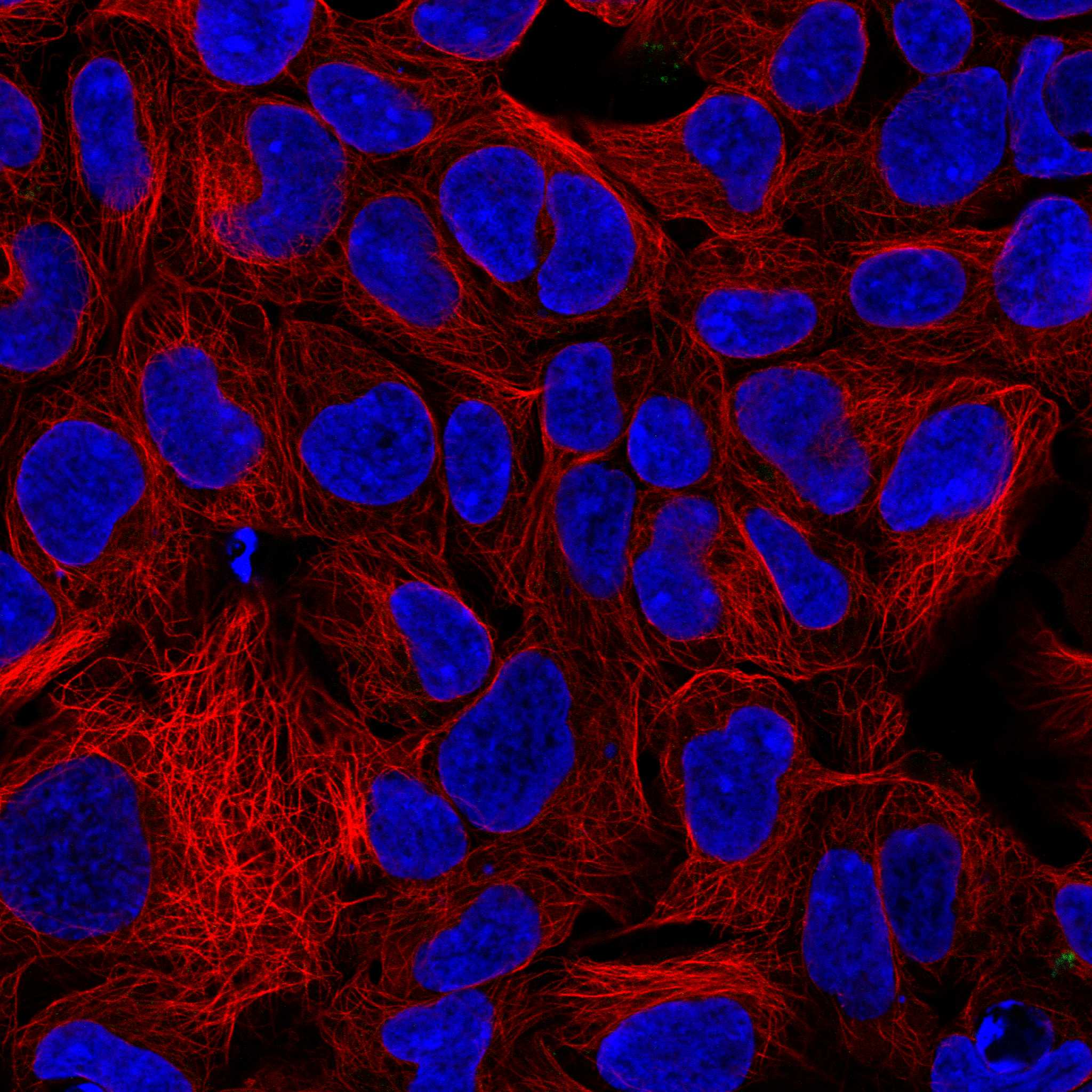
ICC-IF cell lines (HPA subcellular resource): HPA ICC-IF images show AIFM1 in HEK293, U2OS, Sperm, with annotated localisation: Mitochondria (supported) (HPA subcellular).
Technical controls: Include no-primary (secondary-only), host-species-matched isotype, and AIFM1 knockout or validated peptide-block controls (standard IHC practice). With the caption's biotin-based DAB detection (PB9366 tissue-IHC caption), check endogenous peroxidase and endogenous biotin background in kidney sections (standard IHC practice).
⚠️Feasibility: A target-specific fixation window is unreported, and the selected PB9366 paraffin-section caption does not state a fixative (PB9366 tissue-IHC caption). That caption uses citrate retrieval at pH 6 for 20 minutes in rat cardiac muscle; it does not establish that retrieval is required in kidney (PB9366 tissue-IHC caption). Whether frozen sections or IF are easier is unreported; endogenous kidney biotin can complicate the caption's biotin-based detection (standard IHC practice; PB9366 tissue-IHC caption).

HPA tissue IHC evidence for AIFM1

Comprehensive Human Protein Atlas IHC scoring per tissue (reliability: Enhanced — High consistency between antibody staining and RNA expression data.). Rows are taken directly from the HPA tissue chart — click any row's HPA link to view the source.

Positive expression · recommended positive controls

TissueCell typeLevelEvidenceSource
Adrenal gland Glandular cells High Protein (IHC) HPA →
Endometrium Glandular cells High Protein (IHC) HPA →
Epididymis Glandular cells High Protein (IHC) HPA →
Fallopian tube Glandular cells High Protein (IHC) HPA →
Kidney Bowman's capsule High Protein (IHC) HPA →

Undetected expression · recommended negative controls

TissueCell typeLevelEvidenceSource
Adipose tissue Adipocytes Not detected Protein (IHC) HPA →
Bone marrow Hematopoietic cells Not detected Protein (IHC) HPA →
Caudate Glial cells Not detected Protein (IHC) HPA →
Cerebral cortex Endothelial cells Not detected Protein (IHC) HPA →
Hippocampus Glial cells Not detected Protein (IHC) HPA →
Section 3

Advanced AIFM1 IHC Tips

Troubleshoot AIFM1 staining in paraffin sections by checking retrieval, cell type and compartment before interpreting chromogenic signal.

What retrieval should I try first when AIFM1 staining is weak?
Start with heat-mediated retrieval in citrate buffer at pH 6 for 20 minutes (datasheet PB9366). The catalog antibody was used on a paraffin section with this retrieval, followed by 1 µg/mL primary antibody overnight at 4°C (datasheet PB9366). Keep section thickness, heating and cooling consistent across the comparison, because uneven retrieval can mimic differences in staining (standard IHC practice). If signal remains weak, check the positive control and primary-antibody dilution before changing retrieval conditions (standard IHC practice). Compare any revised condition with the citrate-treated section on the same staining run, and reject conditions that damage morphology (standard IHC practice).
Could fixation explain variable AIFM1 staining between paraffin sections?
Target-specific fixation sensitivity is unknown: the catalog antibody’s paraffin-section caption does not state a fixative (datasheet PB9366). Record the fixative, fixation duration and tissue processing for each specimen before attributing differences to AIFM1 biology (standard IHC practice). For a controlled pilot, compare matched material fixed in 10% neutral buffered formalin for 6 and 24 hours, while holding retrieval at citrate pH 6 for 20 minutes (standard IHC practice; datasheet PB9366). Those fixation times are comparison conditions, not validated AIFM1 requirements (standard IHC practice). Check morphology and a same-run positive control; neither the tissue staining profile nor protein processing establishes fixation sensitivity (HPA tissue IHC; UniProt O95831).
How should I assess granular cytoplasmic and nuclear AIFM1 staining?
Granular cytoplasmic staining fits the reported tissue IHC profile and AIFM1’s mainly mitochondrial localisation (HPA tissue IHC; HPA subcellular). Score cytoplasmic and nuclear staining separately, using the same counterstain and exposure to DAB development across sections (standard IHC practice). AIFM1 can move from the mitochondrial intermembrane space through the cytoplasm to the nucleus after apoptotic signalling (UniProt O95831). Nuclear DAB alone does not establish that sequence; assess cell morphology and an independent apoptosis readout before assigning a death-related interpretation (standard IHC practice; UniProt O95831). Recheck diffuse edge staining or signal over damaged cells against an internal negative area and a no-primary control (standard IHC practice).
Can the antibody distinguish processed AIFM1 or its six isoforms?
Do not assign an isoform or processed form from chromogenic staining unless the antibody epitope and its specificity have been established (standard IHC practice). AIFM1 has 6 listed isoforms, and processing yields a mature chain spanning residues 102–613 (UniProt O95831). The supplied catalog caption gives an IHC concentration of 1 µg/mL but does not map the antibody epitope (datasheet PB9366). Ask whether the epitope lies within a sequence shared by the forms under study, then validate any form-specific claim with an independent method (standard IHC practice). Changes near annotated modified residues, including positions 105 and 109, cannot be inferred from staining intensity alone (UniProt O95831; standard IHC practice).
How can IF help resolve an ambiguous AIFM1 IHC pattern?
Use IF as a separate localisation check, with an expected-cell-type marker and a mitochondrial marker in distinct channels (standard IF practice; HPA subcellular). For a glandular-cell comparison, the HPA reports high AIFM1 staining in pancreatic exocrine glandular cells (HPA tissue IHC). Choose a far-red AIFM1 fluorophore if the specimen has strong shorter-wavelength autofluorescence, and inspect unstained controls in every channel (standard IF practice). Because the supplied antibody evidence does not map its epitope, compare no-permeabilisation and mild-detergent conditions before interpreting access to the mitochondrial intermembrane space (datasheet PB9366; UniProt O95831; standard IF practice). Keep single-stain controls for spectral bleed-through and compare nuclear signal with cell morphology rather than IHC DAB intensity (standard IF practice).
How do I reduce diffuse brown signal without losing AIFM1 staining?
Begin with a no-primary control to locate signal from detection reagents, endogenous enzyme activity or tissue pigment (standard IHC practice). Block endogenous peroxidase before DAB development and match the block across test and control sections (standard chromogenic IHC practice). The catalog example used 10% goat serum, 1 µg/mL primary antibody overnight at 4°C, and biotin-based detection with DAB (datasheet PB9366). If background persists, titrate the primary antibody and shorten chromogen development while retaining a same-run positive control (standard IHC practice). For a biotin-based workflow, assess endogenous biotin with the appropriate detection control before calling widespread brown staining AIFM1-positive (datasheet PB9366; standard IHC practice).
How should I quantify AIFM1 IHC across specimens? ⚠ ANSWER MARKED FOR VERIFICATION
Choose the cell population and scoring compartment before reviewing outcomes, because tissue IHC commonly shows granular cytoplasmic staining while AIFM1 can also enter nuclei (HPA tissue IHC; UniProt O95831). For cytoplasmic signal, report the percentage of positive target cells and an H-score from 0–300 using consistent intensity thresholds (standard IHC practice). Score nuclear positivity separately as a percentage of intact target-cell nuclei, with morphology recorded alongside it (standard IHC practice). Normalise counts to the number of evaluable target cells, or report positive-cell density per mm² of viable tissue when cell counting is impractical (standard IHC practice). Exclude folds, edges and necrosis using the same rules for every section (standard IHC practice).
What makes an apparent AIFM1-positive result convincing?
Look first for granular cytoplasmic signal in intact cells, consistent with the tissue profile and mainly mitochondrial localisation (HPA tissue IHC; HPA subcellular). The HPA reports high staining in pancreatic exocrine glandular cells but no detection in adipocytes, offering cell-type comparisons within appropriate specimens (HPA tissue IHC). Treat isolated nuclear staining cautiously: nuclear translocation is biologically possible, yet requires morphology and an independent apoptosis measure for interpretation (UniProt O95831; standard IHC practice). Reject patterns confined to section edges, folds or necrotic areas, and compare them with a no-primary control (standard IHC practice). Rule out endogenous peroxidase and detection-system background before scoring diffuse DAB as AIFM1 (standard chromogenic IHC practice).
Boster reagents

Best AIFM1 / Apoptosis-inducing factor 1, mitochondrial IHC Antibodies

Anti-AIFM1 antibodies have IHC images from human, mouse, and rat tissues and IF images from cultured cells (catalog image captions).

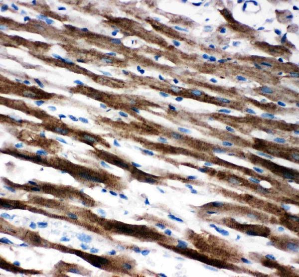
Real IHC data IHC analysis of AIF using anti-AIF antibody (PB9366). AIF was detected in paraffin-embedded section of Rat Cardiac Muscle Tissue. Heat mediated antigen retrieval was performed in citrate buffer (pH6, epitope retrieval solution) for 20 mins. The tissue section was blocked with 10% goat serum. The tissue section was then incubated with 1μg/ml rabbit anti-AIF Antibody (PB9366) overnight at 4°C. Biotinylated goat anti-rabbit IgG was used as secondary antibody and incubated for 30 minutes at 37°C. The tissue section was developed using Strepavidin-Biotin-Complex (SABC)(Catalog # SA1022) with DAB as the chromogen.
Anti-AIF/AIFM1 Antibody ®
Cat # PB9366
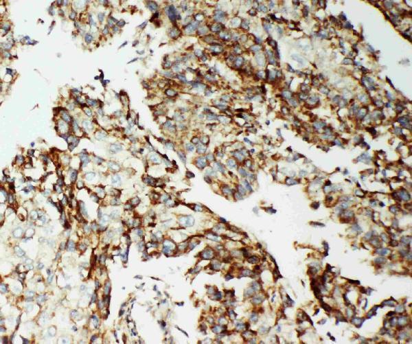
Real IHC data IHC analysis of AIF using anti-AIF antibody (M01571-1). AIF was detected in paraffin-embedded section of human mammary cancer tissue. Heat mediated antigen retrieval was performed in EDTA buffer (pH8.0, epitope retrieval solution). The tissue section was blocked with 10% goat serum. The tissue section was then incubated with 1μg/ml mouse anti-AIF Antibody (M01571-1) overnight at 4°C. Biotinylated goat anti-mouse IgG was used as secondary antibody and incubated for 30 minutes at 37°C. The tissue section was developed using Strepavidin-Biotin-Complex (SABC) (Catalog # SA1021) with DAB as the chromogen.
Anti-AIF/AIFM1 Antibody ® (monoclonal, 2I5)
Cat # M01571-1
Real IHC data Anti-AIF antibody, PA2232, IHC(P) IHC(P): Human Lung Cancer Tissue
Anti-AIF/AIFM1 Antibody ®
Cat # PA2232
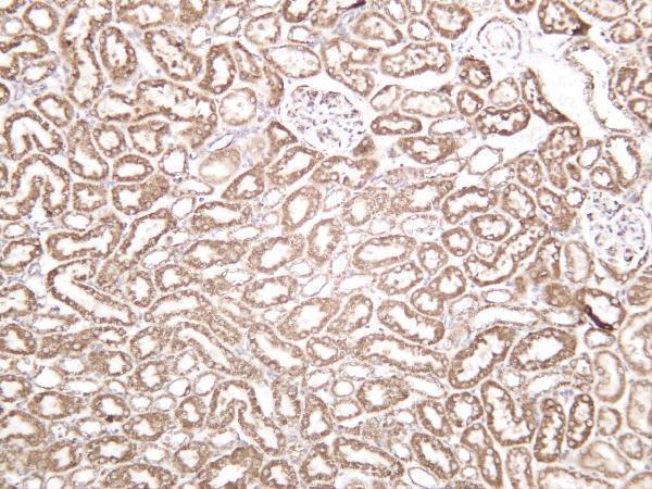
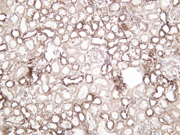
Real IHC data Mousekidney was stained with anti-AIF-M1 rabbit antibody
Anti-AIF-M1 Rabbit Monoclonal Antibody
Cat # M01571-4
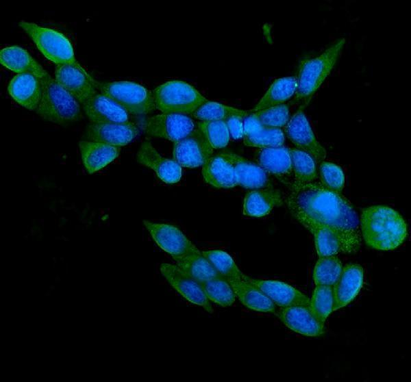
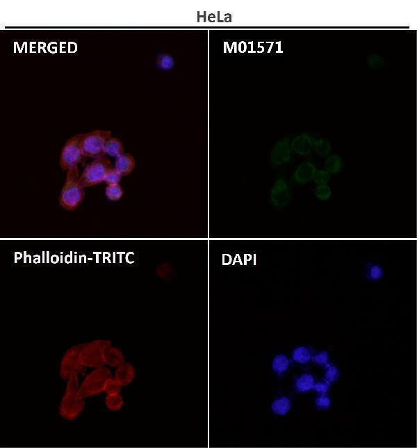
Real IF data Immunofluorescent analysis using the Antibody at 1:50 dilution.
Anti-AIF AIFM1 Rabbit Monoclonal Antibody
Cat # M01571

PB9366 was imaged in rat cardiac muscle, M01571-1 in human mammary cancer, and PA2232 in human lung cancer paraffin sections (catalog IHC captions). M01571-4 has a mouse kidney IHC image, while M01571 has an IF image of HeLa cells (catalog image captions).

Which to pick: For paraffin-section IHC, choose PB9366 for rat cardiac muscle with citrate pH 6 retrieval or M01571-1 for human mammary cancer with EDTA pH 8 retrieval; neither caption reports the fixative (catalog IHC captions). For IF/ICC, M01571 lists both applications and has an IF image of HeLa cells (catalog applications and IF caption). For cross-species tissue IHC, M01571-4 lists human, mouse, and rat reactivity and has kidney IHC images from all three species (catalog reactivity and IHC captions).

Each figure is that product's own IHC / IF validation image from its datasheet.

References

  1. UniProt Consortium. UniProt entry O95831 (AIFM1_HUMAN, Apoptosis-inducing factor 1, mitochondrial).
  2. Human Protein Atlas. AIFM1 tissue IHC expression (reliability: Enhanced).
  3. Human Protein Atlas. AIFM1 subcellular location (ICC-IF): Mainly localized to the mitochondria. In addition localized to the connecting piece..
  4. Human Protein Atlas. AIFM1 antibody validation summary (2 antibodies).
  5. Case Report: A Novel Intronic Mutation in AIFM1 Associated With Fatal Encephalomyopathy and Mitochondrial Disease in Infant. Frontiers in pediatrics 2022 — PMC9194441.
  6. Identification and validation of a prognostic signature of drug resistance and mitochondrial energy metabolism-related differentially expressed genes for breast cancer. Journal of translational medicine 2025 — PMC11780791.
  7. Construction and Validation of a Necroptosis-Related Gene Signature for Predicting Prognosis and Tumor Microenvironment of Pancreatic Cancer. Disease markers 2022 — PMC9214653.
  8. Establishment and validation of a novel risk model based on PANoptosis-related genes to predict prognosis in head and neck squamous cell carcinoma. Medicine 2025 — PMC12055095.
  9. PubMed PMID:9989411 — UniProt-cited evidence.
  10. PubMed PMID:16365034 — UniProt-cited evidence.
  11. PubMed PMID:16644725 — UniProt-cited evidence.