CDK4 / Cyclin-dependent kinase 4 · IHC design guide

Design Immunohistochemistry for CDK4

Plan CDK4 staining in paraffin sections around mainly nuclear, sometimes cytoplasmic signal (HPA tissue IHC). Start with 0.5–1 μg/mL catalog antibody (datasheet: PB9535), then assess staining by cell type with a matched negative control.

Evidence assembled Oct 2026 · For research use; verify linked source records and product datasheet before use
Immunohistochemistry protocol sheet for CDK4 (IHC for CDK4): expected localisation Mainly nuclear, also cytoplasmic in tissue (HPA tissue IHC), antibody PB9535, validated IHC image, and IHC protocol steps
Printable CDK4 IHC protocol sheet — expected localisation Mainly nuclear, also cytoplasmic in tissue (HPA tissue IHC), antibody PB9535, controls and protocol steps. Open the full CDK4 IHC guide →

CDK4 Immunohistochemistry Experimental Design Guide

Expected localisation, validated protocols, controls and antibodies — the at-a-glance facts below, then the full design guide.

Must know before staining
Expected localisation Mainly nuclear, also cytoplasmic in tissue (HPA tissue IHC)
Staining pattern Variable nuclear and cytoplasmic staining in most tissues (HPA tissue IHC)
Antigen retrieval EDTA pH 8.0 HIER, heat-mediated (datasheet PB9535)
Positive control ⓘ Bronchus+4 more · see all
Negative control ⓘ Adipose tissue+4 more · see all
Important caveats
Reasons your staining may differ from the expected pattern.
Fixation Keep formalin fixation consistent across sections. (standard IHC practice; not target-specific)
Caveat Presumed off-target binding can mimic CDK4 staining (HPA tissue IHC)
Regulation Nuclear accumulation at G1/S transition (UniProt)
Isoform / epitope Two isoforms; epitope differences are unspecified (UniProt)
Section 1

Recommended CDK4 IHC & IF Protocols

The catalog antibody’s IHC-P protocol (datasheet: PB9535) is followed by published CDK4 staining protocols for ossifying fibroma, soft tissue tumors, and liposarcoma (PMC8760962; PMC10066853; PMC4133208).

Recommended immunohistochemistry (IHC-P) protocol parameters
SampleParaffin-embedded mouse intestine tissue; fixative not specified (datasheet PB9535)
FixationImage fixative and duration unreported (datasheet PB9535); verify before use.
Sectioning4–5 µm sections on charged slides (standard)
DeparaffinisationXylene, graded ethanol series to water (standard)
Antigen retrievalHeat retrieval: EDTA pH 8.0 (datasheet PB9535); 20 min, 95–100 °C (standard)
Peroxidase block3% H2O2, 10 min, room temperature (standard)
Blocking10% goat serum (datasheet PB9535)
Primary antibodyRabbit anti-CDK4, 0.5-1μg/ml (datasheet PB9535)
Primary incubationOvernight at 4 °C (datasheet PB9535)
DetectionStreptavidin-biotin complex (SABC), DAB chromogen (datasheet PB9535)
CounterstainHematoxylin, blue, dehydrate and mount (standard)
Expected resultCDK4-positive staining in ciliated cells (cell body) of bronchus (HPA tissue IHC: High). HPA tissue profile: Mainly nuclear but also cytoplasmic expression at variable levels in most tissues. No signal in the no-primary control.
💡Decision noteStart with heat-mediated EDTA pH 8.0 retrieval for the catalog antibody (datasheet: PB9535); the published protocols describe their own preparation conditions (PMC8760962; PMC4133208).
Section 2

What Is the Expected CDK4 Staining Pattern?

CDK4 staining should be mainly nuclear, with variable cytoplasmic signal across many tissues (HPA: tissue IHC, Supported). UniProt also places CDK4 in the cytoplasm, nucleus and nuclear membrane as cyclin D complexes move during G1/S; CDK4 has no transmembrane segment (UniProt P11802: subcellular location and topology). Expect staining in the identified positive cell populations, such as ovarian stroma cells and bronchial ciliated cells, rather than uniformly across every cell in a section (HPA: tissue IHC).

What am I looking at on my slide?
Nuclear staining, with some cytoplasmic staining, in ovarian stroma cells or bronchial ciliated cells.This fits the reported compartment pattern and High staining in those cell populations (HPA: tissue IHC). Judge the named cells, since adjacent cell types may have different staining levels. HPA rates the tissue pattern Supported, with medium consistency between staining and RNA data (HPA: reliability description).
Signal is concentrated at an unexpected boundary or outside recognizable cells, without convincing nuclear or cytoplasmic staining.That distribution does not match the mainly nuclear, sometimes cytoplasmic tissue pattern (HPA: tissue IHC). CDK4 has no transmembrane segment, although nuclear membrane localization is reported (UniProt P11802: topology and subcellular location). Check morphology and detection controls before calling boundary staining CDK4.
Strong staining occurs in a cell population listed as not detected, such as adipocytes or lung alveolar cells.Treat this as a specificity question for those specific cells, not as proof that their entire tissue is negative (HPA: adipocytes and alveolar cells, Not detected). Compare a matched negative control and a supported positive cell population; HPA reports that presumed off-target binding was observed and disregarded (HPA: reliability description).
Diffuse chromogen covers many structures, including areas without plausible cells, and obscures compartment boundaries.A diffuse deposit cannot establish the reported nuclear and cytoplasmic pattern (HPA: tissue IHC). General IHC practice is to inspect reagent-only controls and the distribution of background before scoring cells; nonspecific antibody binding or endogenous detection activity are possible explanations, not CDK4-specific findings.
No convincing staining appears in bronchial ciliated cells or ovarian stroma cells.These are reported High cell populations, so absence of signal warrants a run-level check (HPA: bronchus and ovary, High). Confirm that the expected cells are present and assess control performance, antibody use and chromogenic detection. A single negative section does not establish that the listed HPA pattern is wrong.
💡Expected CDK4 appearanceCall a section positive when identifiable cells show predominantly nuclear staining, with permissible cytoplasmic signal and cell-dependent intensity; High signal is reported in ovarian stroma and bronchial ciliated cells, while strong staining in cells HPA lists as Not detected should prompt a specificity check (HPA: tissue IHC).
How each factor affects the staining
Cell-cycle-associated localizationNon-complexed CDK4 is cytoplasmic; cyclin D-CDK4 accumulates at the nuclear membrane and enters the nucleus around G1/S (UniProt P11802: subcellular location). Thus a mixed nuclear and cytoplasmic pattern can be biologically plausible, but localization alone cannot establish cell-cycle phase.
Cell population and tissue contextHPA reports variable expression in most tissues and low RNA tissue specificity (HPA: tissue IHC). Its High, Medium, Low and Not detected entries refer to named cell populations; use those cells when comparing a section with the reference pattern.
Antibody evidenceThe tissue IHC assessment is Supported, with medium staining-to-RNA consistency and presumed off-target binding disregarded (HPA: reliability description). CAB013116 and CAB069405 are listed as IHC Supported; HPA006024 is listed as ICC Supported, with no IHC status supplied (HPA: antibodies).
Protein form and epitope limitsUniProt lists 2 isoforms, a protein kinase domain at residues 6–295, no signal peptide or propeptide, and no glycosylation sites (UniProt P11802). The supplied sources do not identify the antibody epitope or show an isoform-specific staining difference, so these features cannot predict antibody performance.
Why is my staining missing, weak or wrong?
SituationLikely causeNext action
Positive cell populations stain weakly or not at all in IHC.The expected cells may be absent from the examined field, or the staining run may have failed; HPA lists bronchial ciliated cells and ovarian stroma cells as High (HPA: tissue IHC).Locate the named cells first. Then check the run's positive control, antibody preparation and chromogenic detection using the established IHC workflow (general IHC practice). Target-specific fixation sensitivity is not reported in the supplied sources.
Staining is mainly cytoplasmic, with little convincing nuclear signal.Cytoplasmic CDK4 is plausible, especially when non-complexed (UniProt P11802: subcellular location), but HPA describes tissue staining as mainly nuclear (HPA: tissue IHC). A cytoplasm-only result therefore needs review.Inspect nuclear morphology and compare a supported positive population with controls. Record nuclear and cytoplasmic staining separately; avoid assigning a cell-cycle phase from one section.
Strong signal appears in HPA-listed negative cells.Unexpected binding or detection background is possible; HPA notes presumed off-target binding in its tissue assessment (HPA: reliability description). The Not detected calls apply to the listed cells, such as adipocytes, rather than all cells in those tissues (HPA: tissue IHC).Verify cell identity and compare with a matched negative control and an HPA-listed positive cell population. If the unexpected pattern persists, evaluate specificity before scoring it as CDK4.
Background obscures nuclei and cell borders.Nonspecific reagent binding or endogenous chromogenic detection activity can make localization unreadable (general IHC practice); diffuse deposit is not the reported compartment pattern (HPA: tissue IHC).Review negative and reagent-only controls, then adjust blocking, washes or detection conditions according to the established assay (general IHC practice). Score only cells whose compartments remain interpretable.
A section shows patchy staining across a tissue.HPA reports variable staining across most tissues and different levels in named cell populations (HPA: tissue IHC). Patchiness alone cannot distinguish biological distribution from an uneven staining run.Map staining to identifiable cell types, compare several fields and check control uniformity (general IHC practice). Report the cell population and compartment alongside intensity.
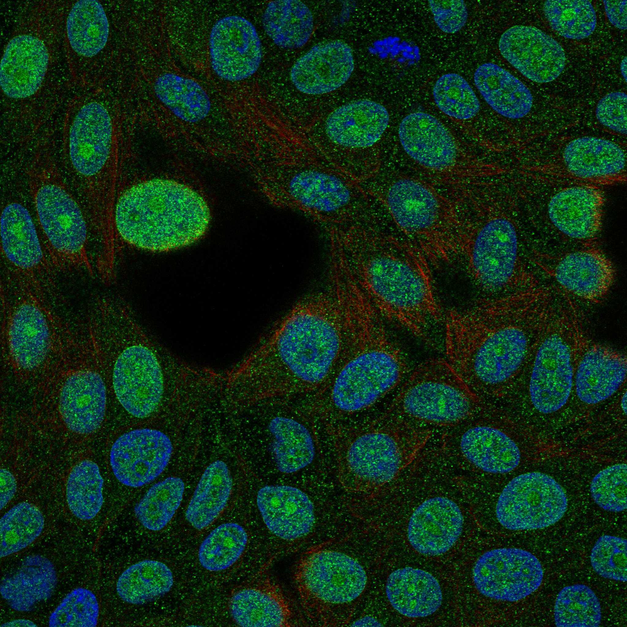
IF/ICC Q&A: Where should CDK4 fluorescence appear?HPA supports nucleoplasm and cytosol as main locations, with additional nucleolar and nuclear membrane localization (HPA: subcellular ICC-IF). These are ICC-IF observations, separate from tissue IHC intensity calls.Compare fluorescence with nuclear and cell outlines when interpreting localization (general IF practice). Use the HPA subcellular pattern for this question; consult the separate IF/ICC guide for assay setup.

Sample controls for CDK4 IHC & IF

🧪Run bronchus first and look for staining in ciliated cells (cell body), scored High by HPA (HPA: bronchus, ciliated cells, High). Use adipose tissue adipocytes as the negative tissue (HPA: adipose tissue, adipocytes, Not detected); on the bronchus slide, use adjacent cells outside the ciliated population as a background comparator, without assuming they are CDK4-negative (HPA: bronchus row specifies ciliated cells only).
Positive control tissue: Bronchus (Ciliated cells (cell body), HPA High)
Negative control tissue: Adipose tissue (HPA Not detected)
ICC-IF cell lines (HPA subcellular resource): HPA ICC-IF images show CDK4 in MCF-7, Rh30, U2OS, hTERT-RPE1 (serum starved), KOLF2.1J, with annotated localisation: Nucleoplasm (supported), Cytosol (supported) (HPA subcellular).
Technical controls: Include no-primary (secondary-only), a host-species- and clonality-matched isotype control, and CDK4-knockout tissue or cells as a biological negative (standard IHC control practice; selected-SKU caption: rabbit primary antibody). For bronchus, quench endogenous peroxidase and check background from endogenous biotin because the caption uses biotin-based DAB detection (selected-SKU tissue-IHC caption: biotinylated secondary, SABC, DAB; standard IHC control practice).
⚠️Feasibility: A target-specific fixation window or fixation effect is unreported, and the exact selected-SKU caption does not state a fixative (selected-SKU tissue-IHC caption: fixative not stated). The caption demonstrates heat retrieval in EDTA at pH 8.0 for a paraffin-embedded mouse intestine section, but does not establish that bronchus requires the same retrieval or that the section was paraffin-section (selected-SKU tissue-IHC caption: mouse intestine, EDTA pH 8.0). Whether frozen sections or IF are easier is unestablished; for bronchus, check ciliated-cell staining against local DAB background, while IF localisation can be assessed against the reported nucleoplasmic and cytosolic pattern (HPA: bronchus, ciliated cells, High; HPA subcellular: nucleoplasm and cytosol supported; standard IHC control practice). The selected PB9535 tissue-IHC caption documents paraffin sections, but does not specify the fixative (selected-SKU IHC image PB9535).

HPA tissue IHC evidence for CDK4

Comprehensive Human Protein Atlas IHC scoring per tissue (reliability: Supported — Medium consistency between antibody staining and RNA expression data. Presumed off target binding observed and disregarded.). Rows are taken directly from the HPA tissue chart — click any row's HPA link to view the source.

Positive expression · recommended positive controls

TissueCell typeLevelEvidenceSource
Bronchus Ciliated cells (cell body) High Protein (IHC) HPA →
Fallopian tube Ciliated cells (cell body) High Protein (IHC) HPA →
Ovary Ovarian stroma cells High Protein (IHC) HPA →
Rectum Endocrine cells High Protein (IHC) HPA →
Adrenal gland Glandular cells Medium Protein (IHC) HPA →

Undetected expression · recommended negative controls

TissueCell typeLevelEvidenceSource
Adipose tissue Adipocytes Not detected Protein (IHC) HPA →
Bone marrow Hematopoietic cells Not detected Protein (IHC) HPA →
Caudate Glial cells Not detected Protein (IHC) HPA →
Cerebellum Cells in granular layer Not detected Protein (IHC) HPA →
Cerebral cortex Endothelial cells Not detected Protein (IHC) HPA →
Section 3

Advanced CDK4 IHC Tips

Troubleshoot CDK4 staining in paraffin sections by checking retrieval, controls, compartmental localisation and cell-level scoring before interpreting chromogenic signal.

What should I change when CDK4 staining is weak after antigen retrieval?
Begin with heat-mediated retrieval in EDTA at pH 8.0 for paraffin sections (datasheet PB9535). The selected image documents this buffer but gives no heating duration, so record the instrument, temperature and time used before comparing runs (caption PB9535). If staining remains weak, compare shorter and longer heating on adjacent sections while keeping the catalog antibody at the documented 1 μg/mL and overnight 4°C incubation (caption PB9535; standard IHC practice). Judge any gain in nuclear and cytoplasmic signal against tissue damage and staining in negative controls; retrieval that raises all staining has not improved CDK4 specificity (HPA tissue IHC; standard IHC practice).
Could fixation explain inconsistent CDK4 staining between paraffin sections?
CDK4-specific fixation sensitivity is unknown: the selected paraffin-section image does not state its fixative (caption PB9535). Record fixative, fixation duration, specimen thickness and processing history for each section, then compare similarly processed material before changing antibody concentration (standard IHC practice). If a fixation effect is suspected, run a controlled pilot with, for example, 12 and 24 hours of the same fixative, followed by identical EDTA retrieval at pH 8.0 (standard IHC practice; datasheet PB9535). Compare cell-level signal and morphology together, because stronger chromogen alone cannot establish that a fixation change better preserves CDK4 detection (standard IHC practice).
Is cytoplasmic CDK4 staining plausible when I expected a nuclear signal?
Yes: CDK4 is cytoplasmic when non-complexed, while cyclin D–CDK4 can accumulate at the nuclear membrane and enter the nucleus during G1/S progression (UniProt P11802). Tissue IHC is mainly nuclear but also variably cytoplasmic, so score the two compartments separately rather than calling every cytoplasmic cell negative (HPA tissue IHC). Confirm that chromogen sits within intact cell boundaries and compare the distribution with a counterstain and a no-primary control on adjacent sections (standard IHC practice). A compartment shift alone does not establish kinase activation; complex assembly and Thr-172 phosphorylation have separate biological roles (UniProt P11802).
Can this stain distinguish CDK4 isoforms or its phosphorylated form?
CDK4 has 2 annotated isoforms, but the supplied catalog image does not map this antibody’s epitope or establish isoform selectivity (UniProt P11802; caption PB9535). The protein kinase domain spans residues 6–295, and Thr-172 is a modified residue; neither fact identifies what the catalog antibody binds (UniProt P11802). Treat a positive DAB signal as CDK4 immunoreactivity, without assigning it to a specific isoform or phosphorylation state (standard IHC interpretation). If that distinction drives the experiment, obtain epitope documentation and validate a suitably specific reagent on matched paraffin sections with appropriate controls (standard IHC practice).
How can I check CDK4 localisation by multiplex immunofluorescence?
Use this as a separate IF/ICC validation: the selected antibody evidence here is paraffin-section chromogenic IHC, while independent subcellular data place CDK4 mainly in nucleoplasm and cytosol (caption PB9535; HPA subcellular). Pair CDK4 with a validated marker for the expected cell type and a nuclear counterstain, checking single-stain controls before interpreting overlap (standard IF practice). Choose a red or far-red fluorophore after inspecting unstained tissue for autofluorescence in each channel (standard IF practice). CDK4 has no transmembrane segment, so optimise permeabilisation for intracellular nuclear and cytosolic epitopes and verify that cell boundaries remain intact (UniProt P11802 topology; standard IF practice).
How do I reduce diffuse DAB or nonspecific CDK4 staining?
First compare no-primary and secondary-only sections with the stained section to locate background from detection reagents or tissue chemistry (standard IHC practice). The selected procedure used 10% goat serum, 1 μg/mL primary antibody overnight at 4°C, and a biotin-based DAB detection system (caption PB9535). Include a peroxidase block, then check whether endogenous biotin or inadequate washing contributes to staining outside intact cells (standard IHC practice). If controls remain clean but staining is diffuse, titrate the primary around the documented concentration and retain settings that preserve nuclear and plausible cytoplasmic signal (HPA tissue IHC; standard IHC practice).
How should I score CDK4 across heterogeneous tissue sections? ⚠ ANSWER MARKED FOR VERIFICATION
Define the cell population and compartment before scoring, because CDK4 tissue staining is mainly nuclear with variable cytoplasmic expression (HPA tissue IHC). For each compartment, report the percentage of positive target cells and, if intensity is reproducible, an H-score using 0–3 intensity grades for a 0–300 range (standard IHC scoring practice). Normalise positive counts to all eligible cells of that type, or area-based counts to analysed viable tissue in mm², and exclude folds and necrosis (standard IHC practice). Keep retrieval, detection and image thresholds constant across groups, and report nuclear and cytoplasmic results separately (standard IHC practice).
When should apparent CDK4 positivity be treated as artefact?
Expect principally nuclear staining with possible cytoplasmic signal, rather than treating every DAB deposit as CDK4 (HPA tissue IHC). Check cell identity: ciliated bronchial cells and ovarian stroma cells are reported high, whereas adipocytes and alveolar cells are reported not detected in the listed observations (HPA tissue IHC). Suspect artefact when signal clusters at section edges, folds or necrosis, appears outside cells, or persists in no-primary controls after peroxidase blocking (standard IHC practice). HPA rates tissue IHC support as medium consistency and notes disregarded presumed off-target binding, so resolve discordant patterns with matched controls or independent validation before interpreting biology (HPA tissue IHC).
Boster reagents

Best CDK4 / Cyclin-dependent kinase 4 IHC Antibodies

Anti-CDK4 antibodies have paraffin-section IHC images from human, mouse and rat tissues (catalog IHC image captions), plus IF images from MCF7 and NIH/3T3 cells (catalog IF image captions).

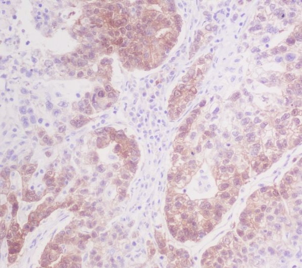
Real IHC data IHC analysis of Cdk4 using anti-Cdk4 antibody (PB9535). Cdk4 was detected in a paraffin-embedded section of mouse intestine tissue. Heat mediated antigen retrieval was performed in EDTA buffer (pH 8.0, epitope retrieval solution). The tissue section was blocked with 10% goat serum. The tissue section was then incubated with 1 μg/ml rabbit anti-Cdk4 Antibody (PB9535) overnight at 4°C. Biotinylated goat anti-rabbit IgG was used as secondary antibody and incubated for 30 minutes at 37°C. The tissue section was developed using Strepavidin-Biotin-Complex (SABC) (Catalog # SA1022) with DAB as the chromogen.
Anti-Cdk4 Antibody ®
Cat # PB9535
Real IHC data Immunohistochemical analysis of paraffin-embedded Human Breast cancer section using Pink1. M00159-2 was diluted at 1:300 dilution. A undiluted biotinylated goat polyvalent antibody was used as the secondary, followed by DAB staining.
Anti-CDK4 Antibody
Cat # M00159-2
Real IHC data Immunohistochemical analysis of paraffin-embedded human stomach cancer, using CDK4 Antibody.
Anti-CDK4 Rabbit Monoclonal Antibody
Cat # M00159-1
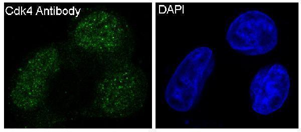
Real IF data Immunofluorescent analysis of NIH/3T3 cells, using CDK4 Antibody.
Anti-CDK4 Rabbit Monoclonal Antibody
Cat # M00159

PB9535 has IHC images from mouse and rat intestine and human lung cancer sections (PB9535 image captions); M00159-2 has a human breast carcinoma IHC-P caption, although its card image caption names Pink1 (M00159-2 image captions). M00159-1 has a human stomach cancer IHC image and an MCF7 IF image (M00159-1 image captions); M00159 has a NIH/3T3 IF image and no listed IHC application (M00159 image caption; catalog applications).

Which to pick: For tissue IHC across human, mouse and rat, choose PB9535: its own captions document paraffin sections, EDTA retrieval at pH 8.0 and 1 μg/ml primary antibody; the fixative is unreported (PB9535 image captions). For human IF/ICC, consider rabbit monoclonal M00159-1, which lists both applications and has an MCF7 IF image; rabbit monoclonal M00159 offers an additional NIH/3T3 IF image (catalog applications, host and clone; M00159-1 and M00159 IF image captions). M00159-2 is a mouse monoclonal IHC-P option for human or mouse samples, but its card caption names Pink1, so check the CDK4-specific breast carcinoma caption before relying on that figure (M00159-2 catalog applications, reactivity, host and clone; M00159-2 image captions).

Each figure is that product's own IHC / IF validation image from its datasheet.

References

  1. UniProt Consortium. UniProt entry P11802 (CDK4_HUMAN, Cyclin-dependent kinase 4).
  2. Human Protein Atlas. CDK4 tissue IHC expression (reliability: Supported).
  3. Human Protein Atlas. CDK4 subcellular location (ICC-IF): Mainly localized to the nucleoplasm and cytosol. In addition localized to the nucleoli and nuclear membrane..
  4. Human Protein Atlas. CDK4 antibody validation summary (3 antibodies).
  5. Juvenile trabecular ossifying fibroma: Immunohistochemical expression of MDM2, CDK4 and p53 compared to conventional ossifying fibroma. Journal of clinical and experimental dentistry 2022 — PMC8760962.
  6. Immunohistochemical Expression of p16 and CDK4 in Soft Tissue Tumors. Cureus 2023 — PMC10066853.
  7. CDK4 amplification predicts recurrence of well-differentiated liposarcoma of the abdomen. PloS one 2014 — PMC4133208.
  8. Angiomatoid fibrous histiocytoma with CDK4 and MDM2 expression misdiagnosed as dedifferentiated liposarcoma: The first case report and literature review. SAGE open medical case reports 2026 — PMC13305618.
  9. PubMed PMID:9192850 — UniProt-cited evidence.
  10. PubMed PMID:7652577 — UniProt-cited evidence.
  11. PubMed PMID:8528263 — UniProt-cited evidence.