Click image to see more details
Product Info Summary
| SKU: | M00334-3 |
|---|---|
| Size: | 100 μl |
| Reactive Species: | Human |
| Host: | Rabbit |
| Application: | IF, IHC, ICC, WB |
Customers Who Bought This Also Bought

Product info
Product Name
Anti-active Caspase-3 CASP3 Rabbit Monoclonal Antibody
SKU/Catalog Number
M00334-3
Size
100 μl
Form
Liquid
Description
Boster Bio Anti-active Caspase-3 CASP3 Rabbit Monoclonal Antibody catalog # M00334-3. Tested in WB, IHC, ICC/IF applications. This antibody reacts with Human.
Storage & Handling
Store at -20°C for one year. For short term storage and frequent use, store at 4°C for up to one month. Avoid repeated freeze-thaw cycles.
Cite This Product
Anti-active Caspase-3 CASP3 Rabbit Monoclonal Antibody (Boster Biological Technology, Pleasanton CA, USA, Catalog # M00334-3)
Host
Rabbit
Contents
Rabbit IgG in phosphate buffered saline, pH 7.4, 150mM NaCl, 0.02% sodium azide and 50% glycerol, 0.4-0.5mg/ml BSA.
Clonality
Monoclonal
Clone Number
HE-3
Isotype
Rabbit IgG
Immunogen
A synthesized peptide derived from human active Caspase-3
*Blocking peptide can be purchased. Costs vary based on immunogen length. Contact us for pricing.
Reactive Species
M00334-3 is reactive to CASP3 in Human
Applications
M00334-3 is guaranteed for IF, IHC, ICC, WB Boster Guarantee
Observed Molecular Weight
47 kDa
Calculated molecular weight
31.608kDa
Background of Caspase-3
C3 plays a central role in the activation of the complement system. Its processing by C3 convertase is the central reaction in both classical and alternative complement pathways. After activation C3b can bind covalently, via its reactive thioester, to cell surface carbohydrates or immune aggregates.
Antibody Validation
Boster validates all antibodies on WB, IHC, ICC, Immunofluorescence, and ELISA with known positive control and negative samples to ensure specificity and high affinity, including thorough antibody incubations.
Innovating Scientists Reward
If you are the first to review this product, or if you have results for a special sample, species or application this product is not validated in, share your results with us and receive product credits you can use towards any Boster products! Applicable to all scientists worldwide.
Submit A Review
Assay dilution & Images
Reconsitution
Restore with deionized water (or equivalent) for reconstitution volume of 1.0 mL
Assay Dilutions Recommendation
The recommendations below provide a starting point for assay optimization. The actual working concentration varies and should be decided by the user.
WB 1:500-1:2000
IHC 1:50-1:100
ICC/IF 1:50-1:100
Validation Images & Assay Conditions
Click image to see more details
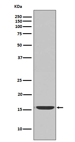
Western blot analysis of active Caspase-3 expression in Jurkat cell lysate treated with Camptothecin.

Click image to see more details
Immunofluorescent analysis of HepG2 cells, using active Caspase-3 Antibody .
Protein Target Info & Infographic
Gene/Protein Information For CASP3 (Source: Uniprot.org, NCBI)
Gene Name
CASP3
Full Name
Caspase-3
Weight
31.608kDa
Superfamily
peptidase C14A family
Alternative Names
Apopain; apoptosis-related cysteine protease; CASP3; CASP-3; caspase 3, apoptosis-related cysteine peptidase; Caspase3; Caspase-3; CPP32; CPP-32; CPP32B; CPP32SREBP cleavage activity 1; Cysteine protease CPP32; EC 3.4.22; EC 3.4.22.56; LICE-1; PARP cleavage protease; procaspase3; Protein Yama; SCA-1; YAMA CASP3 CPP32, CPP32B, SCA-1 caspase 3 caspase-3|CASP-3|CPP-32|PARP cleavage protease|SREBP cleavage activity 1|apopain|caspase 3, apoptosis-related cysteine peptidase|caspase 3, apoptosis-related cysteine protease|cysteine protease CPP32|procaspase3|protein Yama
*If product is indicated to react with multiple species, protein info is based on the gene entry specified above in "Species".For more info on CASP3, check out the CASP3 Infographic

We have 30,000+ of these available, one for each gene! Check them out.
In this infographic, you will see the following information for CASP3: database IDs, superfamily, protein function, synonyms, molecular weight, chromosomal locations, tissues of expression, subcellular locations, post-translational modifications, and related diseases, research areas & pathways. If you want to see more information included, or would like to contribute to it and be acknowledged, please contact [email protected].
Specific Publications For Anti-active Caspase-3 CASP3 Rabbit Monoclonal Antibody (M00334-3)
Hello CJ!
M00334-3 has been cited in 18 publications:
*The publications in this section are manually curated by our staff scientists. They may differ from Bioz's machine gathered results. Both are accurate. If you find a publication citing this product but is missing from this list, please let us know we will issue you a thank-you coupon.
Chen H,Sheng H,Zhao Y,Zhu G.Piperine Inhibits Cell Proliferation and Induces Apoptosis of Human Gastric Cancer Cells by Downregulating Phosphatidylinositol 3-Kinase (PI3K)/Akt Pathway.Med Sci Monit.2020 Dec 31;26:e928403.doi:10.12659/MSM.928403.PMID:33382
Species: Human,Mouse
Wang P,Wang C,Liu C. Antitumor effects of dioscin in A431 cells via adjusting ATM/p53-mediated cell apoptosis, DNA damage and migration. Oncol Lett. 2021 Jan;21(1):59.doi:10.3892/ol.2020.12321.Epub 2020 Nov 19.PMID:33281970;PMCID:PMC7709553.
Species: Human
Maternal separation exacerbates Alzheimer's disease-like behavioral and pathological changes in adult APPswe/PS1dE9 mice
MicroRNA-744 promotes prostate cancer progression through aberrantly activating Wnt/%u03B2-catenin signaling
In vitro and in vivo antitumor effects of acetylshikonin isolated from Arnebia euchroma (Royle) Johnst (Ruanzicao) cell suspension cultures
Enhanced anti-apoptosis and gut epithelium protection function of acidic fibroblast growth factor after cancelling of its mitogenic activity
Antenatal taurine reduces cerebral cell apoptosis in fetal rats with intrauterine growth restriction
In vivo?and?in vitro?evaluation of the cytotoxic effects of Photosan-loaded hollow silica nanoparticles on liver cancer
An J, Liu T, She R, Wu Q, Tian J, Shi R, Hao W, Ren X, Yang Y, Lu Y, Yang Y, Wu Y. Oncotarget. 2017 Dec 17;9(4):4475-4484. doi: 10.18632/oncotarget.23381. eCollection 2018 Jan 12. Replication of hepatitis E virus in the ovary and promotion of oocy...
Liu Nb, Peng T, Pan C, Yao Yy, Shen B, Leng J. World J Gastroenterol. 2005 Oct 28;11(40):6281-7. Overexpression Of Cyclooxygenase-2 In Human Hepg2, Bel-7402 And Smmc-7721 Hepatoma Cell Lines And Mechanism Of Cyclooxygenase-2 Selective Inhibitor Ce...
Recommended Resources
Here are featured tools and databases that you might find useful.
- Boster's Pathways Library
- Protein Databases
- Bioscience Research Protocol Resources
- Data Processing & Analysis Software
- Photo Editing Software
- Scientific Literature Resources
- Research Paper Management Tools
- Molecular Biology Software
- Primer Design Tools
- Bioinformatics Tools
- Phylogenetic Tree Analysis
Customer Reviews
Have you used Anti-active Caspase-3 CASP3 Rabbit Monoclonal Antibody?
Submit a review and receive an Amazon gift card.
- $30 for a review with an image
Be the first to review Anti-active Caspase-3 CASP3 Rabbit Monoclonal Antibody
*The first user to submit a review for a product is eligible for Boster's Innovating Scientists Reward, which gives product credits. This is in addition to the gift card reward.
Customer Q&As
Have a question?
Find answers in Q&As, reviews.
Can't find your answer?
Submit your question
5 Customer Q&As for Anti-active Caspase-3 CASP3 Rabbit Monoclonal Antibody
Question
Our team were happy with the WB result of your anti-active Caspase-3 Rabbit Monoclonal antibody. However we have observed positive staining in lymph cytoplasm. using this antibody. Is that expected? Could you tell me where is CASP3 supposed to be expressed?
Verified Customer
Verified customer
Asked: 2020-02-06
Answer
According to literature, lymph does express CASP3. Generally CASP3 expresses in cytoplasm. Regarding which tissues have CASP3 expression, here are a few articles citing expression in various tissues:
Cervix carcinoma, and Erythroleukemia, Pubmed ID: 23186163
Lymph, Pubmed ID: 15489334
T-cell, Pubmed ID: 7983002, 7774019
Tongue, Pubmed ID: 14702039
Boster Scientific Support
Answered: 2020-02-06
Question
We ordered your anti-active Caspase-3 Rabbit Monoclonal antibody for ICC on lymph in the past. I am using human, and We want to use the antibody for IHC next. I was wanting to use examining lymph as well as cervix carcinoma erythroleukemia in our next experiment. Could you please give me some suggestion on which antibody would work the best for IHC?
Verified Customer
Verified customer
Asked: 2019-12-31
Answer
I viewed the website and datasheets of our anti-active Caspase-3 Rabbit Monoclonal antibody and it seems that M00334-3 has been tested on human in both ICC and IHC. Thus M00334-3 should work for your application. Our Boster satisfaction guarantee will cover this product for IHC in human even if the specific tissue type has not been validated. We do have a comprehensive range of products for IHC detection and you can check out our website bosterbio.com to find out more information about them.
Boster Scientific Support
Answered: 2019-12-31
Question
you antibody using your anti-active Caspase-3 Rabbit Monoclonal antibody for hippocampus development studies. Has this antibody been tested with western blotting on jurkat cell lysate? We would like to see some validation images before ordering.
Verified Customer
Verified customer
Asked: 2019-10-16
Answer
We appreciate your inquiry. This M00334-3 anti-active Caspase-3 Rabbit Monoclonal antibody is tested on jurkat cell lysate. It is guaranteed to work for IF, IHC, ICC, WB in human. Our Boster guarantee will cover your intended experiment even if the sample type has not been be directly tested.
Boster Scientific Support
Answered: 2019-10-16
Question
We are currently using anti-active Caspase-3 Rabbit Monoclonal antibody M00334-3 for human tissue, and we are content with the WB results. The species of reactivity given in the datasheet says human. Is it likely that the antibody can work on goat tissues as well?
Verified Customer
Verified customer
Asked: 2019-07-03
Answer
The anti-active Caspase-3 Rabbit Monoclonal antibody (M00334-3) has not been tested for cross reactivity specifically with goat tissues, though there is a good chance of cross reactivity. We have an innovator award program that if you test this antibody and show it works in goat you can get your next antibody for free. Please contact me if I can help you with anything.
Boster Scientific Support
Answered: 2019-07-03
Question
We have observed staining in human lymph. Any tips? Is anti-active Caspase-3 Rabbit Monoclonal antibody supposed to stain lymph positively?
Verified Customer
Verified customer
Asked: 2018-08-20
Answer
According to literature lymph does express CASP3. According to Uniprot.org, CASP3 is expressed in jejunal mucosa, t-cell, tongue, lymph, cervix carcinoma erythroleukemia, among other tissues. Regarding which tissues have CASP3 expression, here are a few articles citing expression in various tissues:
Cervix carcinoma, and Erythroleukemia, Pubmed ID: 23186163
Lymph, Pubmed ID: 15489334
T-cell, Pubmed ID: 7983002, 7774019
Tongue, Pubmed ID: 14702039
Boster Scientific Support
Answered: 2018-08-20
