Click image to see more details
-

-
-
-
-
+7
Product Info Summary
| SKU: | M01080 |
|---|---|
| Size: | 100ug/vial |
| Reactive Species: | Human, Mouse |
| Host: | Mouse |
| Application: | Flow Cytometry, IF, IHC, WB |
Customers Who Bought This Also Bought


Product info
Product Name
Anti-CD63 (Late Endosomes Marker) Monoclonal Antibody
SKU/Catalog Number
M01080
Size
100ug/vial
Form
Liquid
Description
Boster Bio Anti-CD63 (Late Endosomes Marker) Monoclonal Antibody (Catalog # M01080). Tested in Flow Cytometry, IF, WB, IHC applications. This antibody reacts with Human, Mouse.
Storage & Handling
Antibody with azide - store at 2 to 8 °C. Antibody without azide - store at -20 to -80 °C. Antibody is stable for 24 months. Non-hazardous. No MSDS required.
Cite This Product
Anti-CD63 (Late Endosomes Marker) Monoclonal Antibody (Boster Biological Technology, Pleasanton CA, USA, Catalog # M01080)
Host
Mouse
Contents
Prepared in 10mM PBS with 0.05% BSA & 0.05% azide. Also available WITHOUT BSA & azide at 1.0mg/ml.
Clonality
Monoclonal
Clone Number
Clone: MX-49.129.5
Isotype
IgG1, kappa
Immunogen
Full length CD63 of human origin
*Blocking peptide can be purchased. Costs vary based on immunogen length. Contact us for pricing.
Cross-reactivity
Does not cross-react with primate, avian or amphibian GR.
Reactive Species
M01080 is reactive to CD63 in Human, Mouse
Applications
M01080 is guaranteed for Flow Cytometry, IF, IHC, WB Boster Guarantee
Observed Molecular Weight
68 kDa
Calculated molecular weight
25.637kDa
Background of CD63
This monoclonal antibody recognizes protein of 26kDa-60kDa, which is identified as CD63. Its epitope is different from that of MAb LAMP3/529. The tetraspanins are integral membrane proteins expressed on cell surface and granular membranes of hematopoietic cells and are components of multi-molecular complexes with specific integrins. The tetraspanin CD63 is a lysosomal membrane glycoprotein that translocates to the plasma membrane after platelet activation. CD63 is expressed on activated platelets, monocytes and macrophages, and is weakly expressed on granulocytes, T cell and B cells. It is located on the basophilic granule membranes and on the plasma membranes of lymphocytes and granulocytes. CD63 is a member of the TM4 superfamily of leukocyte glycoproteins that includes CD9, CD37 and CD53, which contain four transmembrane regions. CD63 may play a role in phagocytic and intracellular lysosome-phagosome fusion events. CD63 deficiency is associated with Hermansky-Pudlak syndrome and is strongly expressed during the early stages of melanoma progression.
Antibody Validation
Boster validates all antibodies on WB, IHC, ICC, Immunofluorescence, and ELISA with known positive control and negative samples to ensure specificity and high affinity, including thorough antibody incubations.
Innovating Scientists Reward
If you are the first to review this product, or if you have results for a special sample, species or application this product is not validated in, share your results with us and receive product credits you can use towards any Boster products! Applicable to all scientists worldwide.
Submit A Review
Assay dilution & Images
Reconsitution
Reconstitute with distilled water.
Assay Dilutions Recommendation
The recommendations below provide a starting point for assay optimization. The actual working concentration varies and should be decided by the user.
Flow Cytometry (0.5-1ug/million cells)
Immunofluorescence (0.5-1ug/ml)
Western Blot (0.5-1ug/ml)
Immunohistochemistry (Formalin-fixed) (1-2ug/ml for 30 minutes at RT) (Staining of formalin-fixed tissues requires heating tissue sections in 10mM Tris with 1mM EDTA, pH 9.0, for 45 min at 95 °C followed by cooling at RT for 20 minutes)
Optimal dilution for a specific application should be determined.
Validation Images & Assay Conditions
Click image to see more details
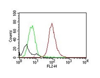
Flow cytometric analysis of NIH/3T3 cells. Black: Cells alone; Green: Isotype Control; Red: PE-labeled Anti-CD63 Mouse Monoclonal Antibody (MX-49.129.5).

Click image to see more details
Flow cytometric analysis of MCF-7 cells. Anti-CD63 Mouse Monoclonal Antibody (MX-49.129.5) followed by goat anti-mouse IgG-CF488 (green); unstained cells (gray)

Click image to see more details
Flow cytometric analysis of bead-bound exosomes derived from MCF-7 cells. Anti-CD63 Mouse Monoclonal Antibody (MX-49.129.5) followed by goat anti-mouse IgG-CF488 (green); unstained exosomes (gray).

Click image to see more details
Flow cytometric analysis of MCF-7 cells. Anti-CD63 Mouse Monoclonal Antibody (MX-49.129.5) followed by goat anti-mouse IgG-CF568 (orange); unstained cells (gray).

Click image to see more details
Flow cytometric analysis of bead-bound exosomes derived from MCF-7 cells. Anti-CD63 Mouse Monoclonal Antibody (MX-49.129.5) followed by goat anti-mouse IgG-CF568 (orange); unstained exosomes (gray).

Click image to see more details
Immunofluorescence analysis of HeLa cells stained using CF ®488-labeled-CD63 Monoclonal Antibody (MX-49.129.5) (green). F-actin filaments labeled with phalloidin (red), nuclei stained with DAPI (blue).

Click image to see more details
Immunofluorescence analysis of PFA-fixed U87MG cells stained using CD63 Mouse Monoclonal Antibody (MX-49.129.5) followed by goat anti-mouse IgG-CF488 (green). CF640R phalloidin (red).

Click image to see more details
Formalin-fixed, paraffin-embedded human melanoma stained with Anti-CD63 Mouse Monoclonal Antibody (MX-49.129.5).

Click image to see more details
Formalin-fixed, paraffin-embedded mouse spleen stained with Anti-CD63 Mouse Monoclonal Antibody (MX-49.129.5).

Click image to see more details
Formalin-fixed, paraffin-embedded human prostate carcinoma stained with Anti-CD63 Mouse Monoclonal Antibody (MX-49.129.5).

Click image to see more details
Western blot analysis of human spleen tissue lysate using Anti-CD63 Mouse Monoclonal Antibody (MX-49.129.5).
Protein Target Info & Infographic
Gene/Protein Information For CD63 (Source: Uniprot.org, NCBI)
Gene Name
CD63
Full Name
CD63 antigen
Weight
25.637kDa
Superfamily
tetraspanin (TM4SF) family
Alternative Names
CD63 antigen (melanoma 1 antigen); CD63 antigen; CD63 molecule; CD63; Granulophysin; Lamp-3; Lysosomal-associated membrane protein 3; ME491; melanoma 1 antigen; Melanoma-associated antigen ME491; melanoma-associated antigen MLA1; MLA1lysosome-associated membrane glycoprotein 3; Ocular melanoma-associated antigen; OMA81H; tetraspanin-30; Tspan30; tspan-30; TSPAN30granulophysin CD63 LAMP-3, ME491, MLA1, OMA81H, TSPAN30 CD63 molecule CD63 antigen|CD63 antigen (melanoma 1 antigen)|granulophysin|lysosomal-associated membrane protein 3|lysosome-associated membrane glycoprotein 3|melanoma-associated antigen ME491|melanoma-associated antigen MLA1|ocular melanoma-associated antigen|tetraspanin-30|tspan-30
*If product is indicated to react with multiple species, protein info is based on the gene entry specified above in "Species".For more info on CD63, check out the CD63 Infographic

We have 30,000+ of these available, one for each gene! Check them out.
In this infographic, you will see the following information for CD63: database IDs, superfamily, protein function, synonyms, molecular weight, chromosomal locations, tissues of expression, subcellular locations, post-translational modifications, and related diseases, research areas & pathways. If you want to see more information included, or would like to contribute to it and be acknowledged, please contact [email protected].
Specific Publications For Anti-CD63 (Late Endosomes Marker) Monoclonal Antibody (M01080)
Hello CJ!
M01080 has been cited in 1 publications:
*The publications in this section are manually curated by our staff scientists. They may differ from Bioz's machine gathered results. Both are accurate. If you find a publication citing this product but is missing from this list, please let us know we will issue you a thank-you coupon.
Du Hy, Dong Lh, Zhao Bj, Fu J, Wang Qq, Chen F, Ou L, Li N, Sun X, Tang Zm, Song Hf. Acta Pharmacol Sin. 2012 Aug;33(8):1047-54. Doi: 10.1038/Aps.2012.54. Epub 2012 Jun 25. Immunostimulatory And Anti-Neoplasm Effects Of A Novel Palindrome Cpg Olig...
Recommended Resources
Here are featured tools and databases that you might find useful.
- Boster's Pathways Library
- Protein Databases
- Bioscience Research Protocol Resources
- Data Processing & Analysis Software
- Photo Editing Software
- Scientific Literature Resources
- Research Paper Management Tools
- Molecular Biology Software
- Primer Design Tools
- Bioinformatics Tools
- Phylogenetic Tree Analysis
Customer Reviews
Have you used Anti-CD63 (Late Endosomes Marker) Monoclonal Antibody?
Submit a review and receive an Amazon gift card.
- $30 for a review with an image
Be the first to review Anti-CD63 (Late Endosomes Marker) Monoclonal Antibody
*The first user to submit a review for a product is eligible for Boster's Innovating Scientists Reward, which gives product credits. This is in addition to the gift card reward.
Customer Q&As
Have a question?
Find answers in Q&As, reviews.
Can't find your answer?
Submit your question
3 Customer Q&As for Anti-CD63 (Late Endosomes Marker) Monoclonal Antibody
Question
We were satisfied with the WB result of your anti-CD63 (Late Endosomes Marker) Monoclonal antibody. However we have observed positive staining in skin cell membrane using this antibody. Is that expected? Could you tell me where is CD63 supposed to be expressed?
Verified Customer
Verified customer
Asked: 2020-03-16
Answer
According to literature, skin does express CD63. Generally CD63 expresses in cell membrane. Regarding which tissues have CD63 expression, here are a few articles citing expression in various tissues:
Liver, Pubmed ID: 19159218
Lung, and Muscle, Pubmed ID: 15489334
Ovary, Pubmed ID: 2171551
Placenta, Pubmed ID: 17897319
Platelet, Pubmed ID: 7682577
Skeletal muscle, Pubmed ID: 14702039
Boster Scientific Support
Answered: 2020-03-16
Question
We have observed staining in human platelet. What should we do? Is anti-CD63 (Late Endosomes Marker) Monoclonal antibody supposed to stain platelet positively?
Verified Customer
Verified customer
Asked: 2018-02-20
Answer
Based on literature platelet does express CD63. Based on Uniprot.org, CD63 is expressed in upper lobe of lung, ovary, skin, skeletal muscle, lung muscle, platelet, placenta, liver, among other tissues. Regarding which tissues have CD63 expression, here are a few articles citing expression in various tissues:
Liver, Pubmed ID: 19159218
Lung, and Muscle, Pubmed ID: 15489334
Ovary, Pubmed ID: 2171551
Placenta, Pubmed ID: 17897319
Platelet, Pubmed ID: 7682577
Skeletal muscle, Pubmed ID: 14702039
Boster Scientific Support
Answered: 2018-02-20
Question
We are currently using anti-CD63 (Late Endosomes Marker) Monoclonal antibody M01080 for mouse tissue, and we are happy with the Flow Cytometry results. The species of reactivity given in the datasheet says human, mouse. Is it possible that the antibody can work on feline tissues as well?
H. Dhar
Verified customer
Asked: 2017-05-18
Answer
The anti-CD63 (Late Endosomes Marker) Monoclonal antibody (M01080) has not been tested for cross reactivity specifically with feline tissues, though there is a good chance of cross reactivity. We have an innovator award program that if you test this antibody and show it works in feline you can get your next antibody for free. Please contact me if I can help you with anything.
Boster Scientific Support
Answered: 2017-05-18
