Click image to see more details
-

-
-
-
-
+1
Product Info Summary
| SKU: | PB9071 |
|---|---|
| Size: | 100 μg/vial |
| Reactive Species: | Human, Mouse, Rat |
| Host: | Rabbit |
| Application: | Flow Cytometry, IHC, WB |
Customers Who Bought This Also Bought


Product info
Product Name
Anti-VEGF/VEGFA Antibody Picoband™
SKU/Catalog Number
PB9071
Size
100 μg/vial
Form
Lyophilized
Description
Boster Bio Anti-VEGF/VEGFA Antibody Picoband™ catalog # PB9071. Tested in Flow Cytometry, IHC, WB applications. This antibody reacts with Human, Mouse, Rat.
Storage & Handling
Store at -20˚C for one year from date of receipt. After reconstitution, at 4˚C for one month. It can also be aliquotted and stored frozen at -20˚C for six months. Avoid repeated freeze-thaw cycles.
Cite This Product
Anti-VEGF/VEGFA Antibody Picoband™ (Boster Biological Technology, Pleasanton CA, USA, Catalog # PB9071)
Host
Rabbit
Contents
Each vial contains 4mg Trehalose, 0.9mg NaCl, 0.2mg Na2HPO4, 0.05mg NaN3.
Clonality
Polyclonal
Isotype
Rabbit IgG
Immunogen
E.coli-derived human VEGF recombinant protein (Position: A27-R191). Human VEGF shares 78% amino acid (aa) sequence identity with both mouse and rat VEGF.
*Blocking peptide can be purchased. Costs vary based on immunogen length. Contact us for pricing.
Cross-reactivity
No cross-reactivity with other proteins
Reactive Species
PB9071 is reactive to VEGFA in Human, Mouse, Rat
Applications
PB9071 is guaranteed for Flow Cytometry, IHC, WB Boster Guarantee
Observed Molecular Weight
27 kDa
Calculated molecular weight
27.042kDa
Background of VEGFA
VEGF, a homodimeric glycoprotein of relative molecular mass 45,000, is the only mitogen that specifically acts on endothelial cells. It may be a major regulator of tumor angiogenesis in vivo. It is, however, structurally related to platelet-derived growth factor. VEGF shares homology with the PDGF A chain and B chain, including conservation of all 8 cysteines found in PDGFA and PDGFB. VEGF gene contains 8 exons. VEGF induces remodeling and enhances TH2-mediated sensitization and inflammation in the lung. And this gene also can regulate haematopoietic stem cell survival by an internal autocrine loop mechanism. What’s more, it also stimulates neurogenesis in vitro and in vivo.
Antibody Validation
Boster validates all antibodies on WB, IHC, ICC, Immunofluorescence, and ELISA with known positive control and negative samples to ensure specificity and high affinity, including thorough antibody incubations.
Innovating Scientists Reward
If you are the first to review this product, or if you have results for a special sample, species or application this product is not validated in, share your results with us and receive product credits you can use towards any Boster products! Applicable to all scientists worldwide.
Submit A Review
Assay dilution & Images
Reconsitution
Add 0.2ml of distilled water will yield a concentration of 500ug/ml.
Assay Dilutions Recommendation
The recommendations below provide a starting point for assay optimization. The actual working concentration varies and should be decided by the user.
Western blot, 0.1-0.5μg/ml, Human, Mouse, Rat
Immunohistochemistry (Paraffin-embedded Section), 0.5-1μg/ml, Human, Rat, By Heat
Flow Cytometry, 1-3μg/1x106 cells, Human
Validation Images & Assay Conditions
Click image to see more details
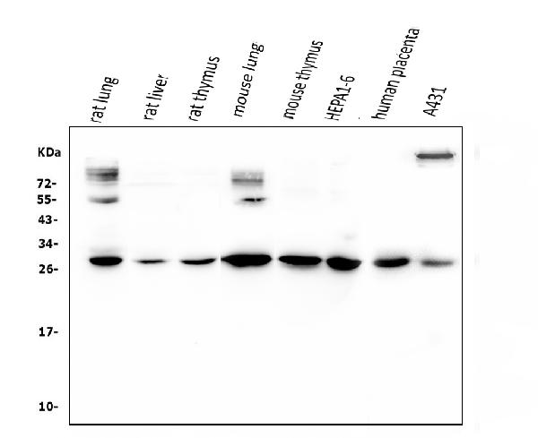
Figure 1. Western blot analysis of VEGF using anti-VEGF antibody (PB9071).
Electrophoresis was performed on a 5-20% SDS-PAGE gel at 70V (Stacking gel) / 90V (Resolving gel) for 2-3 hours. The sample well of each lane was loaded with 50ug of sample under reducing conditions.
Lane 1: rat lung tissue lysate,
Lane 2: rat lver tissue lysate,
Lane 3: rat thymus tissue lysate,
Lane 4: mouse lung tissue lysate,
Lane 5: mouse thymus tissue lysate,
Lane 6: HEPA1-6 whole cell lysate,
Lane 7: human placenta tissue lysate,
Lane 8: A431 whole cell lysate,
After Electrophoresis, proteins were transferred to a Nitrocellulose membrane at 150mA for 50-90 minutes. Blocked the membrane with 5% Non-fat Milk/ TBS for 1.5 hour at RT. The membrane was incubated with rabbit anti-VEGF antigen affinity purified polyclonal antibody (Catalog # PB9071) at 0.5 μg/mL overnight at 4°C, then washed with TBS-0.1%Tween 3 times with 5 minutes each and probed with a goat anti-rabbit IgG-HRP secondary antibody at a dilution of 1:10000 for 1.5 hour at RT. The signal is developed using an Enhanced Chemiluminescent detection (ECL) kit (Catalog # EK1002) with Tanon 5200 system. A specific band was detected for VEGF at approximately 27KD. The expected band size for VEGF is at 27KD.

Click image to see more details
Figure 2. IHC analysis of VEGF using anti-VEGF antibody (PB9071).
VEGF was detected in paraffin-embedded section of human rectal cancer tissue. Heat mediated antigen retrieval was performed in EDTA buffer (pH8.0, epitope retrieval solution). The tissue section was blocked with 10% goat serum. The tissue section was then incubated with 1μg/ml rabbit anti-VEGF Antibody (PB9071) overnight at 4°C. Biotinylated goat anti-rabbit IgG was used as secondary antibody and incubated for 30 minutes at 37°C. The tissue section was developed using Strepavidin-Biotin-Complex (SABC)(Catalog # SA1022) with DAB as the chromogen.

Click image to see more details
Figure 3. IHC analysis of VEGF using anti-VEGF antibody (PB9071).
VEGF was detected in paraffin-embedded section of human rectal cancer tissue. Heat mediated antigen retrieval was performed in EDTA buffer (pH8.0, epitope retrieval solution). The tissue section was blocked with 10% goat serum. The tissue section was then incubated with 1μg/ml rabbit anti-VEGF Antibody (PB9071) overnight at 4°C. Biotinylated goat anti-rabbit IgG was used as secondary antibody and incubated for 30 minutes at 37°C. The tissue section was developed using Strepavidin-Biotin-Complex (SABC)(Catalog # SA1022) with DAB as the chromogen.

Click image to see more details
Figure 4. IHC analysis of VEGF using anti-VEGF antibody (PB9071).
VEGF was detected in paraffin-embedded section of rat brain tissue. Heat mediated antigen retrieval was performed in EDTA buffer (pH8.0, epitope retrieval solution). The tissue section was blocked with 10% goat serum. The tissue section was then incubated with 1μg/ml rabbit anti-VEGF Antibody (PB9071) overnight at 4°C. Biotinylated goat anti-rabbit IgG was used as secondary antibody and incubated for 30 minutes at 37°C. The tissue section was developed using Strepavidin-Biotin-Complex (SABC)(Catalog # SA1022) with DAB as the chromogen.

Click image to see more details
Figure 5. Flow Cytometry analysis of SiHa cells using anti-VEGF antibody (PB9071).
Overlay histogram showing SiHa cells stained with PB9071 (Blue line).The cells were blocked with 10% normal goat serum. And then incubated with rabbit anti-VEGF Antibody (PB9071, 1μg/1x106 cells) for 30 min at 20°C. DyLight®488 conjugated goat anti-rabbit IgG (BA1127, 5-10μg/1x106 cells) was used as secondary antibody for 30 minutes at 20°C. Isotype control antibody (Green line) was rabbit IgG (1μg/1x106) used under the same conditions. Unlabelled sample (Red line) was also used as a control.
Protein Target Info & Infographic
Gene/Protein Information For VEGFA (Source: Uniprot.org, NCBI)
Gene Name
VEGFA
Full Name
Vascular endothelial growth factor A
Weight
27.042kDa
Superfamily
PDGF/VEGF growth factor family
Alternative Names
MVCD1; VAS; vascular endothelial growth factor A; Vascular permeability factor; Vasculotropin; VEGF; VEGFA; VEGF-A; VEGFMGC70609; VPF; VPFvascular endothelial growth factor VEGFA MVCD1, VEGF, VPF vascular endothelial growth factor A vascular endothelial growth factor A|vascular endothelial growth factor A121|vascular endothelial growth factor A165|vascular permeability factor
*If product is indicated to react with multiple species, protein info is based on the gene entry specified above in "Species".For more info on VEGFA, check out the VEGFA Infographic

We have 30,000+ of these available, one for each gene! Check them out.
In this infographic, you will see the following information for VEGFA: database IDs, superfamily, protein function, synonyms, molecular weight, chromosomal locations, tissues of expression, subcellular locations, post-translational modifications, and related diseases, research areas & pathways. If you want to see more information included, or would like to contribute to it and be acknowledged, please contact [email protected].
Specific Publications For Anti-VEGF/VEGFA Antibody Picoband™ (PB9071)
Hello CJ!
PB9071 has been cited in 118 publications:
*The publications in this section are manually curated by our staff scientists. They may differ from Bioz's machine gathered results. Both are accurate. If you find a publication citing this product but is missing from this list, please let us know we will issue you a thank-you coupon.
Buyanghuanwu decoction promotes angiogenesis after cerebral ischemia/reperfusion injury: mechanisms of brain tissue repair
Induction stage-dependent expression of vascular endothelial growth factor and aquaporin-1 in diethylstilbestrol-treated rat pituitary
Ginsenoside protects against AKI via activation of HIF‑1α and VEGF‑A in the kidney‑brain axis
Curcumin Derivative Cur20 Attenuated Cerebral Ischemic Injury by Antioxidant Effect and HIF-1α/VEGF/TFEB-Activated Angiogenesis
Decreased TIP30 Expression Promotes Tumor Metastasis in Lung Cancer
Extract of Ginkgo Biloba Promotes the Expression of VEGF Following Subarachnoid Hemorrhage in Rats
Construction of AuHQ nano-sensitizer for enhanced radiotherapy efficacy through remolding tumor vasculature
An oxidation responsive nano-radiosensitizer increases radiotherapy efficacy by remolding tumor vasculature
A Comparison of Ramipril and Bevacizumab to Mitigate Radiation-Induced Brain Necrosis: An Experimental Study
An Experimental Study on Repeated Brief Ischemia in Promoting Sciatic Nerve Repair and Regeneration in Rats
Recommended Resources
Here are featured tools and databases that you might find useful.
- Boster's Pathways Library
- Protein Databases
- Bioscience Research Protocol Resources
- Data Processing & Analysis Software
- Photo Editing Software
- Scientific Literature Resources
- Research Paper Management Tools
- Molecular Biology Software
- Primer Design Tools
- Bioinformatics Tools
- Phylogenetic Tree Analysis
Customer Reviews
Have you used Anti-VEGF/VEGFA Antibody Picoband™?
Submit a review and receive an Amazon gift card.
- $30 for a review with an image
Be the first to review Anti-VEGF/VEGFA Antibody Picoband™
*The first user to submit a review for a product is eligible for Boster's Innovating Scientists Reward, which gives product credits. This is in addition to the gift card reward.
Customer Q&As
Have a question?
Find answers in Q&As, reviews.
Can't find your answer?
Submit your question
5 Customer Q&As for Anti-VEGF/VEGFA Antibody Picoband™
Question
We need using your anti-VEGF/VEGFA antibody for positive regulation of cell proliferation by vegf-activated platelet derived growth factor receptor signaling pathway studies. Has this antibody been tested with western blotting on tissue lysate? We would like to see some validation images before ordering.
Verified Customer
Verified customer
Asked: 2020-03-13
Answer
I appreciate your inquiry. This PB9071 anti-VEGF/VEGFA antibody is tested on human placenta tissue, lung cancer tissue, tissue lysate, rat thymus tissue, brain tissue. It is guaranteed to work for IHC, WB in human, rat. Our Boster guarantee will cover your intended experiment even if the sample type has not been be directly tested.
Boster Scientific Support
Answered: 2020-03-13
Question
Our lab were satisfied with the WB result of your anti-VEGF/VEGFA antibody. However we have observed positive staining in left lobe of thyroid gland secreted using this antibody. Is that expected? Could you tell me where is VEGFA supposed to be expressed?
Verified Customer
Verified customer
Asked: 2020-01-17
Answer
According to literature, left lobe of thyroid gland does express VEGFA. Generally VEGFA expresses in secreted. Regarding which tissues have VEGFA expression, here are a few articles citing expression in various tissues:
Heart, Pubmed ID: 19054851
Kidney, Pubmed ID: 9878851, 12124351
Lung, Pubmed ID: 15489334
Mammary gland, Pubmed ID: 9450968
Renal glomerulus, Pubmed ID: 10464055
Retina, Pubmed ID: 10067980, 11563986
Boster Scientific Support
Answered: 2020-01-17
Question
We are currently using anti-VEGF/VEGFA antibody PB9071 for rat tissue, and we are satisfied with the WB results. The species of reactivity given in the datasheet says human, rat. Is it true that the antibody can work on dog tissues as well?
Verified Customer
Verified customer
Asked: 2019-11-13
Answer
The anti-VEGF/VEGFA antibody (PB9071) has not been validated for cross reactivity specifically with dog tissues, though there is a good chance of cross reactivity. We have an innovator award program that if you test this antibody and show it works in dog you can get your next antibody for free. Please contact me if I can help you with anything.
Boster Scientific Support
Answered: 2019-11-13
Question
We have been able to see staining in human retina. Do you have any suggestions? Is anti-VEGF/VEGFA antibody supposed to stain retina positively?
R. Jha
Verified customer
Asked: 2019-11-11
Answer
According to literature retina does express VEGFA. According to Uniprot.org, VEGFA is expressed in left lobe of thyroid gland, kidney, mammary gland, renal glomerulus, hemangioendothelioma, heart, lung, retina, among other tissues. Regarding which tissues have VEGFA expression, here are a few articles citing expression in various tissues:
Heart, Pubmed ID: 19054851
Kidney, Pubmed ID: 9878851, 12124351
Lung, Pubmed ID: 15489334
Mammary gland, Pubmed ID: 9450968
Renal glomerulus, Pubmed ID: 10464055
Retina, Pubmed ID: 10067980, 11563986
Boster Scientific Support
Answered: 2019-11-11
Question
We purchased anti-VEGF/VEGFA antibody for IHC on mammary gland in the past. I am using human, and We want to use the antibody for WB next. you antibody examining mammary gland as well as retina in our next experiment. Could you please give me some suggestion on which antibody would work the best for WB?
R. Anderson
Verified customer
Asked: 2018-03-08
Answer
I looked at the website and datasheets of our anti-VEGF/VEGFA antibody and it appears that PB9071 has been validated on human in both IHC and WB. Thus PB9071 should work for your application. Our Boster satisfaction guarantee will cover this product for WB in human even if the specific tissue type has not been validated. We do have a comprehensive range of products for WB detection and you can check out our website bosterbio.com to find out more information about them.
Boster Scientific Support
Answered: 2018-03-08
