Click image to see more details
Product Info Summary
| SKU: | PB9062 |
|---|---|
| Size: | 100 μg/vial |
| Reactive Species: | Human |
| Host: | Rabbit |
| Application: | Flow Cytometry, IHC, IHC-F, ICC, WB |
Customers Who Bought This Also Bought


Product info
Product Name
Anti-Von Willebrand Factor/VWF Antibody Picoband™
SKU/Catalog Number
PB9062
Size
100 μg/vial
Form
Lyophilized
Description
Boster Bio Anti-Von Willebrand Factor/VWF Antibody Picoband™ catalog # PB9062. Tested in Flow Cytometry, IHC, IHC-F, ICC, WB applications. This antibody reacts with Human.
Storage & Handling
Store at -20˚C for one year from date of receipt. After reconstitution, at 4˚C for one month. It can also be aliquotted and stored frozen at -20˚C for six months. Avoid repeated freeze-thaw cycles.
Cite This Product
Anti-Von Willebrand Factor/VWF Antibody Picoband™ (Boster Biological Technology, Pleasanton CA, USA, Catalog # PB9062)
Host
Rabbit
Contents
Each vial contains 5mg BSA, 0.9mg NaCl, 0.2mg Na2HPO4, 0.05mg NaN3.
Clonality
Polyclonal
Isotype
Rabbit IgG
Immunogen
E.coli-derived human VWF recombinant protein (Position: R2535-K2813). Human VWF shares 79% amino acid (aa) sequence identity with mouse VWF.
*Blocking peptide can be purchased. Costs vary based on immunogen length. Contact us for pricing.
Cross-reactivity
No cross-reactivity with other proteins
Reactive Species
PB9062 is reactive to VWF in Human
Applications
PB9062 is guaranteed for Flow Cytometry, IHC, IHC-F, ICC, WB Boster Guarantee
Observed Molecular Weight
309 kDa
Calculated molecular weight
309.265kDa
Background of VWF
Von Willebrand factor (VWF) is a blood glycoprotein involved in hemostasis. It is mapped to 12p13.31. The VWF gene encodes von Willebrand factor (VWF), a large multimeric glycoprotein that plays a central role in the blood coagulation system, serving both as a major mediator of platelet-vessel wall interaction and platelet adhesion, and as a carrier for coagulation factor VIII. VWF released from endothelial cell Weibel-Palade bodies bound particularly avidly to the extracellular matrix. VWF deficiency or dysfunction (von Willebrand disease) leads to a bleeding tendency, which is most apparent in tissues having high blood flow shear in narrow vessels.
Antibody Validation
Boster validates all antibodies on WB, IHC, ICC, Immunofluorescence, and ELISA with known positive control and negative samples to ensure specificity and high affinity, including thorough antibody incubations.
Innovating Scientists Reward
If you are the first to review this product, or if you have results for a special sample, species or application this product is not validated in, share your results with us and receive product credits you can use towards any Boster products! Applicable to all scientists worldwide.
Submit A Review
Assay dilution & Images
Reconsitution
Add 0.2ml of distilled water will yield a concentration of 500ug/ml.
Assay Dilutions Recommendation
The recommendations below provide a starting point for assay optimization. The actual working concentration varies and should be decided by the user.
Western blot, 0.1-0.5μg/ml
Immunohistochemistry (Paraffin-embedded Section), 0.5-1μg/ml, By Heat
Immunohistochemistry (Frozen Section), 0.5-1μg/ml
Immunocytochemistry, 0.5-1μg/ml
Flow Cytometry, 1-3μg/1x106 cells
Validation Images & Assay Conditions
Click image to see more details
Figure 1. IHC analysis of VWF using anti-VWF antibody (PB9062).
VWF was detected in paraffin-embedded section of Human Lung Cancer Tissue . Heat mediated antigen retrieval was performed in citrate buffer (pH6, epitope retrieval solution) for 20 mins. The tissue section was blocked with 10% goat serum. The tissue section was then incubated with 1μg/ml rabbit anti-VWF Antibody (PB9062) overnight at 4°C. Biotinylated goat anti-rabbit IgG was used as secondary antibody and incubated for 30 minutes at 37°C. The tissue section was developed using Strepavidin-Biotin-Complex (SABC)(Catalog # SA1022) with DAB as the chromogen.

Click image to see more details
Figure 2. Western blot analysis of VWF using anti-VWF antibody (PB9062).
Electrophoresis was performed on a 5-20% SDS-PAGE gel at 70V (Stacking gel) / 90V (Resolving gel) for 2-3 hours. The sample well of each lane was loaded with 50ug of sample under reducing conditions.
Lane 1: HT1080 Whole Cell Lysate.
After Electrophoresis, proteins were transferred to a Nitrocellulose membrane at 150mA for 50-90 minutes. Blocked the membrane with 5% Non-fat Milk/ TBS for 1.5 hour at RT. The membrane was incubated with rabbit anti-VWF antigen affinity purified polyclonal antibody (Catalog # PB9062) at 0.5 μg/mL overnight at 4°C, then washed with TBS-0.1%Tween 3 times with 5 minutes each and probed with a goat anti-rabbit IgG-HRP secondary antibody at a dilution of 1:10000 for 1.5 hour at RT. The signal is developed using an Enhanced Chemiluminescent detection (ECL) kit (Catalog # EK1002) with Tanon 5200 system. A specific band was detected for VWF at approximately 309KD. The expected band size for VWF is at 309KD.

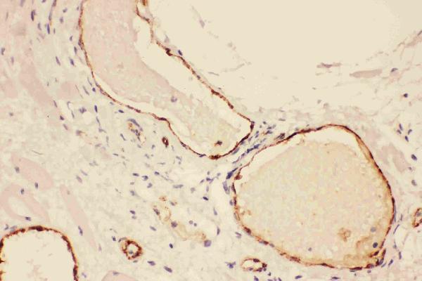
Click image to see more details
Figure 3. IHC analysis of VWF using anti-VWF antibody (PB9062).
VWF was detected in frozen section of human placenta tissue . The tissue section was blocked with 10% goat serum. The tissue section was then incubated with 1μg/ml rabbit anti-VWF Antibody (PB9062) overnight at 4°C. Biotinylated goat anti-rabbit IgG was used as secondary antibody and incubated for 30 minutes at 37°C. The tissue section was developed using Strepavidin-Biotin-Complex (SABC)(Catalog # SA1022) with DAB as the chromogen.

Click image to see more details
Figure 4. Flow Cytometry analysis of A431 cells using anti-VWF antibody (PB9062).
Overlay histogram showing A431 cells stained with PB9062 (Blue line).The cells were blocked with 10% normal goat serum. And then incubated with rabbit anti-VWF Antibody (PB9062, 1μg/1x106 cells) for 30 min at 20°C. DyLight®488 conjugated goat anti-rabbit IgG (BA1127, 5-10μg/1x106 cells) was used as secondary antibody for 30 minutes at 20°C. Isotype control antibody (Green line) was rabbit IgG (1μg/1x106) used under the same conditions. Unlabelled sample (Red line) was also used as a control.
Protein Target Info & Infographic
Gene/Protein Information For VWF (Source: Uniprot.org, NCBI)
Gene Name
VWF
Full Name
von Willebrand factor
Weight
309.265kDa
Alternative Names
coagulation factor VIII VWF; F8; F8VWF; von Willebrand factor; VWD; vWF VWF F8VWF, VWD von Willebrand factor von Willebrand factor|coagulation factor VIII VWF
*If product is indicated to react with multiple species, protein info is based on the gene entry specified above in "Species".For more info on VWF, check out the VWF Infographic

We have 30,000+ of these available, one for each gene! Check them out.
In this infographic, you will see the following information for VWF: database IDs, superfamily, protein function, synonyms, molecular weight, chromosomal locations, tissues of expression, subcellular locations, post-translational modifications, and related diseases, research areas & pathways. If you want to see more information included, or would like to contribute to it and be acknowledged, please contact [email protected].
Specific Publications For Anti-Von Willebrand Factor/VWF Antibody Picoband™ (PB9062)
Hello CJ!
PB9062 has been cited in 34 publications:
*The publications in this section are manually curated by our staff scientists. They may differ from Bioz's machine gathered results. Both are accurate. If you find a publication citing this product but is missing from this list, please let us know we will issue you a thank-you coupon.
Fibroblast growth factor 21 delayed endothelial replicative senescence and protected cells from H2O2-induced premature senescence through SIRT1
Identification and characterization of cancer stem-like cells from primary carcinoma of the cervix uteri
Histological type of oncogenity and expression of cell cycle genes in tumor cells from human mesenchymal stem cells
Paxillin regulates vascular endothelial growth factor A-induced in vitro angiogenesis of human umbilical vein endothelial cells
Effects of propranolol and isoproterenol on infantile hemangioma endothelial cells in vitro
PlexinA1 expression in gastric carcinoma and its relationship with tumor angiogenesis and proliferation
A Three-Dimensional Printed Polycaprolactone Scaffold Combined with Co-Axially Electrospun Vancomycin/Ceftazidime/Bone Morphological Protein-2 Sheath-Core Nanofibers for the Repair of Segmental Bone Defects During the Masquelet Procedure
IFI6 Inhibits Apoptosis via Mitochondrial-Dependent Pathway in Dengue Virus 2 Infected Vascular Endothelial Cells
The protective effects of angelica organic acid against ox-LDL-induced autophagy dysfunction of HUVECs
RoY Peptide-Modified Chitosan-Based Hydrogel to Improve Angiogenesis and Cardiac Repair under Hypoxia
Recommended Resources
Here are featured tools and databases that you might find useful.
- Boster's Pathways Library
- Protein Databases
- Bioscience Research Protocol Resources
- Data Processing & Analysis Software
- Photo Editing Software
- Scientific Literature Resources
- Research Paper Management Tools
- Molecular Biology Software
- Primer Design Tools
- Bioinformatics Tools
- Phylogenetic Tree Analysis
Customer Reviews
Have you used Anti-Von Willebrand Factor/VWF Antibody Picoband™?
Submit a review and receive an Amazon gift card.
- $30 for a review with an image
Be the first to review Anti-Von Willebrand Factor/VWF Antibody Picoband™
*The first user to submit a review for a product is eligible for Boster's Innovating Scientists Reward, which gives product credits. This is in addition to the gift card reward.
Customer Q&As
Have a question?
Find answers in Q&As, reviews.
Can't find your answer?
Submit your question
16 Customer Q&As for Anti-Von Willebrand Factor/VWF Antibody Picoband™
Question
We are currently using anti-Von Willebrand Factor/VWF antibody PB9062 for human tissue, and we are happy with the ICC results. The species of reactivity given in the datasheet says human. Is it likely that the antibody can work on canine tissues as well?
Verified Customer
Verified customer
Asked: 2020-04-20
Answer
The anti-Von Willebrand Factor/VWF antibody (PB9062) has not been tested for cross reactivity specifically with canine tissues, but there is a good chance of cross reactivity. We have an innovator award program that if you test this antibody and show it works in canine you can get your next antibody for free. Please contact me if I can help you with anything.
Boster Scientific Support
Answered: 2020-04-20
Question
Is there a BSA free version of anti-Von Willebrand Factor/VWF antibody PB9062 available?
V. Lewis
Verified customer
Asked: 2020-02-06
Answer
We appreciate your recent telephone inquiry. I can confirm that some lots of this anti-Von Willebrand Factor/VWF antibody PB9062 are BSA free. For now, these lots are available and we can make a BSA free formula for you free of charge. It will take 3 extra days to prepare. If you require this antibody BSA free again in future, please do not hesitate to contact me and I will be pleased to check which lots we have in stock that are BSA free.
Boster Scientific Support
Answered: 2020-02-06
Question
Our team were content with the WB result of your anti-Von Willebrand Factor/VWF antibody. However we have been able to see positive staining in liver secreted. using this antibody. Is that expected? Could you tell me where is VWF supposed to be expressed?
V. Evans
Verified customer
Asked: 2019-10-15
Answer
Based on literature, liver does express VWF. Generally VWF expresses in secreted. Regarding which tissues have VWF expression, here are a few articles citing expression in various tissues:
Liver, Pubmed ID: 19159218
Lung, Pubmed ID: 15489334
Plasma, Pubmed ID: 14760718
Platelet, Pubmed ID: 12665801
Umbilical vein endothelial cell, Pubmed ID: 3495266
Boster Scientific Support
Answered: 2019-10-15
Question
I have a question about product PB9062, anti-Von Willebrand Factor/VWF antibody. I was wondering if it would be possible to conjugate this antibody with biotin. I would need it to be without BSA or sodium azide. I am planning on using a buffer exchange of sodium azide with PBS only. Would there be problems for me to conjugate the antibody and store it in -20 degrees in small aliquots?
Verified Customer
Verified customer
Asked: 2019-09-25
Answer
We suggest not storing this antibody with PBS buffer only in -20 degrees. If you want to store it in -20 degrees it is best to add some cryoprotectant like glycerol. If you want carrier free PB9062 anti-Von Willebrand Factor/VWF antibody, we can provide it to you in a special formula with trehalose and/or glycerol. These molecules will not interfere with conjugation chemistry and provide a good level of protection for the antibody from degradation. Please be sure to specify this in your purchase order.
Boster Scientific Support
Answered: 2019-09-25
Question
We ordered your anti-Von Willebrand Factor/VWF antibody for IHC-P on plasma a few months ago. I am using human, and I plan to use the antibody for Flow Cytometry next. I was wanting to use examining plasma as well as lung in our next experiment. Do you have any suggestion on which antibody would work the best for Flow Cytometry?
Verified Customer
Verified customer
Asked: 2019-08-23
Answer
I took a look at the website and datasheets of our anti-Von Willebrand Factor/VWF antibody and it seems that PB9062 has been validated on human in both IHC-P and Flow Cytometry. Thus PB9062 should work for your application. Our Boster satisfaction guarantee will cover this product for Flow Cytometry in human even if the specific tissue type has not been validated. We do have a comprehensive range of products for Flow Cytometry detection and you can check out our website bosterbio.com to find out more information about them.
Boster Scientific Support
Answered: 2019-08-23
Question
I see that the anti-Von Willebrand Factor/VWF antibody PB9062 works with IHC-P, what is the protocol used to produce the result images on the product page?
Verified Customer
Verified customer
Asked: 2019-08-23
Answer
You can find protocols for IHC-P on the "support/technical resources" section of our navigation menu. If you have any further questions, please send an email to [email protected]
Boster Scientific Support
Answered: 2019-08-23
Question
Is a blocking peptide available for product anti-Von Willebrand Factor/VWF antibody (PB9062)?
Verified Customer
Verified customer
Asked: 2019-06-13
Answer
We do provide the blocking peptide for product anti-Von Willebrand Factor/VWF antibody (PB9062). If you would like to place an order for it please contact [email protected] and make a special request.
Boster Scientific Support
Answered: 2019-06-13
Question
Would anti-Von Willebrand Factor/VWF antibody PB9062 work for IHC-P with liver?
Verified Customer
Verified customer
Asked: 2019-03-21
Answer
According to the expression profile of liver, VWF is highly expressed in liver. So, it is likely that anti-Von Willebrand Factor/VWF antibody PB9062 will work for IHC-P with liver.
Boster Scientific Support
Answered: 2019-03-21
Question
My question regards to test anti-Von Willebrand Factor/VWF antibody PB9062 on human liver for research purposes, then I may be interested in using anti-Von Willebrand Factor/VWF antibody PB9062 for diagnostic purposes as well. Is the antibody suitable for diagnostic purposes?
Verified Customer
Verified customer
Asked: 2018-05-23
Answer
The products we sell, including anti-Von Willebrand Factor/VWF antibody PB9062, are only intended for research use. They would not be suitable for use in diagnostic work. If you have the means to develop a product into diagnostic use, and are interested in collaborating with us and develop our product into an IVD product, please contact us for more discussions.
Boster Scientific Support
Answered: 2018-05-23
Question
I was wanting to use using your anti-Von Willebrand Factor/VWF antibody for gp1b-ix-v activation signalling studies. Has this antibody been tested with western blotting on lung cancer tissue? We would like to see some validation images before ordering.
Verified Customer
Verified customer
Asked: 2018-05-04
Answer
I appreciate your inquiry. This PB9062 anti-Von Willebrand Factor/VWF antibody is tested on human placenta tissue, lung cancer tissue, ht1080 whole cell lysate, a431 cells. It is guaranteed to work for Flow Cytometry, IHC-P, IHC-F, ICC, WB in human. Our Boster guarantee will cover your intended experiment even if the sample type has not been be directly tested.
Boster Scientific Support
Answered: 2018-05-04
Question
I was wanting to use your anti-Von Willebrand Factor/VWF antibody for IHC-P for human liver on frozen tissues, but I want to know if it has been tested for this particular application. Has this antibody been tested and is this antibody a good choice for human liver identification?
Verified Customer
Verified customer
Asked: 2017-09-11
Answer
You can see on the product datasheet, PB9062 anti-Von Willebrand Factor/VWF antibody has been validated for Flow Cytometry, IHC-P, IHC-F, ICC, WB on human tissues. We have an innovator award program that if you test this antibody and show it works in human liver in IHC-frozen, you can get your next antibody for free.
Boster Scientific Support
Answered: 2017-09-11
Question
We have been able to see staining in human plasma. Do you have any suggestions? Is anti-Von Willebrand Factor/VWF antibody supposed to stain plasma positively?
J. Moore
Verified customer
Asked: 2016-08-02
Answer
Based on literature plasma does express VWF. Based on Uniprot.org, VWF is expressed in lung, umbilical vein endothelial cell, platelet, plasma, liver, among other tissues. Regarding which tissues have VWF expression, here are a few articles citing expression in various tissues:
Liver, Pubmed ID: 19159218
Lung, Pubmed ID: 15489334
Plasma, Pubmed ID: 14760718
Platelet, Pubmed ID: 12665801
Umbilical vein endothelial cell, Pubmed ID: 3495266
Boster Scientific Support
Answered: 2016-08-02
Question
Is this PB9062 anti-Von Willebrand Factor/VWF antibody reactive to the isotypes of VWF?
P. Singh
Verified customer
Asked: 2016-04-29
Answer
The immunogen of PB9062 anti-Von Willebrand Factor/VWF antibody is E.coli-derived human VWF recombinant protein (Position: R2535-K2813). Human VWF shares 79% amino acid (aa) sequence identity with mouse VWF. Could you tell me which isotype you are interested in so I can help see if the immunogen is part of this isotype?
Boster Scientific Support
Answered: 2016-04-29
Question
Thanks for helping with my inquiry over the phone. Here are the WB image, lot number and protocol we used for liver using anti-Von Willebrand Factor/VWF antibody PB9062. Let me know if you need anything else.
D. Singh
Verified customer
Asked: 2014-06-18
Answer
I appreciate the data. You have provided everything we needed. Our lab team are working to resolve your inquiry as quickly as possible, and we appreciate your patience and understanding! Please let me know if there is anything you need in the meantime.
Boster Scientific Support
Answered: 2014-06-18
Question
See attached the WB image, lot number and protocol we used for liver using anti-Von Willebrand Factor/VWF antibody PB9062. Please let me know if you require anything else.
E. Li
Verified customer
Asked: 2013-10-22
Answer
Thank you very much for the data. Our lab team are working to resolve this as quickly as possible, and we appreciate your patience and understanding! You have provided everything we needed. Please let me know if there is anything you need in the meantime.
Boster Scientific Support
Answered: 2013-10-22
Question
Will PB9062 anti-Von Willebrand Factor/VWF antibody work on parafin embedded sections? If so, which fixation method do you recommend we use (PFA, paraformaldehyde, other)?
G. Wu
Verified customer
Asked: 2013-08-27
Answer
As indicated on the product datasheet, PB9062 anti-Von Willebrand Factor/VWF antibody as been tested on IHC-P. It is best to use PFA for fixation because it has better tissue penetration ability. PFA needs to be prepared fresh before use. Long term stored PFA turns into formalin, as the PFA molecules congregate and become formalin.
Boster Scientific Support
Answered: 2013-08-27
