This website uses cookies to ensure you get the best experience on our website.


- Table of Contents
Sodium citrate buffer, also known as citrate buffer, is a common reagent used for antigen retrieval in immunohistochemistry. Immunohistochemistry (IHC) is a technique that enables high-quality visualization of proteins in a variety of tissues. IHC is a multi-step process that comprises fixation, embedding, and sectioning of a target tissue followed by incubation with an antibody targeting a protein of interest.
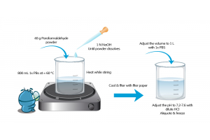
Once the tissue is fixed, embedded in paraffin, and sectioned on slides, researchers can utilize antibodies to target and identify their protein of interest. However, fixation and embedding of tissues, often with 10% formalin and paraffin respectively, initiates cross-linking of proteins that can mask antigen epitopes. Antigen masking may reduce the sensitivity of antibodies and decrease the detection of certain proteins of interest. A process known as antigen retrieval breaks the bonds between formalin and proteins, removing the effects of cross-linking, thereby enabling better detection of proteins. Sodium citrate buffer is highly used in the process of antigen retrieval, specifically for heat-induced antigen retrieval. Boster Bio offers a simple-to-use Sodium Citrate Buffer (Catalog# AR0024) for heat-induced antigen retrieval to remove the effects of antigen masking in your IHC experiments.
Antigen retrieval is a process of breaking the bonds from cross-linking, enabling proteins to interact with antibodies during immunohistochemistry. The purpose of antigen retrieval in immunohistochemistry is to unmask antigens in fixed tissues, which can significantly improve the quality of staining and detection of proteins. While optimization of this method may add additional steps to an already multi-step, complex procedure, antigen retrieval has improved IHC...

Western blotting (also called Protein Immunoblotting) is an analytical technique used to detect specific proteins in the given sample. A deep understanding of the western blot principle provides the essential foundation for applying this method successfully in protein detection. It uses SDS-polyacrylamide gel electrophoresis (SDS-PAGE) to separate various proteins contained in the sample. The separated proteins are then transferred or blotted onto a matrix, where they are stained with antibodies specific to the target protein. Expression details of the target proteins in the given cells or tissue homogenate can then be obtained through analyzing the location and intensity of the specific reaction. Western blotting analysis can detect target protein as low as 1 ng due to high resolution of the gel electrophoresis and strong specificity and high sensitivity of the immunoassay. This method is used in the fields of molecular biology, biochemistry, immunogenetics and other molecular biology disciplines for various experiments. Researchers who prefer standardized, high-sensitivity workflows often rely on a professional western blot service to ensure consistent and reproducible results.
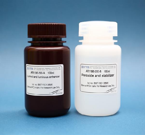
Enhanced Chemiluminescence (ECL) Western Blot Substrate is a very sensitive, non-radioactive, enhanced luminol-based chemiluminescent substrate that allows for easy detection of horseradish peroxidase (HRP) on immunoblots. HRP is a common molecule conjugated to antibodies. ECL Western Blot Substrate has the capability...
Western blot filter paper stabilizes the transfer stack and regulates the buffer flow, helping proteins transfer efficiently from the gel to the membrane. Correct paper choice, hydration, and stacking pressure reduce artifacts and improve band clarity.
Western blotting (also called Protein Immunoblotting) is used to detect specific proteins separated by SDS-polyacrylamide gel electrophoresis (SDS-PAGE) and transferred to a membrane where antibodies bind the target. The workflow includes transfer, blocking, antibody incubation, and detection—each step affecting signal quality and background. For researchers looking to streamline or standardize their workflow, a credible Western Blotting Service can help ensure consistent, high-quality results.
Western blotting works best when the basics are solid and the sample is prepared well. After SDS-PAGE separates the proteins, they’re transferred to a membrane and probed with antibodies that recognize the target. The band position and intensity indicate expression levels in cells or tissue lysates, with sensitivity down to about 1 ng under optimized conditions. Because of its resolution and antibody specificity, the technique is widely used in molecular biology, biochemistry, and immunogenetics for both qualitative and quantitative protein analysis.
Filter paper (or blotting paper) is important to ensure quick and efficient transfer of molecules from the electrophoresis gel to the matrix membrane. It is used for transfer sandwiches and cassettes during Western blotting and assists with transferring proteins unto polyvinylidene fluoride (PVDF membranes), nitrocellulose membranes, and other types of transfer membranes for accurate protein detection. Western blot filter paper is a semi-permeable paper barrier used to separate fine solid particles from liquids or gases.
It can be purchased as pre-cut blotting paper sheets for wet, semi-dry, passive, or electrophoretic transfer of proteins. The thickness of the paper determines the flow rate of the protein or nucleic acid.
During the process of western blotting, Mahmood et al., 2012 des...

According to a report on GEN, 41% of researchers admit that their Western blots are unsuccessful at least 25% of the time.
Yikes! Western blotting (WB) is a widely practiced analytical technique to detect target proteins within samples using antigen-specific antibodies, and when properly optimized, it can be significantly enhanced by a professional Western Blotting Service that ensures reproducibility and high-quality results. A solid grasp of the western blot principle is essential to maximize the success rate and minimize common experimental failures. , while meticulous western blot sample preparation lays the foundation for accurate and reliable protein detection. When it fails to perform as expected, it can really be a downer. Common issues like high background or weak signals often stem from early-stage variables related to western blot optimization.
We’re here to help you succeed. Next time you encounter another problem with Western blot, we’ve compiled a checklist to help you troubleshoot your experiment.
High background frequently occurs due to...

Following protein transfer, it is important to block the unreacted sites on the membrane using inert proteins and/or nonionic detergent to reduce levels of nonspecific protein binding during the assay. For labs aiming to reduce background and streamline results, using a professionally...

Antigen retrieval is essential in immunohistochemistry (IHC) due to protein cross-linking caused by formalin fixation, which masks antigenic sites and hinders antibody binding. Formaldehyde fixation usually generates methylene bridges which cross-link proteins and therefore mask the epitope of interest. It is essential to unmask the antibody...
Nitrocellulose membranes are one of the top matrices used in protein blotting in Western Blotting, offering significant advantages over simple filter paper in terms of protein retention and signal clarity. They have high protein-binding capacity, strong affinity for proteins of varying protein size, compatibility with a variety of protein detection methods, and the ability to immobilize proteins, glycoproteins, or nucleic acids. This variety of detection methods include chemiluminescence, chromogenic, and fluorescence. It is proven to produce excellent signal-to-noise results when used for amino acid analysis, protein sequencing and western, northern, and Southern blotting.
Western blotting (also called Protein Immunoblotting) is an analytical technique used to detect specific proteins in the given sample. A thorough understanding of the western blot principle is crucial to successfully applying this method and accurately interpreting the results, while proper western...

In order to get the best results from your ELISA assay, the dilution factors of the sample and the detection antibodies must be optimized—this principle also applies broadly across many types of assay services where precision and optimization are key. If your sample or antibodies are too concentrated, you risk saturating the a...
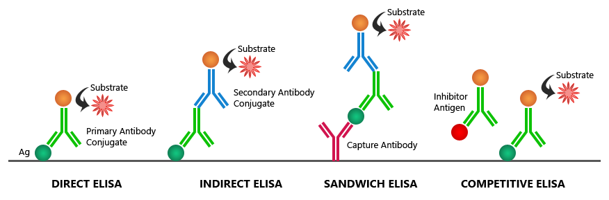
Are you familiar with the multiple methods you could use to perform an ELISA, including the wide range of commercially available elisa kits designed for different targets and sensitivity requirements? Among the standard
If you are having trouble with saturated signals in your ELISA data results, check out this table for Boster’s possible solutions to your problem:
| Possible Causes | Possible Solutions |
|---|---|
| High sample concentration | Use higher sample dilutions (Determine the optimal dilutions by titration assay). In parallel, construct an ELISA standard curve using known concentrations to more accurately map signal intensities to antigen levels. |
| Excessive substrate | Decrease concentration or amount of substrate: Follow manufacturer guidelines (The substrate provided with the ELISA kit might require further dilution) |
| Substrate color changed before use | Make substrate immediately before use |
| Non-specific antibody binding | Try different formulations in coating solutions; Ensure wells are pre-processed to prevent non-specific binding; Use affinity-purified antibody and preferably one that is pre- adsorbed; Use serum (5-10%) from same species as secondary antibody (bovine serum is also recommended). |
| Incubation time too long | Follow the manufacturer guidelines (If the problem persists, try incubating samples at 4°C overnight) |
| Excess antibody | Repeat the assay with lower antibody concentrations to find the optimal one for your experiment |
| Contaminated buffers with metals or HRP | Make and use fresh buffers |