Click image to see more details
Product Info Summary
| SKU: | MA1045 |
|---|---|
| Size: | 100 μg/vial |
| Reactive Species: | Human, Mouse, Pig, Rat |
| Host: | Mouse |
| Application: | IF, IHC, IHC-F, WB |
Customers Who Bought This Also Bought

Product info
Product Name
Anti-GFAP Antibody (Monoclonal, G-A-5)
SKU/Catalog Number
MA1045
Size
100 μg/vial
Form
Lyophilized
Description
Boster Bio Anti-GFAP Antibody (Monoclonal, G-A-5) catalog # MA1045. Tested in IF, IHC, IHC-F, WB applications. This antibody reacts with Human, Mouse, Pig, Rat.
Storage & Handling
Store at -20˚C for one year from date of receipt. After reconstitution, at 4˚C for one month. It can also be aliquotted and stored frozen at -20˚C for six months. Avoid repeated freeze-thaw cycles.
Cite This Product
Anti-GFAP Antibody (Monoclonal, G-A-5) (Boster Biological Technology, Pleasanton CA, USA, Catalog # MA1045)
Host
Mouse
Contents
Mouse ascites fluid, 1.2% sodium acetate, 2mg BSA, with 0.01mg NaN3 as preservative.
Clonality
Monoclonal
Clone Number
G-A-5
Isotype
Mouse IgG1
Immunogen
GFAP from pig spinal cord.
*Blocking peptide can be purchased. Costs vary based on immunogen length. Contact us for pricing.
Cross-reactivity
No cross-reactivity with other proteins
Reactive Species
MA1045 is reactive to GFAP in Human, Mouse, Pig, Rat
Applications
MA1045 is guaranteed for IF, IHC, IHC-F, WB Boster Guarantee
Observed Molecular Weight
50 kDa
Calculated molecular weight
49.88kDa
Background of GFAP
Glial fibrillary acidic protein (GFAP) is an intermediate filament protein of 52Kda. GFAP gene is mapped to human 17q21. GFAP is a useful marker of astroglia in the brain. Mutations in GFAP, encoding glial fibrillary acidic protein, are associated with Alexander disease.
Antibody Validation
Boster validates all antibodies on WB, IHC, ICC, Immunofluorescence, and ELISA with known positive control and negative samples to ensure specificity and high affinity, including thorough antibody incubations.
Innovating Scientists Reward
If you are the first to review this product, or if you have results for a special sample, species or application this product is not validated in, share your results with us and receive product credits you can use towards any Boster products! Applicable to all scientists worldwide.
Submit A Review
Assay dilution & Images
Reconsitution
Add 1ml of PBS buffer will yield a concentration of 100ug/ml.
Assay Dilutions Recommendation
The recommendations below provide a starting point for assay optimization. The actual working concentration varies and should be decided by the user.
Western blot, 0.5-1μg/ml, Human, mouse, pig, rat
Immunohistochemistry (Paraffin-embedded Section), 0.4-1μg/ml, Human, pig, rat, By Heat
Immunohistochemistry (Frozen Section), 0.5-1μg/ml, Human, pig, rat, -
Immunofluorescence, 2μg/ml, Rat
Validation Images & Assay Conditions
Click image to see more details
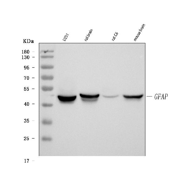
Figure 1. Western blot analysis of GFAP using anti-GFAP antibody (MA1045).
Electrophoresis was performed on a 5-20% SDS-PAGE gel at 70V (Stacking gel) / 90V (Resolving gel) for 2-3 hours. The sample well of each lane was loaded with 30 ug of sample under reducing conditions.
Lane 1: human U251 whole cell lysates,
Lane 2: rat brain tissue lysates,
Lane 3: rat C6 whole cell lysates,
Lane 4: mouse brain tissue lysates.
After electrophoresis, proteins were transferred to a nitrocellulose membrane at 150 mA for 50-90 minutes. Blocked the membrane with 5% non-fat milk/TBS for 1.5 hour at RT. The membrane was incubated with mouse anti-GFAP antigen affinity purified monoclonal antibody (Catalog # MA1045) at 1 μg/mL overnight at 4°C, then washed with TBS-0.1%Tween 3 times with 5 minutes each and probed with a goat anti-mouse IgG-HRP secondary antibody at a dilution of 1:10000 for 1.5 hour at RT. The signal is developed using an Enhanced Chemiluminescent detection (ECL) kit (Catalog # EK1001) with Tanon 5200 system. A specific band was detected for GFAP at approximately 50 kDa. The expected band size for GFAP is at 54 kDa.

Click image to see more details
Figure 2. IF analysis of GFAP using anti-GFAP antibody (MA1045) and anti-MBP antibody (PA1050)
GFAP was detected in paraffin-embedded section of rat brain tissues. Heat mediated antigen retrieval was performed in citrate buffer (pH6 epitope retrieval solution ) for 20 mins. The tissue section was blocked with 10% goat serum. The tissue section was then incubated with 1μg/mL mouse anti-GFAP Antibody (MA1045)and anti-MBP Antibody (PA1050) overnight at 4°C. DyLight®488 Conjugated Goat Anti-Mouse IgG (BA1126) and Cy3 Conjugated Goat Anti-Rabbit IgG (BA1032) were used as secondary antibody at 1:100 dilution and incubated for 30 minutes at 37°C. The section was counterstained with DAPI. Visualize using a fluorescence microscope and filter sets appropriate for the label used.

Click image to see more details
Figure 3. IHC analysis of GFAP using anti-GFAP antibody (MA1045).
GFAP was detected in a paraffin-embedded section of rat brain tissue. Heat mediated antigen retrieval was performed in EDTA buffer (pH 8.0, epitope retrieval solution). The tissue section was blocked with 10% goat serum. The tissue section was then incubated with 1 μg/ml mouse anti-GFAP Antibody (MA1045) overnight at 4°C. Peroxidase Conjugated Goat Anti-mouse IgG was used as secondary antibody and incubated for 30 minutes at 37°C. The tissue section was developed using HRP Conjugated Mouse IgG Super Vision Assay Kit (Catalog # SV0001) with DAB as the chromogen.
Protein Target Info & Infographic
Gene/Protein Information For GFAP (Source: Uniprot.org, NCBI)
Gene Name
GFAP
Full Name
Glial fibrillary acidic protein
Weight
49.88kDa
Superfamily
intermediate filament family
Alternative Names
FLJ45472; GFAP astrocytes; GFAP immunohistochemistry; GFAP mouse; GFAP rabbit; GFAP stain; GFAP; glial fibrillary acidic protein GFAP ALXDRD glial fibrillary acidic protein glial fibrillary acidic protein
*If product is indicated to react with multiple species, protein info is based on the gene entry specified above in "Species".For more info on GFAP, check out the GFAP Infographic

We have 30,000+ of these available, one for each gene! Check them out.
In this infographic, you will see the following information for GFAP: database IDs, superfamily, protein function, synonyms, molecular weight, chromosomal locations, tissues of expression, subcellular locations, post-translational modifications, and related diseases, research areas & pathways. If you want to see more information included, or would like to contribute to it and be acknowledged, please contact [email protected].
Specific Publications For Anti-GFAP Antibody (Monoclonal, G-A-5) (MA1045)
Hello CJ!
MA1045 has been cited in 103 publications:
*The publications in this section are manually curated by our staff scientists. They may differ from Bioz's machine gathered results. Both are accurate. If you find a publication citing this product but is missing from this list, please let us know we will issue you a thank-you coupon.
Label-retaining assay enriches tumor-initiating cells in glioblastoma spheres cultivated in serum-free medium
Xiang Z,Jiang X,Ji R,Yuan H.Enhanced expression of P2X4 purinoceptors in pyramidal neurons of the rat hippocampal CA1 region may be involved ischemia-reperfusion injury.Purinergic Signal.2021 May 9.doi:10.1007/s11302-021-09780-z.Epub ahead of print.PMID:33966147.
Species: Rat
MA1045 usage in article: APP:IF, SAMPLE:BRAIN TISSUE, DILUTION: 1:400
Zhu X,Yao Y, Yang J,Zhang C,Li X,Zhang A,Liu X,Zhang C,Gan G.ADAM10 suppresses demyelination and reduces seizure susceptibility in cuprizone-induced demyelination model.Free Radic Biol Med.2021 May 6:S0891-584 9(21)00282-3.doi:10.1016/j.freeradbiomed.2021.05.001.Epub ahead of print.PMID:33965566.
Species: Mouse
MA1045 usage in article: APP:ICC, SAMPLE:CORPUS CALLOSUM TISSUE, DILUTION:1:100
Xiang S,Zhao D,Hao H,Wang XU,Li L,Yang T.α-Helical protein absorption at post-traumatic epileptic foci monitored by Fourier transform infrared mapping.J Biosci.2020;45:55.PMID:32345781.
Species: Rat
MA1045 usage in article: APP:IHC, SAMPLE:FRONTAL BRAIN LOBE, DILUTION:1:100
Tauroursodeoxycholic Acid Alleviates Secondary Injury in Spinal Cord Injury Mice Through Reducing Oxidative Stress
Authors:Yonghui Hou,Jiyao Luan,Tiancheng Deng et al.
Species: Mouse
MA1045 usage in article: APP:IF, SAMPLE:CORTICAL NEURONS, DILUTION:1:500
Moraes TR,Elisei LS,Malta IH,Galdino G.Participation of CXCL1 in the glial cells during neuropathic pain.Eur J Pharmacol.2020 May 15;875:173039.doi:10.1016/j.ejphar.2020.173039.Epub 2020 Feb 28.PMID:32119843.
Species: Rat
Zhang Y,Zhang M,Zhu W,Pan X,Wang Q,Gao X,Wang C,Zhang X,Liu Y,Li S,Sun H.Role of Elevated Thrombospondin-1 in Kainic Acid-Induced Status Epilepticus.Neurosci Bull.2020 Mar;36(3):263-276.doi:10.1007/s12264-019-00437-x.Epub 2019 Oct 29.PMID:31664678;PMCID:P
Species: Rat
MA1045 usage in article: APP:WB, SAMPLE:HIPPOCAMPUS AND CORTEX, DILUTION:1:1000
Zhang LY,Jin QQ,Hölscher C,Li L.Glucagon-like peptide-1/glucose-dependent insulinotropic polypeptide dual receptor agonist DA-CH5 is superior to exendin-4 in protecting neurons in the 6-hydroxydopamine rat Parkinson model. Neural Regen Res 2021;16:1660-70
Species: Human,Rat
MA1045 usage in article: APP:IF, SAMPLE:SN TISSUE, DILUTION:1:100
Jiangong Wang,Bin Liu,Yong Xu et al. Inhibition of Histamine H3 receptor Attenuates Neuroinflammation and Cognitive Impairments in Alzheimer’s Disease via activating CREB Pathway,16 December 2020, PREPRINT (Version 1) available at Research Square [https:/
Species: Mouse
MA1045 usage in article: APP:IHC, SAMPLE:BRAIN TISSUE, DILUTION:1:200
Ahmed S. Ahmed,JAK-1/STAT-3 pathway mediated role in aging cerebellar cortex degenerative changes of albino wistar rats, Translational Research in Anatomy, 2020,100089,ISSN 2214-854X, https://doi.org/10. 1016/j.tria.2020.100089.
Species: Rat
MA1045 usage in article: APP:IHC, SAMPLE:CEREBELLAR CORTEX, DILUTION:1:20
Recommended Resources
Here are featured tools and databases that you might find useful.
- Boster's Pathways Library
- Protein Databases
- Bioscience Research Protocol Resources
- Data Processing & Analysis Software
- Photo Editing Software
- Scientific Literature Resources
- Research Paper Management Tools
- Molecular Biology Software
- Primer Design Tools
- Bioinformatics Tools
- Phylogenetic Tree Analysis
Customer Reviews
Have you used Anti-GFAP Antibody (Monoclonal, G-A-5)?
Submit a review and receive an Amazon gift card.
- $30 for a review with an image
Be the first to review Anti-GFAP Antibody (Monoclonal, G-A-5)
*The first user to submit a review for a product is eligible for Boster's Innovating Scientists Reward, which gives product credits. This is in addition to the gift card reward.
Customer Q&As
Have a question?
Find answers in Q&As, reviews.
Can't find your answer?
Submit your question
16 Customer Q&As for Anti-GFAP Antibody (Monoclonal, G-A-5)
Question
We are currently using anti-GFAP antibody (Monoclonal, G-A-5) MA1045 for pig tissue, and we are happy with the WB results. The species of reactivity given in the datasheet says human, pig, rat. Is it true that the antibody can work on dog tissues as well?
Verified Customer
Verified customer
Asked: 2020-01-31
Answer
The anti-GFAP antibody (Monoclonal, G-A-5) (MA1045) has not been validated for cross reactivity specifically with dog tissues, though there is a good chance of cross reactivity. We have an innovator award program that if you test this antibody and show it works in dog you can get your next antibody for free. Please contact me if I can help you with anything.
Boster Scientific Support
Answered: 2020-01-31
Question
Is this MA1045 anti-GFAP antibody (Monoclonal, G-A-5) reactive to the isotypes of GFAP?
Verified Customer
Verified customer
Asked: 2020-01-09
Answer
The immunogen of MA1045 anti-GFAP antibody (Monoclonal, G-A-5) is GFAP from pig spinal cord. Could you tell me which isotype you are interested in so I can help see if the immunogen is part of this isotype?
Boster Scientific Support
Answered: 2020-01-09
Question
Thank you for helping with my inquiry over the phone. Here are the WB image, lot number and protocol we used for blood using anti-GFAP antibody (Monoclonal, G-A-5) MA1045. Let me know if you need anything else.
Verified Customer
Verified customer
Asked: 2019-12-23
Answer
We appreciate the data. You have provided everything we needed. Our lab team are working to resolve your inquiry as quickly as possible, and we appreciate your patience and understanding! Please let me know if there is anything you need in the meantime.
Boster Scientific Support
Answered: 2019-12-23
Question
you antibody to test anti-GFAP antibody (Monoclonal, G-A-5) MA1045 on rat blood for research purposes, then I may be interested in using anti-GFAP antibody (Monoclonal, G-A-5) MA1045 for diagnostic purposes as well. Is the antibody suitable for diagnostic purposes?
Verified Customer
Verified customer
Asked: 2019-11-29
Answer
The products we sell, including anti-GFAP antibody (Monoclonal, G-A-5) MA1045, are only intended for research use. They would not be suitable for use in diagnostic work. If you have the means to develop a product into diagnostic use, and are interested in collaborating with us and develop our product into an IVD product, please contact us for more discussions.
Boster Scientific Support
Answered: 2019-11-29
Question
Is a blocking peptide available for product anti-GFAP antibody (Monoclonal, G-A-5) (MA1045)?
Verified Customer
Verified customer
Asked: 2019-10-03
Answer
We do provide the blocking peptide for product anti-GFAP antibody (Monoclonal, G-A-5) (MA1045). If you would like to place an order for it please contact [email protected] and make a special request.
Boster Scientific Support
Answered: 2019-10-03
Question
We have seen staining in rat brain thalamus. Any tips? Is anti-GFAP antibody (Monoclonal, G-A-5) supposed to stain brain thalamus positively?
Verified Customer
Verified customer
Asked: 2019-09-26
Answer
From what I have seen in literature brain thalamus does express GFAP. From what I have seen in Uniprot.org, GFAP is expressed in dorsal motor nucleus of vagus nerve, brain thalamus, brain, kidney, fetal brain cortex, fetal brain, blood, among other tissues. Regarding which tissues have GFAP expression, here are a few articles citing expression in various tissues:
Blood, Pubmed ID: 12837269
Brain, Pubmed ID: 15489334
Brain, and Thalamus, Pubmed ID: 14702039
Fetal brain, Pubmed ID: 12058025
Fetal brain cortex, Pubmed ID: 2780570
Kidney, Pubmed ID: 17974005
Boster Scientific Support
Answered: 2019-09-26
Question
I was wanting to use your anti-GFAP antibody (Monoclonal, G-A-5) for IHC-P for rat blood on frozen tissues, but I want to know if it has been validated for this particular application. Has this antibody been validated and is this antibody a good choice for rat blood identification?
Verified Customer
Verified customer
Asked: 2019-08-05
Answer
It shows on the product datasheet, MA1045 anti-GFAP antibody (Monoclonal, G-A-5) has been tested for IF, IHC-P, IHC-F, WB on human, pig, rat tissues. We have an innovator award program that if you test this antibody and show it works in rat blood in IHC-frozen, you can get your next antibody for free.
Boster Scientific Support
Answered: 2019-08-05
Question
I have attached the WB image, lot number and protocol we used for blood using anti-GFAP antibody (Monoclonal, G-A-5) MA1045. Please let me know if you require anything else.
Verified Customer
Verified customer
Asked: 2019-07-03
Answer
Thank you very much for the data. Our lab team are working to resolve this as quickly as possible, and we appreciate your patience and understanding! You have provided everything we needed. Please let me know if there is anything you need in the meantime.
Boster Scientific Support
Answered: 2019-07-03
Question
We bought anti-GFAP antibody (Monoclonal, G-A-5) for IF on kidney a few years ago. I am using rat, and We intend to use the antibody for IHC-P next. My question regards examining kidney as well as blood in our next experiment. Could you please give me some suggestion on which antibody would work the best for IHC-P?
H. Roberts
Verified customer
Asked: 2018-10-25
Answer
I have checked the website and datasheets of our anti-GFAP antibody (Monoclonal, G-A-5) and it seems that MA1045 has been validated on rat in both IF and IHC-P. Thus MA1045 should work for your application. Our Boster satisfaction guarantee will cover this product for IHC-P in rat even if the specific tissue type has not been validated. We do have a comprehensive range of products for IHC-P detection and you can check out our website bosterbio.com to find out more information about them.
Boster Scientific Support
Answered: 2018-10-25
Question
My question regarding product MA1045, anti-GFAP antibody (Monoclonal, G-A-5). I was wondering if it would be possible to conjugate this antibody with biotin. I would need it to be without BSA or sodium azide. I am planning on using a buffer exchange of sodium azide with PBS only. Would there be problems for me to conjugate the antibody and store it in -20 degrees in small aliquots?
Verified Customer
Verified customer
Asked: 2017-10-05
Answer
We suggest not storing this antibody with PBS buffer only in -20 degrees. If you want to store it in -20 degrees it is best to add some cryoprotectant like glycerol. If you want carrier free MA1045 anti-GFAP antibody (Monoclonal, G-A-5), we can provide it to you in a special formula with trehalose and/or glycerol. These molecules will not interfere with conjugation chemistry and provide a good level of protection for the antibody from degradation. Please be sure to specify this in your purchase order.
Boster Scientific Support
Answered: 2017-10-05
Question
Is there a BSA free version of anti-GFAP antibody (Monoclonal, G-A-5) MA1045 available?
Verified Customer
Verified customer
Asked: 2017-09-18
Answer
I appreciate your recent telephone inquiry. I can confirm that some lots of this anti-GFAP antibody (Monoclonal, G-A-5) MA1045 are BSA free. For now, these lots are available and we can make a BSA free formula for you free of charge. It will take 3 extra days to prepare. If you require this antibody BSA free again in future, please do not hesitate to contact me and I will be pleased to check which lots we have in stock that are BSA free.
Boster Scientific Support
Answered: 2017-09-18
Question
We are interested in using your anti-GFAP antibody (Monoclonal, G-A-5) for r-rno-9613829; chaperone mediated autophagy studies. Has this antibody been tested with western blotting on rat brain tissue? We would like to see some validation images before ordering.
Verified Customer
Verified customer
Asked: 2017-08-03
Answer
I appreciate your inquiry. This MA1045 anti-GFAP antibody (Monoclonal, G-A-5) is validated on rat brain tissue. It is guaranteed to work for IF, IHC-P, IHC-F, WB in human, pig, rat. Our Boster guarantee will cover your intended experiment even if the sample type has not been be directly tested.
Boster Scientific Support
Answered: 2017-08-03
Question
Would MA1045 anti-GFAP antibody (Monoclonal, G-A-5) work on parafin embedded sections? If so, which fixation method do you recommend we use (PFA, paraformaldehyde, other)?
T. Jackson
Verified customer
Asked: 2017-07-13
Answer
It shows on the product datasheet, MA1045 anti-GFAP antibody (Monoclonal, G-A-5) as been validated on IHC-P. It is best to use PFA for fixation because it has better tissue penetration ability. PFA needs to be prepared fresh before use. Long term stored PFA turns into formalin, as the PFA molecules congregate and become formalin.
Boster Scientific Support
Answered: 2017-07-13
Question
I see that the anti-GFAP antibody (Monoclonal, G-A-5) MA1045 works with IHC-P, what is the protocol used to produce the result images on the product page?
Verified Customer
Verified customer
Asked: 2017-05-17
Answer
You can find protocols for IHC-P on the "support/technical resources" section of our navigation menu. If you have any further questions, please send an email to [email protected]
Boster Scientific Support
Answered: 2017-05-17
Question
My colleagues were happy with the WB result of your anti-GFAP antibody (Monoclonal, G-A-5). However we have seen positive staining in fetal brain cortex cytoplasm. using this antibody. Is that expected? Could you tell me where is GFAP supposed to be expressed?
A. Li
Verified customer
Asked: 2016-10-11
Answer
From literature, fetal brain cortex does express GFAP. Generally GFAP expresses in cytoplasm. Regarding which tissues have GFAP expression, here are a few articles citing expression in various tissues:
Blood, Pubmed ID: 12837269
Brain, Pubmed ID: 15489334
Brain, and Thalamus, Pubmed ID: 14702039
Fetal brain, Pubmed ID: 12058025
Fetal brain cortex, Pubmed ID: 2780570
Kidney, Pubmed ID: 17974005
Boster Scientific Support
Answered: 2016-10-11
Question
Will anti-GFAP antibody (Monoclonal, G-A-5) MA1045 work for IHC-P with blood?
L. Wu
Verified customer
Asked: 2015-12-30
Answer
According to the expression profile of blood, GFAP is highly expressed in blood. So, it is likely that anti-GFAP antibody (Monoclonal, G-A-5) MA1045 will work for IHC-P with blood.
Boster Scientific Support
Answered: 2015-12-30
