
Click image to see more details
-

-
-
-
-
+8
Product Info Summary
| SKU: | PB9148 |
|---|---|
| Size: | 100 μg/vial |
| Reactive Species: | Human, Mouse, Rat |
| Host: | Rabbit |
| Application: | ELISA, Flow Cytometry, IF, IHC, IHC-F, ICC, WB |
Customers Who Bought This Also Bought

Product info
Product Name
Anti-MyD88 Antibody Picoband™
SKU/Catalog Number
PB9148
Size
100 μg/vial
Form
Lyophilized
Description
Boster Bio Anti-MyD88 Antibody Picoband™ catalog # PB9148. Tested in ELISA, Flow Cytometry, IF, IHC, IHC-F, ICC, WB applications. This antibody reacts with Human, Mouse, Rat.
Storage & Handling
Store at -20˚C for one year from date of receipt. After reconstitution, at 4˚C for one month. It can also be aliquotted and stored frozen at -20˚C for six months. Avoid repeated freeze-thaw cycles.
Cite This Product
Anti-MyD88 Antibody Picoband™ (Boster Biological Technology, Pleasanton CA, USA, Catalog # PB9148)
Host
Rabbit
Contents
Each vial contains 5mg BSA, 0.9mg NaCl, 0.2mg Na2HPO4, 0.05mg NaN3.
Clonality
Polyclonal
Isotype
Rabbit IgG
Immunogen
E.coli-derived human MyD88 recombinant protein (Position: A44-F264). Human MyD88 shares 84% and 83% amino acid (aa) sequences identity with mouse and rat MyD88, respectively.
*Blocking peptide can be purchased. Costs vary based on immunogen length. Contact us for pricing.
Cross-reactivity
No cross-reactivity with other proteins
Reactive Species
PB9148 is reactive to PTGS2 in Human, Mouse, Rat
Applications
PB9148 is guaranteed for ELISA, Flow Cytometry, IF, IHC, IHC-F, ICC, WB Boster Guarantee
Observed Molecular Weight
33 kDa
Calculated molecular weight
68.996kDa
Background of COX-2
MYD88 (MYELOID DIFFERENTIATION PRIMARY RESPONSE GENE 88), is a protein that, in humans, is encoded by the MYD88 gene. MyD88 is a key downstream adapter for most Toll-like receptors (TLRs) and interleukin-1 receptors (IL1Rs). And it is mapped on 3p22.2. MYD88 encodes a cytosolic adapter protein that plays a central role in the innate and adaptive immune response. This protein functions as an essential signal transducer in the interleukin-1 and Toll-like receptor signaling pathways. Overexpression of MYD88 caused an increase in the level of transcription from the interleukin-8 promoter. The C-terminal domain of MYD88 has significant sequence similarity to the cytoplasmic domain of IL1RAP. Inhibiting the IL1R-MYD88 pathway in vivo could block the damage from acute inflammation that occurs in response to sterile cell death, and do so in a way that might not compromise tissue repair or host defense against pathogens.
Antibody Validation
Boster validates all antibodies on WB, IHC, ICC, Immunofluorescence, and ELISA with known positive control and negative samples to ensure specificity and high affinity, including thorough antibody incubations.
Innovating Scientists Reward
If you are the first to review this product, or if you have results for a special sample, species or application this product is not validated in, share your results with us and receive product credits you can use towards any Boster products! Applicable to all scientists worldwide.
Submit A Review
Assay dilution & Images
Reconsitution
Add 0.2ml of distilled water will yield a concentration of 500ug/ml.
Assay Dilutions Recommendation
The recommendations below provide a starting point for assay optimization. The actual working concentration varies and should be decided by the user.
Western blot, 0.1-0.5μg/ml
Immunohistochemistry (Paraffin-embedded Section), 0.5-1μg/ml
Immunohistochemistry (Frozen Section), 0.5-1μg/ml
Immunocytochemistry, 0.5-1μg/ml
Immunofluorescence, 2μg/ml
Flow Cytometry, 1-3μg/1x106 cells
ELISA (Cap), 1-5μg/ml
Validation Images & Assay Conditions
Click image to see more details
Figure 1. Western blot analysis of MYD88 using anti-MYD88 antibody (PB9148).
Electrophoresis was performed on a 5-20% SDS-PAGE gel at 70V (Stacking gel) / 90V (Resolving gel) for 2-3 hours.
Lane 1: Recombinant Human MYD88 Protein 0.5ng.
After Electrophoresis, proteins were transferred to a Nitrocellulose membrane at 150mA for 50-90 minutes. Blocked the membrane with 5% Non-fat Milk/ TBS for 1.5 hour at RT. The membrane was incubated with rabbit anti-MYD88 antigen affinity purified polyclonal antibody (Catalog # PB9148) at 0.5 μg/mL overnight at 4°C, then washed with TBS-0.1%Tween 3 times with 5 minutes each and probed with a goat anti-rabbit IgG-HRP secondary antibody at a dilution of 1:10000 for 1.5 hour at RT. The signal is developed using an Enhanced Chemiluminescent detection (ECL) kit (Catalog # EK1002) with Tanon 5200 system. A specific band was detected for MYD88 at approximately 49KD. The expected band size for MYD88 is at 49KD.

Click image to see more details
Figure 10. IF analysis of MYD88 using anti-MYD88 antibody (PB9148)
MYD88 was detected in paraffin-embedded section of human mammary cancar tissues. Heat mediated antigen retrieval was performed in citrate buffer (pH6, epitope retrieval solution ) for 20 mins. The tissue section was blocked with 10% goat serum. The tissue section was then incubated with 1μg/mL rabbit anti-MYD88 Antibody (PB9148) overnight at 4°C. Cy3 Conjugated Goat Anti-Rabbit IgG (BA1032) was used as secondary antibody at 1:100 dilution and incubated for 30 minutes at 37°C. The section was counterstained with DAPI. Visualize using a fluorescence microscope and filter sets appropriate for the label used.

Click image to see more details
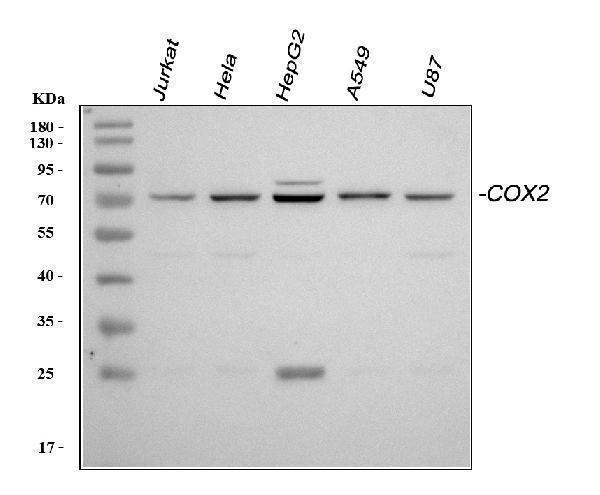
Figure 2. Western blot analysis of MYD88 using anti-MYD88 antibody (PB9148).
Electrophoresis was performed on a 5-20% SDS-PAGE gel at 70V (Stacking gel) / 90V (Resolving gel) for 2-3 hours. The sample well of each lane was loaded with 50ug of sample under reducing conditions.
Lane 1: Rat Cardiac Muscle Tissue Lysate
Lane 2: HELA Whole Cell Lysate
Lane 3: MCF Whole Cell Lysate
Lane 4: HEPG2 Whole Cell Lysate
Lane 5: JURKAT Whole Cell Lysate
Lane 6: RAJI Whole Cell Lysate
After Electrophoresis, proteins were transferred to a Nitrocellulose membrane at 150mA for 50-90 minutes. Blocked the membrane with 5% Non-fat Milk/ TBS for 1.5 hour at RT. The membrane was incubated with rabbit anti-MYD88 antigen affinity purified polyclonal antibody (Catalog # PB9148) at 0.5 μg/mL overnight at 4°C, then washed with TBS-0.1%Tween 3 times with 5 minutes each and probed with a goat anti-rabbit IgG-HRP secondary antibody at a dilution of 1:10000 for 1.5 hour at RT. The signal is developed using an Enhanced Chemiluminescent detection (ECL) kit (Catalog # EK1002) with Tanon 5200 system. A specific band was detected for MYD88 at approximately 33KD. The expected band size for MYD88 is at 33KD.

Click image to see more details
Figure 3. IHC analysis of MYD88 using anti-MYD88 antibody (PB9148).
MYD88 was detected in paraffin-embedded section of rat lung tissue. Heat mediated antigen retrieval was performed in citrate buffer (pH6, epitope retrieval solution) for 20 mins. The tissue section was blocked with 10% goat serum. The tissue section was then incubated with 1μg/ml rabbit anti-MYD88 Antibody (PB9148) overnight at 4°C. Biotinylated goat anti-rabbit IgG was used as secondary antibody and incubated for 30 minutes at 37°C. The tissue section was developed using Strepavidin-Biotin-Complex (SABC)(Catalog # SA1022) with DAB as the chromogen.

Click image to see more details
Figure 4. IHC analysis of MYD88 using anti-MYD88 antibody (PB9148).
MYD88 was detected in paraffin-embedded section of human intestinal cancer tissue. Heat mediated antigen retrieval was performed in citrate buffer (pH6, epitope retrieval solution) for 20 mins. The tissue section was blocked with 10% goat serum. The tissue section was then incubated with 1μg/ml rabbit anti-MYD88 Antibody (PB9148) overnight at 4°C. Biotinylated goat anti-rabbit IgG was used as secondary antibody and incubated for 30 minutes at 37°C. The tissue section was developed using Strepavidin-Biotin-Complex (SABC)(Catalog # SA1022) with DAB as the chromogen.

Click image to see more details
Figure 5. IHC analysis of MYD88 using anti-MYD88 antibody (PB9148).
MYD88 was detected in paraffin-embedded section of mouse spleen tissue. Heat mediated antigen retrieval was performed in citrate buffer (pH6, epitope retrieval solution) for 20 mins. The tissue section was blocked with 10% goat serum. The tissue section was then incubated with 1μg/ml rabbit anti-MYD88 Antibody (PB9148) overnight at 4°C. Biotinylated goat anti-rabbit IgG was used as secondary antibody and incubated for 30 minutes at 37°C. The tissue section was developed using Strepavidin-Biotin-Complex (SABC)(Catalog # SA1022) with DAB as the chromogen.

Click image to see more details
Figure 6. IF analysis of MYD88 using anti-MYD88 antibody (PB9148)
MYD88 was detected in paraffin-embedded section of human tonsil tissues. Heat mediated antigen retrieval was performed in citrate buffer (pH6, epitope retrieval solution ) for 20 mins. The tissue section was blocked with 10% goat serum. The tissue section was then incubated with 1μg/mL rabbit anti-MYD88 Antibody (PB9148) overnight at 4°C. Cy3 Conjugated Goat Anti-Rabbit IgG (BA1032) was used as secondary antibody at 1:100 dilution and incubated for 30 minutes at 37°C. The section was counterstained with DAPI. Visualize using a fluorescence microscope and filter sets appropriate for the label used.

Click image to see more details
Figure 7. IF analysis of MYD88 using anti-MYD88 antibody (PB9148)
MYD88 was detected in paraffin-embedded section of human colon cancar tissues. Heat mediated antigen retrieval was performed in citrate buffer (pH6, epitope retrieval solution ) for 20 mins. The tissue section was blocked with 10% goat serum. The tissue section was then incubated with 1μg/mL rabbit anti-MYD88 Antibody (PB9148) overnight at 4°C. Cy3 Conjugated Goat Anti-Rabbit IgG (BA1032) was used as secondary antibody at 1:100 dilution and incubated for 30 minutes at 37°C. The section was counterstained with DAPI. Visualize using a fluorescence microscope and filter sets appropriate for the label used.

Click image to see more details
Figure 8. IF analysis of MYD88 using anti-MYD88 antibody (PB9148)
MYD88 was detected in paraffin-embedded section of human colon cancar tissues. Heat mediated antigen retrieval was performed in citrate buffer (pH6, epitope retrieval solution ) for 20 mins. The tissue section was blocked with 10% goat serum. The tissue section was then incubated with 1μg/mL rabbit anti-MYD88 Antibody (PB9148) overnight at 4°C. Cy3 Conjugated Goat Anti-Rabbit IgG (BA1032) was used as secondary antibody at 1:100 dilution and incubated for 30 minutes at 37°C. The section was counterstained with DAPI. Visualize using a fluorescence microscope and filter sets appropriate for the label used.

Click image to see more details
Figure 9. IF analysis of MYD88 using anti-MYD88 antibody (PB9148)
MYD88 was detected in paraffin-embedded section of human mammary cancar tissues. Heat mediated antigen retrieval was performed in citrate buffer (pH6, epitope retrieval solution ) for 20 mins. The tissue section was blocked with 10% goat serum. The tissue section was then incubated with 1μg/mL rabbit anti-MYD88 Antibody (PB9148) overnight at 4°C. Cy3 Conjugated Goat Anti-Rabbit IgG (BA1032) was used as secondary antibody at 1:100 dilution and incubated for 30 minutes at 37°C. The section was counterstained with DAPI. Visualize using a fluorescence microscope and filter sets appropriate for the label used.

Click image to see more details
Figure 11. IF analysis of MYD88 using anti-MYD88 antibody (PB9148)
MYD88 was detected in paraffin-embedded section of human placenta tissues. Heat mediated antigen retrieval was performed in citrate buffer (pH6, epitope retrieval solution ) for 20 mins. The tissue section was blocked with 10% goat serum. The tissue section was then incubated with 1μg/mL rabbit anti-MYD88 Antibody (PB9148) overnight at 4°C. Cy3 Conjugated Goat Anti-Rabbit IgG (BA1032) was used as secondary antibody at 1:100 dilution and incubated for 30 minutes at 37°C. The section was counterstained with DAPI. Visualize using a fluorescence microscope and filter sets appropriate for the label used.

Click image to see more details
Figure 12. Flow Cytometry analysis of A549 cells using anti-MYD88 antibody (PB9148).
Overlay histogram showing A549 cells stained with PB9148 (Blue line).The cells were blocked with 10% normal goat serum. And then incubated with rabbit anti-MYD88 Antibody (PB9148, 1μg/1x106 cells) for 30 min at 20°C. DyLight®488 conjugated goat anti-rabbit IgG (BA1127, 5-10μg/1x106 cells) was used as secondary antibody for 30 minutes at 20°C. Isotype control antibody (Green line) was rabbit IgG (1μg/1x106) used under the same conditions. Unlabelled sample (Red line) was also used as a control.
Protein Target Info & Infographic
Gene/Protein Information For PTGS2 (Source: Uniprot.org, NCBI)
Gene Name
PTGS2
Full Name
Prostaglandin G/H synthase 2
Weight
68.996kDa
Superfamily
prostaglandin G/H synthase family
Alternative Names
COX2; COX-2; COX2cyclooxygenase 2b; cyclooxygenase-2; EC 1.14.99; EC 1.14.99.1; GRIPGHS; hCox-2; PGG/HS; PGH synthase 2; PGHS-2; PHS II; PHS-2; PHS-II; prostaglandin G/H synthase 2; prostaglandin G/H synthase and cyclooxygenase; Prostaglandin H2 synthase 2; prostaglandin-endoperoxide synthase 2 (prostaglandin G/H synthase andcyclooxygenase); Prostaglandin-endoperoxide synthase 2; PTGS2 PTGS2 COX-2, COX2, GRIPGHS, PGG/HS, PGHS-2, PHS-2, hCox-2 prostaglandin-endoperoxide synthase 2 prostaglandin G/H synthase 2|PGH synthase 2|PHS II|cyclooxygenase 2|cyclooxygenase 2b|prostaglandin H2 synthase 2|prostaglandin-endoperoxide synthase 2 (prostaglandin G/H synthase and cyclooxygenase)
*If product is indicated to react with multiple species, protein info is based on the gene entry specified above in "Species".For more info on PTGS2, check out the PTGS2 Infographic

We have 30,000+ of these available, one for each gene! Check them out.
In this infographic, you will see the following information for PTGS2: database IDs, superfamily, protein function, synonyms, molecular weight, chromosomal locations, tissues of expression, subcellular locations, post-translational modifications, and related diseases, research areas & pathways. If you want to see more information included, or would like to contribute to it and be acknowledged, please contact [email protected].
Specific Publications For Anti-MyD88 Antibody Picoband™ (PB9148)
Hello CJ!
PB9148 has been cited in 23 publications:
*The publications in this section are manually curated by our staff scientists. They may differ from Bioz's machine gathered results. Both are accurate. If you find a publication citing this product but is missing from this list, please let us know we will issue you a thank-you coupon.
Puerarin protects brain tissue against cerebral ischemia/reperfusion injury by inhibiting the inflammatory response
Gastroprotective Effects of Periplaneta americana L. Extract Against Ethanol-Induced Gastric Ulcer in Mice by Suppressing Apoptosis-Related Pathways.
Combination of fasudil and celecoxib promotes the recovery of injured spinal cord in rats better than celecoxib or fasudil alone
Puerarin protects brain tissue against cerebral ischemia/reperfusion injury by inhibiting the inflammatory response
Effect of cyclooxygenase-2 inhibition on the development of post-traumatic stress disorder in rats
MicroRNA-101 regulates the viability and invasion of cervical cancer cells
Granulocyte Macrophage-Colony Stimulating Factor (GM-CSF) Downregulates the Expression of Protumor Factors Cyclooxygenase-2 and Inducible Nitric Oxide Synthase in a GM-CSF Receptor-Independent Manner in Cervical Cancer Cells
Odontogenic epithelial proliferation is correlated with COX-2 expression in dentigerous cyst and ameloblastoma
Anti-inflammatory Effects of Phyllanthus emblica L on Benzopyrene-Induced Precancerous Lung Lesion by Regulating the IL-1?/miR-101/Lin28B Signaling Pathway
miR-144 and targets, c-fos and cyclooxygenase-2 (COX2), modulate synthesis of PGE2 in the amnion during pregnancy and labor
Recommended Resources
Here are featured tools and databases that you might find useful.
- Boster's Pathways Library
- Protein Databases
- Bioscience Research Protocol Resources
- Data Processing & Analysis Software
- Photo Editing Software
- Scientific Literature Resources
- Research Paper Management Tools
- Molecular Biology Software
- Primer Design Tools
- Bioinformatics Tools
- Phylogenetic Tree Analysis
Customer Reviews
Have you used Anti-MyD88 Antibody Picoband™?
Submit a review and receive an Amazon gift card.
- $30 for a review with an image
Be the first to review Anti-MyD88 Antibody Picoband™
*The first user to submit a review for a product is eligible for Boster's Innovating Scientists Reward, which gives product credits. This is in addition to the gift card reward.
Customer Q&As
Have a question?
Find answers in Q&As, reviews.
Can't find your answer?
Submit your question
5 Customer Q&As for Anti-MyD88 Antibody Picoband™
Question
We have seen staining in rat pancreas. Are there any suggestions? Is anti-MyD88 antibody supposed to stain pancreas positively?
Verified Customer
Verified customer
Asked: 2020-05-04
Answer
From what I have seen in literature pancreas does express MYD88. From what I have seen in Uniprot.org, MYD88 is expressed in blood, dendritic cell, epidermal carcinoma, umbilical cord blood, pancreas, erythroleukemia, among other tissues. Regarding which tissues have MYD88 expression, here are a few articles citing expression in various tissues:
Dendritic cell, Pubmed ID: 8957090
Epidermal carcinoma, Pubmed ID: 9013863
Erythroleukemia, Pubmed ID: 23186163
Pancreas, Pubmed ID: 15489334
Umbilical cord blood, Pubmed ID: 14702039
Boster Scientific Support
Answered: 2020-05-04
Question
We need using your anti-MyD88 antibody for cellular response to lead ion studies. Has this antibody been tested with western blotting on cardiac muscle tissue? We would like to see some validation images before ordering.
Verified Customer
Verified customer
Asked: 2019-11-08
Answer
We appreciate your inquiry. This PB9148 anti-MyD88 antibody is tested on rat lung tissue, cardiac muscle tissue, tissue lysate, hela whole cell lysate, mcf whole cell lysate, hepg2 whole cell lysate, jurkat whole cell lysate, raji whole cell lysate, intestinal cancer tissue, mouse spleen tissue, a549 cells. It is guaranteed to work for ELISA, Flow Cytometry, IF, IHC-P, IHC-F, ICC, WB in human, mouse, rat. Our Boster guarantee will cover your intended experiment even if the sample type has not been be directly tested.
Boster Scientific Support
Answered: 2019-11-08
Question
My team were well pleased with the WB result of your anti-MyD88 antibody. However we have been able to see positive staining in erythroleukemia cytoplasm using this antibody. Is that expected? Could you tell me where is MYD88 supposed to be expressed?
Verified Customer
Verified customer
Asked: 2019-08-26
Answer
Based on literature, erythroleukemia does express MYD88. Generally MYD88 expresses in cytoplasm. Regarding which tissues have MYD88 expression, here are a few articles citing expression in various tissues:
Dendritic cell, Pubmed ID: 8957090
Epidermal carcinoma, Pubmed ID: 9013863
Erythroleukemia, Pubmed ID: 23186163
Pancreas, Pubmed ID: 15489334
Umbilical cord blood, Pubmed ID: 14702039
Boster Scientific Support
Answered: 2019-08-26
Question
We are currently using anti-MyD88 antibody PB9148 for mouse tissue, and we are content with the Flow Cytometry results. The species of reactivity given in the datasheet says human, mouse, rat. Is it possible that the antibody can work on dog tissues as well?
L. Taylor
Verified customer
Asked: 2016-03-04
Answer
The anti-MyD88 antibody (PB9148) has not been tested for cross reactivity specifically with dog tissues, though there is a good chance of cross reactivity. We have an innovator award program that if you test this antibody and show it works in dog you can get your next antibody for free. Please contact me if I can help you with anything.
Boster Scientific Support
Answered: 2016-03-04
Question
We have tried in the past anti-MyD88 antibody for WB on pancreas in a previous project. I am using rat, and We intend to use the antibody for ELISA next. We need examining pancreas as well as epidermal carcinoma in our next experiment. Could give a recommendation on which antibody would work the best for ELISA?
G. Kulkarni
Verified customer
Asked: 2015-06-30
Answer
I took a look at the website and datasheets of our anti-MyD88 antibody and it appears that PB9148 has been tested on rat in both WB and ELISA. Thus PB9148 should work for your application. Our Boster satisfaction guarantee will cover this product for ELISA in rat even if the specific tissue type has not been validated. We do have a comprehensive range of products for ELISA detection and you can check out our website bosterbio.com to find out more information about them.
Boster Scientific Support
Answered: 2015-06-30
