Click image to see more details
-

-
-
-
-
+6
Product Info Summary
| SKU: | M00300-1 |
|---|---|
| Size: | 100 μg/vial |
| Reactive Species: | Human, Mouse, Rat |
| Host: | Mouse |
| Application: | Flow Cytometry, IF, IHC, ICC, WB |
Customers Who Bought This Also Bought


Product info
Product Name
Anti-SQSTM1/p62 Antibody Picoband™ (monoclonal, 3H11)
View all p62/SQSTM1 Antibodies
SKU/Catalog Number
M00300-1
Size
100 μg/vial
Form
Lyophilized
Description
Boster Bio Anti-SQSTM1/p62 Antibody Picoband™ (monoclonal, 3H11) catalog # M00300-1. Tested in Flow Cytometry, IF, IHC, ICC, WB applications. This antibody reacts with Human, Mouse, Rat.
Storage & Handling
Store at -20˚C for one year from date of receipt. After reconstitution, at 4˚C for one month. It can also be aliquotted and stored frozen at -20˚C for six months. Avoid repeated freeze-thaw cycles.
Cite This Product
Anti-SQSTM1/p62 Antibody Picoband™ (monoclonal, 3H11) (Boster Biological Technology, Pleasanton CA, USA, Catalog # M00300-1)
Host
Mouse
Contents
Each vial contains 4mg Trehalose, 0.9mg NaCl, 0.2mg Na2HPO4, 0.05mg NaN3.
Clonality
Monoclonal
Clone Number
3H11
Isotype
Mouse IgG2a
Immunogen
A synthetic peptide corresponding to a sequence at the N-terminus of human SQSTM1/p62, identical to the related mouse and rat sequences.
*Blocking peptide can be purchased. Costs vary based on immunogen length. Contact us for pricing.
Cross-reactivity
No cross-reactivity with other proteins.
Reactive Species
M00300-1 is reactive to SQSTM1 in Human, Mouse, Rat
Applications
M00300-1 is guaranteed for Flow Cytometry, IF, IHC, ICC, WB Boster Guarantee
Observed Molecular Weight
62 kDa
Calculated molecular weight
47.687kDa
Background of p62/SQSTM1
SQSTM1 (Sequestosome-1), also known as Ubiquitin-Binding Protein P62 or P62, is a protein that in humans is encoded by the SQSTM1 gene. The Src homology type 2 (SH2) domain is a highly conserved motif of about 100 amino acids which mediates protein-protein interactions by binding to phosphotyrosine.p56-lck, a T-cell-specific src family tyrosine kinase with an SH2 domain, is involved in T-cell signal transduction. The International Radiation Hybrid Mapping Consortium mapped the p62 gene to chromosome 5q35. Park et al. (1995) found that the p56-lck SH2 domain binds to p62 at the ser59 of p62 only when that serine is phosphorylated. Joung et al. (1996) expressed epitope-tagged p62 in Hela cells and showed that the expressed protein bound to the lck SH2 domain and that this binding was dependent on the N-terminal 50 amino acids of p62 but not on the tyrosine residue in this region.
Antibody Validation
Boster validates all antibodies on WB, IHC, ICC, Immunofluorescence, and ELISA with known positive control and negative samples to ensure specificity and high affinity, including thorough antibody incubations.
Innovating Scientists Reward
If you are the first to review this product, or if you have results for a special sample, species or application this product is not validated in, share your results with us and receive product credits you can use towards any Boster products! Applicable to all scientists worldwide.
Submit A Review
Assay dilution & Images
Reconsitution
Add 0.2ml of distilled water will yield a concentration of 500μg/ml.
Assay Dilutions Recommendation
The recommendations below provide a starting point for assay optimization. The actual working concentration varies and should be decided by the user.
Western blot, 0.1-0.5μg/ml, Human, Mouse, Rat
Immunohistochemistry (Paraffin-embedded Section), 0.5-1μg/ml, Human, Mouse, Rat, By Heat
Immunocytochemistry/Immunofluorescence, 2μg/ml, Human
Flow Cytometry, 1-3μg/1x106 cells, Human
Validation Images & Assay Conditions
Click image to see more details
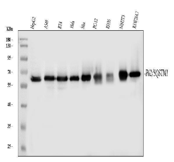
Figure 1. Western blot analysis of SQSTM1 using anti-SQSTM1 antibody (M00300-1).
Electrophoresis was performed on a 5-20% SDS-PAGE gel at 70V (Stacking gel) / 90V (Resolving gel) for 2-3 hours. The sample well of each lane was loaded with 30 ug of sample under reducing conditions.
Lane 1: human HepG2 whole cell lysates,
Lane 2: human A549 whole cell lysates,
Lane 3: human RT4 whole cell lysates,
Lane 4: human Hela whole cell lysates,
Lane 5: human SiHa whole cell lysates,
Lane 6: rat PC-12 whole cell lysates,
Lane 7: rat RH35 whole cell lysates,
Lane 8: mouse NIH/3T3 whole cell lysates,
Lane 9: mouse RAW264.7 whole cell lysates.
After electrophoresis, proteins were transferred to a nitrocellulose membrane at 150 mA for 50-90 minutes. Blocked the membrane with 5% non-fat milk/TBS for 1.5 hour at RT. The membrane was incubated with mouse anti-SQSTM1 antigen affinity purified monoclonal antibody (Catalog # M00300-1) at 0.5 μg/mL overnight at 4°C, then washed with TBS-0.1%Tween 3 times with 5 minutes each and probed with a goat anti-mouse IgG-HRP secondary antibody at a dilution of 1:10000 for 1.5 hour at RT. The signal is developed using an Enhanced Chemiluminescent detection (ECL) kit (Catalog # EK1001) with Tanon 5200 system. A specific band was detected for SQSTM1 at approximately 62 kDa. The expected band size for SQSTM1 is at 48 kDa.

Click image to see more details
Figure 2. IHC analysis of SQSTM1 using anti-SQSTM1 antibody (M00300-1).
SQSTM1 was detected in section of rat intestine tissues. Heat mediated antigen retrieval was performed in citrate buffer (pH6, epitope retrieval solution) for 20 mins. The tissue section was blocked with 10% goat serum. The tissue section was then incubated with μg/ml mouse anti-SQSTM1 Antibody (M00300-1) overnight at 4°C. Biotinylated goat anti-mouse IgG was used as secondary antibody and incubated for 30 minutes at 37°C. The tissue section was developed using Strepavidin-Biotin-Complex (SABC)(Catalog # SA1021) with DAB as the chromogen.

Click image to see more details
Figure 3. IHC analysis of SQSTM1 using anti-SQSTM1 antibody (M00300-1).
SQSTM1 was detected in section of human colon cancer tissues. Heat mediated antigen retrieval was performed in citrate buffer (pH6, epitope retrieval solution) for 20 mins. The tissue section was blocked with 10% goat serum. The tissue section was then incubated with μg/ml mouse anti-SQSTM1 Antibody (M00300-1) overnight at 4°C. Biotinylated goat anti-mouse IgG was used as secondary antibody and incubated for 30 minutes at 37°C. The tissue section was developed using Strepavidin-Biotin-Complex (SABC)(Catalog # SA1021) with DAB as the chromogen.

Click image to see more details
Figure 4. IHC analysis of SQSTM1 using anti-SQSTM1 antibody (M00300-1).
SQSTM1 was detected in section of human mammary cancer tissues. Heat mediated antigen retrieval was performed in citrate buffer (pH6, epitope retrieval solution) for 20 mins. The tissue section was blocked with 10% goat serum. The tissue section was then incubated with μg/ml mouse anti-SQSTM1 Antibody (M00300-1) overnight at 4°C. Biotinylated goat anti-mouse IgG was used as secondary antibody and incubated for 30 minutes at 37°C. The tissue section was developed using Strepavidin-Biotin-Complex (SABC)(Catalog # SA1021) with DAB as the chromogen.

Click image to see more details
Figure 5. IHC analysis of SQSTM1 using anti-SQSTM1 antibody (M00300-1).
SQSTM1 was detected in section of human lung cancer tissues. Heat mediated antigen retrieval was performed in citrate buffer (pH6, epitope retrieval solution) for 20 mins. The tissue section was blocked with 10% goat serum. The tissue section was then incubated with μg/ml mouse anti-SQSTM1 Antibody (M00300-1) overnight at 4°C. Biotinylated goat anti-mouse IgG was used as secondary antibody and incubated for 30 minutes at 37°C. The tissue section was developed using Strepavidin-Biotin-Complex (SABC)(Catalog # SA1021) with DAB as the chromogen.

Click image to see more details
Figure 6. IHC analysis of SQSTM1 using anti-SQSTM1 antibody (M00300-1).
SQSTM1 was detected in section of mouse intestine tissues. Heat mediated antigen retrieval was performed in citrate buffer (pH6, epitope retrieval solution) for 20 mins. The tissue section was blocked with 10% goat serum. The tissue section was then incubated with μg/ml mouse anti-SQSTM1 Antibody (M00300-1) overnight at 4°C. Biotinylated goat anti-mouse IgG was used as secondary antibody and incubated for 30 minutes at 37°C. The tissue section was developed using Strepavidin-Biotin-Complex (SABC)(Catalog # SA1021) with DAB as the chromogen.

Click image to see more details
Figure 7. IF analysis of SQSTM1 using anti- SQSTM1 antibody (M00300-1).
SQSTM1 was detected in immunocytochemical section of MCF7 cells. Enzyme antigen retrieval was performed using IHC enzyme antigen retrieval reagent (AR0022) for 15 mins. The cells were blocked with 10% goat serum. And then incubated with 2μg/mL mouse anti- SQSTM1 Antibody (M00300-1) overnight at 4°C. DyLight®488 Conjugated Goat Anti-Mouse IgG (BA1126) was used as secondary antibody at 1:100 dilution and incubated for 30 minutes at 37°C. The section was counterstained with DAPI. Visualize using a fluorescence microscope and filter sets appropriate for the label used.

Click image to see more details
Figure 8. IF analysis of SQSTM1 using anti-SQSTM1 antibody (M00300-1).
SQSTM1 was detected in immunocytochemical section of A431 cell. Enzyme antigen retrieval was performed using IHC enzyme antigen retrieval reagent (AR0022) for 15 mins. The cells were blocked with 10% goat serum. And then incubated with 2μg/mL mouse anti-SQSTM1 Antibody (M00300-1) overnight at 4°C. DyLight®488 Conjugated Goat Anti-mouse IgG (BA1126) was used as secondary antibody at 1:100 dilution and incubated for 30 minutes at 37°C. The section was counterstained with DAPI. Visualize using a fluorescence microscope and filter sets appropriate for the label used.

Click image to see more details
Figure 9. Flow Cytometry analysis of A549 cells using anti-SQSTM1 antibody (M00300-1).
Overlay histogram showing A549 cells stained with M00300-1 (Blue line).The cells were blocked with 10% normal goat serum. And then incubated with mouse anti-SQSTM1 Antibody (M00300-1,1μg/1x106 cells) for 30 min at 20°C. DyLight®488 conjugated goat anti-mouse IgG (BA1126, 5-10μg/1x106 cells) was used as secondary antibody for 30 minutes at 20°C. Isotype control antibody (Green line) was mouse IgG (1μg/1x106) used under the same conditions. Unlabelled sample (Red line) was also used as a control.

Click image to see more details
Figure 10. Flow Cytometry analysis of PC-3 cells using anti-SQSTM1 antibody (M00300-1).
Overlay histogram showing PC-3 cells stained with M00300-1 (Blue line).The cells were blocked with 10% normal goat serum. And then incubated with mouse anti-SQSTM1 Antibody (M00300-1,1μg/1x106 cells) for 30 min at 20°C. DyLight®488 conjugated goat anti-mouse IgG (BA1126, 5-10μg/1x106 cells) was used as secondary antibody for 30 minutes at 20°C. Isotype control antibody (Green line) was mouse IgG (1μg/1x106) used under the same conditions. Unlabelled sample (Red line) was also used as a control.
Protein Target Info & Infographic
Gene/Protein Information For SQSTM1 (Source: Uniprot.org, NCBI)
Gene Name
SQSTM1
Full Name
Sequestosome-1
Weight
47.687kDa
Alternative Names
A170; Autophagy Receptor P62; DMRV; EBI3-associated protein of 60 kDa; EBI3-associated protein p60; EBIAP; FTDALS3; NADGP; ORCA; OSIL; oxidative stress induced like; p60PDB3; p62; p62B; Paget disease of bone 3; PDB3; phosphotyrosine independent ligand for the Lck SH2 domain p62; Phosphotyrosine-independent ligand for the Lck SH2 domain of 62 kDa; Sequestosome 1; sequestosome-1; SQSTM1; Ubiquitin-binding protein p62; ZIP3 SQSTM1 A170, DMRV, FTDALS3, NADGP, OSIL, PDB3, ZIP3, p60, p62, p62B sequestosome 1 sequestosome-1|EBI3-associated protein of 60 kDa|EBI3-associated protein p60|EBIAP|autophagy receptor p62|oxidative stress induced like|phosphotyrosine independent ligand for the Lck SH2 domain p62|phosphotyrosine-independent ligand for the Lck SH2 domain of 62 kDa|ubiquitin-binding protein p62
*If product is indicated to react with multiple species, protein info is based on the gene entry specified above in "Species".For more info on SQSTM1, check out the SQSTM1 Infographic

We have 30,000+ of these available, one for each gene! Check them out.
In this infographic, you will see the following information for SQSTM1: database IDs, superfamily, protein function, synonyms, molecular weight, chromosomal locations, tissues of expression, subcellular locations, post-translational modifications, and related diseases, research areas & pathways. If you want to see more information included, or would like to contribute to it and be acknowledged, please contact [email protected].
Specific Publications For Anti-SQSTM1/p62 Antibody Picoband™ (monoclonal, 3H11) (M00300-1)
Hello CJ!
M00300-1 has been cited in 9 publications:
*The publications in this section are manually curated by our staff scientists. They may differ from Bioz's machine gathered results. Both are accurate. If you find a publication citing this product but is missing from this list, please let us know we will issue you a thank-you coupon.
Generation and Characterization of Novel Monoclonal Antibodies Targeting p62/sequestosome-1 Across Human Neurodegenerative Diseases
Zhiwei Liao,Suyun Li,Rong Liu,Xiaobo Feng,Yunsong Shi,Kun Wang,Shuai Li,Yukun Zhang,Xinghuo Wu,Cao Yang,"Autophagic Degradation of Gasdermin D Protects against Nucleus Pulposus Cell Pyroptosis and Retards Intervertebral Disc Degeneration In Vivo", Oxidative Medicine and Cellular Longevity,vol.2021,Article ID 5584447,22 pages,2021.https://doi.org/10.1155/2021/5584447
Species: Human,Rat
M00300-1 usage in article: APP:WB, SAMPLE:NP CELL, DILUTION:1:1000
Zhang Tao,Xiaoqing Zhou,Yan Zhang,Wenfeng Pu,Yi Yang,Fuxia Wei,Qian Zhou,Lin Zhang,Zhonghan Du,Ji Wu,"Xi Lei San Attenuates Dextran Sulfate Sodium-Induced Colitis in Rats and TNF-α-Stimulated Colitis in CACO2 Cells: Involvement of the NLRP3 Inflammasome and Autophagy",Mediators of Inflammation,vol. 2021, Article ID 1610251,12 pages,2021.https://doi.org/10.1155/2021/1610251
Species: Human,Rat
M00300-1 usage in article: APP:WB, SAMPLE:COLON TISSUE AND CACO2 CELL, DILUTION:1:1500
Liu, J.,Qin, X.,Ma, W.,Jia, S.,Zhang, X.,Yang, X. ... Jin, F. (2021). Corilagin induces apoptosis and autophagy in NRF2‑addicted U251 glioma cell line. Molecular Medicine Reports,23,320. https://doi.org/10.3892/mmr.2021.11959
Species: Human
M00300-1 usage in article: APP:WB, SAMPLE:U251 CELL, DILUTION:1:1000
Zhong W,Tang X,Liu Y,Zhou C,Liu P,Li E,Zhong P,Lv H,Zou Q,Wang M.Benzoxazole Derivative K313 Induces Cell Cycle Arrest, Apoptosis and Autophagy Blockage and Suppresses mTOR/p70S6K Pathway in Nalm-6 and Daudi Cells.Molecules.2020 Feb 21;25(4):971.doi:10.33
Species: Human
M00300-1 usage in article: APP:WB, SAMPLE:HELA CELL, DILUTION:NA
Liu X,Feng C,Wei G,Kong W,Meng H,Du Y,Li J.Mitofusin1 Is a Major Mediator in Glucose-Induced Epithelial-to-Mesenchymal Transition in Lung Adenocarcinoma Cells.Onco Targets Ther.2020 Apr 24;13:3511-3523.doi:10.2147/OTT.S238714.PMID:32425551;PMCID:PMC718794
Species: Human
M00300-1 usage in article: APP:WB, SAMPLE:A549 CELL, DILUTION:1:500
Yuan H,Wang Y,Chen H,Cai X.Protective effect of flavonoids from Rosa roxburghii Tratt on myocardial cells via autophagy.3 Biotech.2020 Feb;10(2):58.doi:10.1007/s13205-019-2049-1.Epub 2020 Jan 22.PMID: 32015954;PMCID:PMC6976074.
Species: Rat
M00300-1 usage in article: APP:IF, SAMPLE:RAT MYOCARDIAL CELLS, DILUTION:1:500
Song L,Yao L,Zhang L,Piao Z,Lu Y.Schizandrol A protects against Aβ1-42-induced autophagy via activation of PI3K/AKT/mTOR pathway in SH-SY5Y cells and primary hippocampal neurons.Naunyn Schmiedebergs Arch Pharmacol.2020 Sep;393(9):1739-1752.doi:10.1007/s00
Species: Human
M00300-1 usage in article: APP:WB, SAMPLE:SH-SY5Y CELLS, DILUTION:1:400
Regulation of autophagy by AMP-activated protein kinase/sirtuin 1 pathway reduces spinal cord neurons damage
Recommended Resources
Here are featured tools and databases that you might find useful.
- Boster's Pathways Library
- Protein Databases
- Bioscience Research Protocol Resources
- Data Processing & Analysis Software
- Photo Editing Software
- Scientific Literature Resources
- Research Paper Management Tools
- Molecular Biology Software
- Primer Design Tools
- Bioinformatics Tools
- Phylogenetic Tree Analysis
Customer Reviews
Have you used Anti-SQSTM1/p62 Antibody Picoband™ (monoclonal, 3H11)?
Submit a review and receive an Amazon gift card.
- $30 for a review with an image
Be the first to review Anti-SQSTM1/p62 Antibody Picoband™ (monoclonal, 3H11)
*The first user to submit a review for a product is eligible for Boster's Innovating Scientists Reward, which gives product credits. This is in addition to the gift card reward.
Customer Q&As
Have a question?
Find answers in Q&As, reviews.
Can't find your answer?
Submit your question
6 Customer Q&As for Anti-SQSTM1/p62 Antibody Picoband™ (monoclonal, 3H11)
Question
Does M00300-1 work on frozen tissue?
Verified customer
Asked: 2022-06-07
Answer
The Anti-SQSTM1/P62 Antibody Picoband™ (Monoclonal, 3H11) (M00300-1) is not suggested since we have not tested it on frozen tissue.
Boster Scientific Support
Answered: 2022-06-07
Question
Our lab were happy with the WB result of your anti-SQSTM1/p62 antibody (monoclonal, 3H11). However we have observed positive staining in pancreas cytosol using this antibody. Is that expected? Could you tell me where is SQSTM1 supposed to be expressed?
Verified Customer
Verified customer
Asked: 2019-11-26
Answer
According to literature, pancreas does express SQSTM1. Generally SQSTM1 expresses in cytoplasm, cytosol. Regarding which tissues have SQSTM1 expression, here are a few articles citing expression in various tissues:
B-cell, Pubmed ID: 8551575
Caudate nucleus, and Trachea, Pubmed ID: 14702039
Cervix carcinoma, Pubmed ID: 8650207, 16964243, 17081983, 18669648, 18691976, 20068231
Cervix carcinoma, and Erythroleukemia, Pubmed ID: 23186163
Leukemic T-cell, Pubmed ID: 19690332
Liver, Pubmed ID: 24275569
Pancreas, Placenta, Skin, and Uterus, Pubmed ID: 15489334
Boster Scientific Support
Answered: 2019-11-26
Question
We need using your anti-SQSTM1/p62 antibody (monoclonal, 3H11) for positive regulation of long-term synaptic potentiation studies. Has this antibody been tested with western blotting on mouse liver tissue? We would like to see some validation images before ordering.
Verified Customer
Verified customer
Asked: 2019-11-13
Answer
We appreciate your inquiry. This M00300-1 anti-SQSTM1/p62 antibody (monoclonal, 3H11) is validated on human hepg2 whole cell lysates, sw620 whole cell lysates, mouse liver tissue, rat liver tissue, brain tissue, a549 cells. It is guaranteed to work for Flow Cytometry, IF, IHC-P, ICC, WB in human, mouse, rat. Our Boster guarantee will cover your intended experiment even if the sample type has not been be directly tested.
Boster Scientific Support
Answered: 2019-11-13
Question
We are currently using anti-SQSTM1/p62 antibody (monoclonal, 3H11) M00300-1 for mouse tissue, and we are satisfied with the IHC-P results. The species of reactivity given in the datasheet says human, mouse, rat. Is it true that the antibody can work on bovine tissues as well?
Verified Customer
Verified customer
Asked: 2019-06-12
Answer
The anti-SQSTM1/p62 antibody (monoclonal, 3H11) (M00300-1) has not been tested for cross reactivity specifically with bovine tissues, but there is a good chance of cross reactivity. We have an innovator award program that if you test this antibody and show it works in bovine you can get your next antibody for free. Please contact me if I can help you with anything.
Boster Scientific Support
Answered: 2019-06-12
Question
Our lab used your anti-SQSTM1/p62 antibody (monoclonal, 3H11) for Flow Cytometry on left adrenal gland cortex last year. I am using rat, and We intend to use the antibody for IF next. I am interested in examining left adrenal gland cortex as well as skin uterus in our next experiment. Could you please give me some suggestion on which antibody would work the best for IF?
Verified Customer
Verified customer
Asked: 2018-11-14
Answer
I have checked the website and datasheets of our anti-SQSTM1/p62 antibody (monoclonal, 3H11) and I see that M00300-1 has been validated on rat in both Flow Cytometry and IF. Thus M00300-1 should work for your application. Our Boster satisfaction guarantee will cover this product for IF in rat even if the specific tissue type has not been validated. We do have a comprehensive range of products for IF detection and you can check out our website bosterbio.com to find out more information about them.
Boster Scientific Support
Answered: 2018-11-14
Question
We have seen staining in rat pancreas. What should we do? Is anti-SQSTM1/p62 antibody (monoclonal, 3H11) supposed to stain pancreas positively?
R. Edwards
Verified customer
Asked: 2013-02-22
Answer
Based on literature pancreas does express SQSTM1. Based on Uniprot.org, SQSTM1 is expressed in left adrenal gland cortex, b-cell, cervix carcinoma, caudate nucleus trachea, pancreas, placenta, skin uterus, leukemic t-cell, cervix carcinoma erythroleukemia, liver, among other tissues. Regarding which tissues have SQSTM1 expression, here are a few articles citing expression in various tissues:
B-cell, Pubmed ID: 8551575
Caudate nucleus, and Trachea, Pubmed ID: 14702039
Cervix carcinoma, Pubmed ID: 8650207, 16964243, 17081983, 18669648, 18691976, 20068231
Cervix carcinoma, and Erythroleukemia, Pubmed ID: 23186163
Leukemic T-cell, Pubmed ID: 19690332
Liver, Pubmed ID: 24275569
Pancreas, Placenta, Skin, and Uterus, Pubmed ID: 15489334
Boster Scientific Support
Answered: 2013-02-22
