Click image to see more details
-

-
-
-
-
+2
Product Info Summary
| SKU: | A00284-1 |
|---|---|
| Size: | 100 μg/vial |
| Reactive Species: | Human, Mouse, Rat |
| Host: | Rabbit |
| Application: | ELISA, Flow Cytometry, IF, IHC, ICC, WB |
Customers Who Bought This Also Bought

Product info
Product Name
Anti-NF-kB p65/RELA Antibody Picoband™
SKU/Catalog Number
A00284-1
Size
100 μg/vial
Form
Lyophilized
Description
Boster Bio Anti-NF-kB p65/RELA Antibody Picoband™ catalog # A00284-1. Tested in ELISA, Flow Cytometry, IF, IHC, ICC, WB applications. This antibody reacts with Human, Mouse, Rat.
Storage & Handling
Store at -20˚C for one year from date of receipt. After reconstitution, at 4˚C for one month. It can also be aliquotted and stored frozen at -20˚C for six months. Avoid repeated freeze-thaw cycles.
Cite This Product
Anti-NF-kB p65/RELA Antibody Picoband™ (Boster Biological Technology, Pleasanton CA, USA, Catalog # A00284-1)
Host
Rabbit
Contents
Each vial contains 4mg Trehalose, 0.9mg NaCl, 0.2mg Na2HPO4, 0.05mg NaN3.
Clonality
Polyclonal
Isotype
Rabbit IgG
Immunogen
E. coli-derived human NF-kB p65 recombinant protein (Position: F99-S551).
*Blocking peptide can be purchased. Costs vary based on immunogen length. Contact us for pricing.
Cross-reactivity
No cross-reactivity with other proteins.
Reactive Species
A00284-1 is reactive to RELA in Human, Mouse, Rat
Applications
A00284-1 is guaranteed for ELISA, Flow Cytometry, IF, IHC, ICC, WB Boster Guarantee
Observed Molecular Weight
65 kDa
Calculated molecular weight
60.219kDa
Background of RELA
Transcription factor p65, also known as NFKB3 or NF-kB p65, is a protein that in humans is encoded by the RELA gene. It is mapped to 11q13.1. NFKB is an essential transcription factor complex involved in all types of cellular processes, including cellular metabolism, chemotaxis, etc, and it may play a role in inflammatory conditions of the peripheral nervous system. Phosphorylation and acetylation of NFKB3 are crucial post-translational modifications required for NFKB activation. It has also been shown to modulate immune responses, and activation of NFKB3 is positively associated with multiple types of cancer. In addition to that, NFKB3 antagonizes TNFR1-JNK proliferative signals in epidermis and plays a nonredundant role in restraining epidermal growth.
Antibody Validation
Boster validates all antibodies on WB, IHC, ICC, Immunofluorescence, and ELISA with known positive control and negative samples to ensure specificity and high affinity, including thorough antibody incubations.
Innovating Scientists Reward
If you are the first to review this product, or if you have results for a special sample, species or application this product is not validated in, share your results with us and receive product credits you can use towards any Boster products! Applicable to all scientists worldwide.
Submit A Review
Assay dilution & Images
Reconsitution
Add 0.2ml of distilled water will yield a concentration of 500ug/ml.
Assay Dilutions Recommendation
The recommendations below provide a starting point for assay optimization. The actual working concentration varies and should be decided by the user.
Western blot, 0.1-0.5μg/ml
Immunohistochemistry (Paraffin-embedded Section), 0.5-1μg/ml
Immunocytochemistry/Immunofluorescence, 2μg/ml
Flow Cytometry, 1-3μg/1x106 cells
Direct ELISA, 0.1-0.5μg/ml
Validation Images & Assay Conditions
Click image to see more details
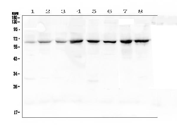
Figure 1. Western blot analysis of NF-kB p65 using anti-NF-kB p65 antibody (A00284-1).
Electrophoresis was performed on a 5-20% SDS-PAGE gel at 70V (Stacking gel) / 90V (Resolving gel) for 2-3 hours. The sample well of each lane was loaded with 50ug of sample under reducing conditions.
Lane 1: rat spleen tissue lysates,
Lane 2: rat lung tissue lysates,
Lane 3: rat kidney tissue lysates,
Lane 4: rat testis tissue lysates,
Lane 5: mouse spleen tissue lysates,
Lane 6: mouse lung tissue lysates,
Lane 7: mouse testis tissue lysates,
Lane 8: mouse NIH3T3 whole cell lysates.
After Electrophoresis, proteins were transferred to a Nitrocellulose membrane at 150mA for 50-90 minutes. Blocked the membrane with 5% Non-fat Milk/ TBS for 1.5 hour at RT. The membrane was incubated with rabbit anti-NF-kB p65 antigen affinity purified polyclonal antibody (Catalog # A00284-1) at 0.5 μg/mL overnight at 4℃, then washed with TBS-0.1%Tween 3 times with 5 minutes each and probed with a goat anti-rabbit IgG-HRP secondary antibody at a dilution of 1:10000 for 1.5 hour at RT. The signal is developed using an Enhanced Chemiluminescent detection (ECL) kit (Catalog # EK1002) with Tanon 5200 system. A specific band was detected for NF-kB p65 at approximately 65KD. The expected band size for NF-kB p65 is at 65KD.

Click image to see more details
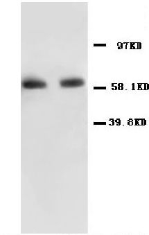
Figure 2. Western blot analysis of NF-kB p65 using anti-NF-kB p65 antibody (A00284-1).
Electrophoresis was performed on a 5-20% SDS-PAGE gel at 70V (Stacking gel) / 90V (Resolving gel) for 2-3 hours. The sample well of each lane was loaded with 50ug of sample under reducing conditions.
Lane 1: human COLO-320 whole cell lysates,
Lane 2: human A549 whole cell lysates,
Lane 3: human HepG2 whole cell lysates,
Lane 4: human MDA-MB-231 whole cell lysates,
Lane 5: human PANC-1 whole cell lysates,
Lane 6: human A375 whole cell lysates.
After Electrophoresis, proteins were transferred to a Nitrocellulose membrane at 150mA for 50-90 minutes. Blocked the membrane with 5% Non-fat Milk/ TBS for 1.5 hour at RT. The membrane was incubated with rabbit anti-NF-kB p65 antigen affinity purified polyclonal antibody (Catalog # A00284-1) at 0.5 μg/mL overnight at 4℃, then washed with TBS-0.1%Tween 3 times with 5 minutes each and probed with a goat anti-rabbit IgG-HRP secondary antibody at a dilution of 1:10000 for 1.5 hour at RT. The signal is developed using an Enhanced Chemiluminescent detection (ECL) kit (Catalog # EK1002) with Tanon 5200 system. A specific band was detected for NF-kB p65 at approximately 65KD. The expected band size for NF-kB p65 is at 65KD.

Click image to see more details
Figure 3. IHC analysis of NF-kB p65 using anti-NF-kB p65 antibody (A00284-1).
NF-kB p65 was detected in paraffin-embedded section of human colon cancer tissue. Heat mediated antigen retrieval was performed in citrate buffer (pH6, epitope retrieval solution) for 20 mins. The tissue section was blocked with 10% goat serum. The tissue section was then incubated with 2μg/ml rabbit anti-NF-kB p65 Antibody (A00284-1) overnight at 4℃. Biotinylated goat anti-rabbit IgG was used as secondary antibody and incubated for 30 minutes at 37℃. The tissue section was developed using Strepavidin-Biotin-Complex (SABC)(Catalog # SA1022) with DAB as the chromogen.

Click image to see more details
Figure 4. IHC analysis of NF-kB p65 using anti-NF-kB p65 antibody (A00284-1).
NF-kB p65 was detected in paraffin-embedded section of rat small intestine tissue. Heat mediated antigen retrieval was performed in citrate buffer (pH6, epitope retrieval solution) for 20 mins. The tissue section was blocked with 10% goat serum. The tissue section was then incubated with 2μg/ml rabbit anti-NF-kB p65 Antibody (A00284-1) overnight at 4°C. Biotinylated goat anti-rabbit IgG was used as secondary antibody and incubated for 30 minutes at 37°C. The tissue section was developed using Strepavidin-Biotin-Complex (SABC)(Catalog # SA1022) with DAB as the chromogen.

Click image to see more details
Figure 5. IHC analysis of NF-kB p65 using anti-NF-kB p65 antibody (A00284-1).
NF-kB p65 was detected in paraffin-embedded section of human mammary cancer tissue. Heat mediated antigen retrieval was performed in citrate buffer (pH6, epitope retrieval solution) for 20 mins. The tissue section was blocked with 10% goat serum. The tissue section was then incubated with 2μg/ml rabbit anti-NF-kB p65 Antibody (A00284-1) overnight at 4°C. Biotinylated goat anti-rabbit IgG was used as secondary antibody and incubated for 30 minutes at 37°C. The tissue section was developed using Strepavidin-Biotin-Complex (SABC)(Catalog # SA1022) with DAB as the chromogen.

Click image to see more details
Figure 6. IHC analysis of NF-kB p65 using anti-NF-kB p65 antibody (A00284-1).
NF-kB p65 was detected in paraffin-embedded section of rat kidney tissue . Heat mediated antigen retrieval was performed in citrate buffer (pH6, epitope retrieval solution) for 20 mins. The tissue section was blocked with 10% goat serum. The tissue section was then incubated with 2μg/ml rabbit anti-NF-kB p65 Antibody (A00284-1) overnight at 4°C. Biotinylated goat anti-rabbit IgG was used as secondary antibody and incubated for 30 minutes at 37°C. The tissue section was developed using Strepavidin-Biotin-Complex (SABC)(Catalog # SA1022) with DAB as the chromogen.
Protein Target Info & Infographic
Gene/Protein Information For RELA (Source: Uniprot.org, NCBI)
Gene Name
RELA
Full Name
Transcription factor p65
Weight
60.219kDa
Alternative Names
NF kB p65; NFkB p65; NF-kB p65; p65; rela p65; RelA; MGC131774; nf kb p65; NFkB p65; NF-kB p65; NFKB3; NFKB3v-rel avian reticuloendotheliosis viral oncogene homolog A (nuclear factor ofkappa light polypeptide gene enhancer in B-cells 3 (p65)); Nuclear factor NF-kappa-B p65 subunit; Nuclear factor of kappa light polypeptide gene enhancer in B-cells 3transcription factor p65; p65 flow cytometry; p65 immunohistochemistry; p65; p65RelA; rela p65; RelA; v-rel reticuloendotheliosis viral oncogene homolog A (avian); v-rel reticuloendotheliosis viral oncogene homolog A, nuclear factor of kappalight po RELA CMCU, NFKB3, p65 RELA proto-oncogene, NF-kB subunit transcription factor p65|NF-kappa-B p65delta3|NF-kappa-B transcription factor p65|nuclear factor NF-kappa-B p65 subunit|nuclear factor of kappa light polypeptide gene enhancer in B-cells 3|v-rel avian reticuloendotheliosis viral oncogene homolog A
*If product is indicated to react with multiple species, protein info is based on the gene entry specified above in "Species".For more info on RELA, check out the RELA Infographic

We have 30,000+ of these available, one for each gene! Check them out.
In this infographic, you will see the following information for RELA: database IDs, superfamily, protein function, synonyms, molecular weight, chromosomal locations, tissues of expression, subcellular locations, post-translational modifications, and related diseases, research areas & pathways. If you want to see more information included, or would like to contribute to it and be acknowledged, please contact [email protected].
Specific Publications For Anti-NF-kB p65/RELA Antibody Picoband™ (A00284-1)
Hello CJ!
A00284-1 has been cited in 116 publications:
*The publications in this section are manually curated by our staff scientists. They may differ from Bioz's machine gathered results. Both are accurate. If you find a publication citing this product but is missing from this list, please let us know we will issue you a thank-you coupon.
Characterization of acetaminophen-induced cytotoxicity in target tissues
Anti-inflammatory and antioxidant effects of curcumin on acute lung injury in a rodent model of intestinal ischemia reperfusion by inhibiting the pathway of NF-Kb
Effects of PDTC on NF-κB expression and apoptosis in rats with severe acute pancreatitis-associated lung injury
Effects of hydroxysafflor yellow A on proliferation and collagen synthesis of rat vascular adventitial fibroblasts induced by angiotensin II
Overexpression of mitogen-activated protein kinase kinase 4 and nuclear factor-κB in laryngeal squamous cell carcinoma: A potential indicator for poor prognosis
Ursolic acid protects against ulcerative colitis via anti-inflammatory and antioxidant effects in mice
Effects of resveratrol and genistein on nuclear factor‑κB, tumor necrosis factor‑α and matrix metalloproteinase‑9 in patients with chronic obstructive pulmonary disease
Heme oxygenase-1 induction by hemin protects liver cells from ischemia/reperfusion injury in cirrhotic rats
Celastrol Attenuates Multiple Sclerosis and Optic Neuritis in an Experimental Autoimmune Encephalomyelitis Model
Diosgenin Attenuates Lipopolysaccharide-Induced Parkinson’s Disease by Inhibiting the TLR/NF- κB Pathway
Recommended Resources
Here are featured tools and databases that you might find useful.
- Boster's Pathways Library
- Protein Databases
- Bioscience Research Protocol Resources
- Data Processing & Analysis Software
- Photo Editing Software
- Scientific Literature Resources
- Research Paper Management Tools
- Molecular Biology Software
- Primer Design Tools
- Bioinformatics Tools
- Phylogenetic Tree Analysis
Customer Reviews
Have you used Anti-NF-kB p65/RELA Antibody Picoband™?
Submit a review and receive an Amazon gift card.
- $30 for a review with an image
Be the first to review Anti-NF-kB p65/RELA Antibody Picoband™
*The first user to submit a review for a product is eligible for Boster's Innovating Scientists Reward, which gives product credits. This is in addition to the gift card reward.
Customer Q&As
Have a question?
Find answers in Q&As, reviews.
Can't find your answer?
Submit your question
1 Customer Q&As for Anti-NF-kB p65/RELA Antibody Picoband™
Question
Can A00284-1 be used in both membrane type pvdf and nitrocellulose?
Verified customer
Asked: 2021-02-03
Answer
The Anti-NF-KB P65/RELA Antibody Picoband™ (A00284-1) is okay to use for pvdf or nitrocellulose membrane.
Boster Scientific Support
Answered: 2021-02-03
