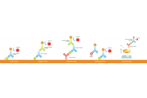
Learn how the high-dose hook effect can cause falsely low ELISA results, how to identify it through serial dilution, and how to distinguish it from signal saturation and other assay interference.
Seeing western blot smiling bands, with edges higher than the center? Learn how to identify possible causes, troubleshoot electrophoresis conditions, and improve band consistency.
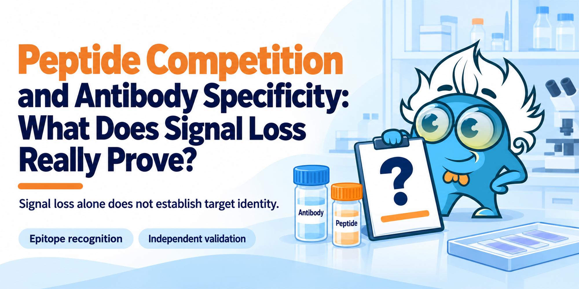
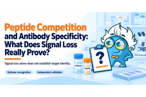
A disappearing signal after peptide blocking supports epitope-dependent antibody binding—but it does not prove that the signal comes from the intended target. Learn what peptide competition can show, where its limits are, and when genetic or orthogonal validation adds stronger evidence.
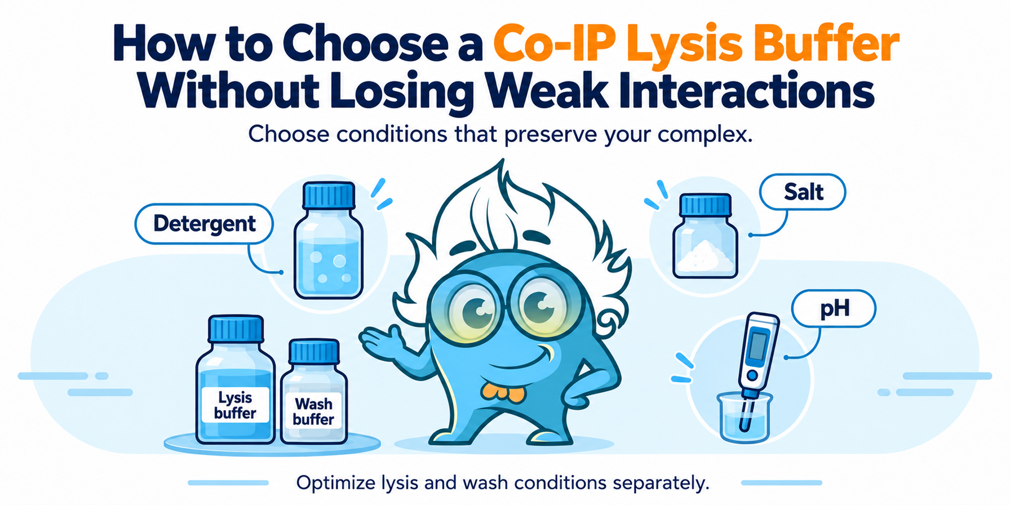
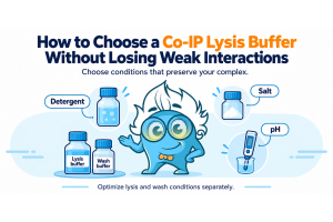
RIPA extracts the bait but loses the partner, while a milder buffer raises background? Learn how to balance Co-IP lysis and wash stringency, reduce nonspecific binding, and preserve weak protein interactions.
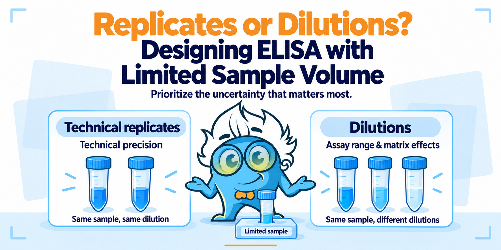
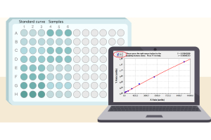
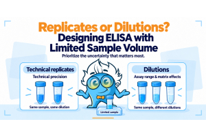
Working with limited ELISA sample volume? Learn how to decide between duplicate wells and multiple dilutions, account for dead volume and matrix effects, and design a practical workflow that makes the most of scarce samples such as CSF or animal serum.
Not sure which genomic regions to use as ChIP-qPCR controls? Learn how to choose positive and negative loci based on your target, cell type, public ChIP-seq data, and primer performance.
A flow cytometry panel that once compensated cleanly can show more spillover weeks later. Learn how tandem dye changes, reagent lots, fixation, and compensation controls can affect the result—and how to tell which factor is responsible.
Bone tissue can look well preserved while IHC staining becomes unexpectedly weak. Learn how decalcification, fixation, and antigen retrieval can affect antigen preservation—and how to tell whether the problem comes from tissue processing or the antibody.
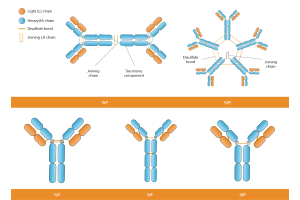
Heterophilic antibodies can create false-positive ELISA signals by interacting with assay antibodies rather than the intended target. Learn how to recognize suspicious results and use dilution, blocking, and alternative antibody pairs to confirm interference.
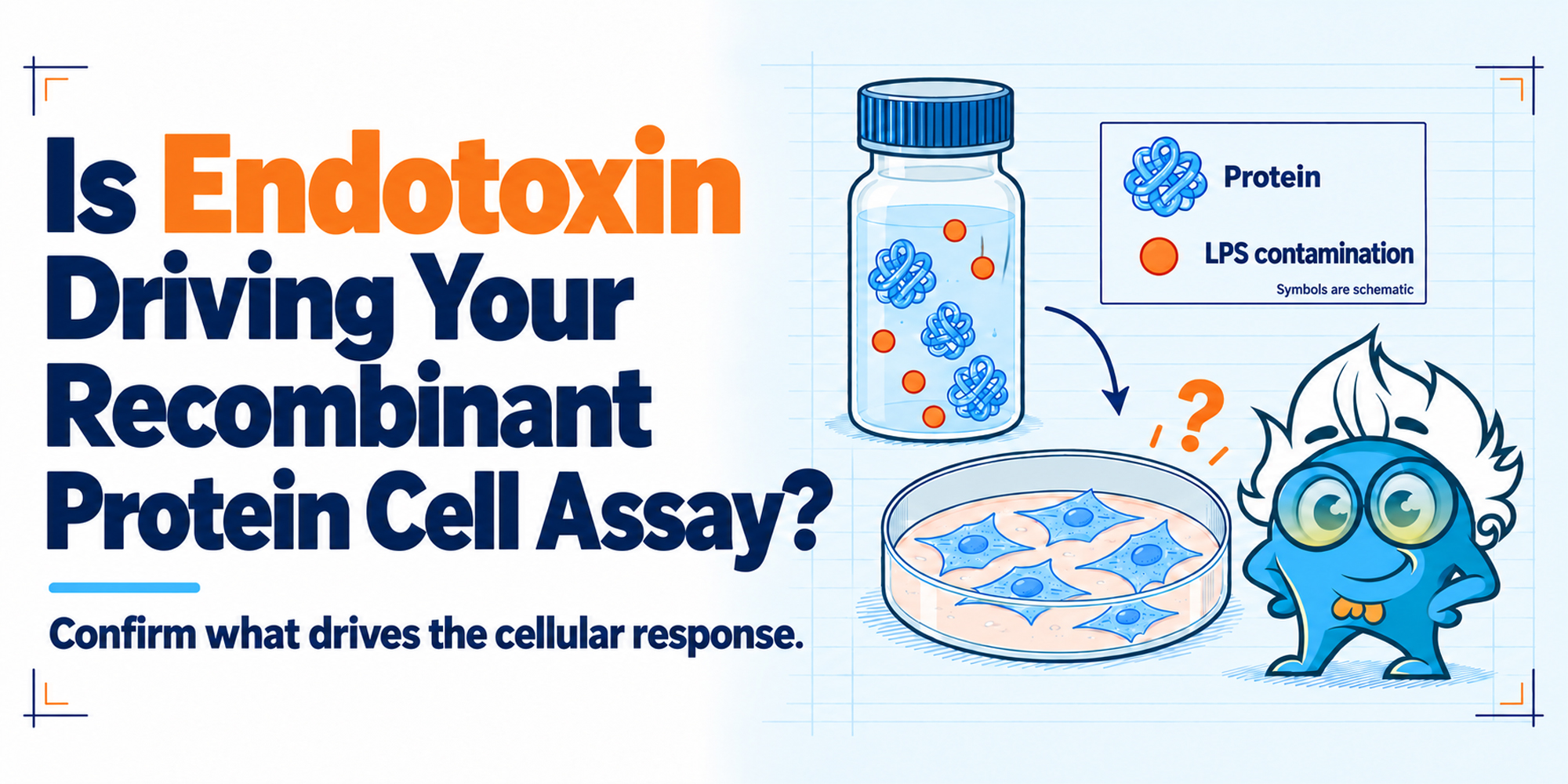
Endotoxin contamination can mimic recombinant protein activity in cell-based assays, especially in inflammation-related models. Learn how to calculate actual endotoxin exposure and use the right controls to distinguish LPS-driven effects from true protein bioactivity.