This website uses cookies to ensure you get the best experience on our website.


- Table of Contents
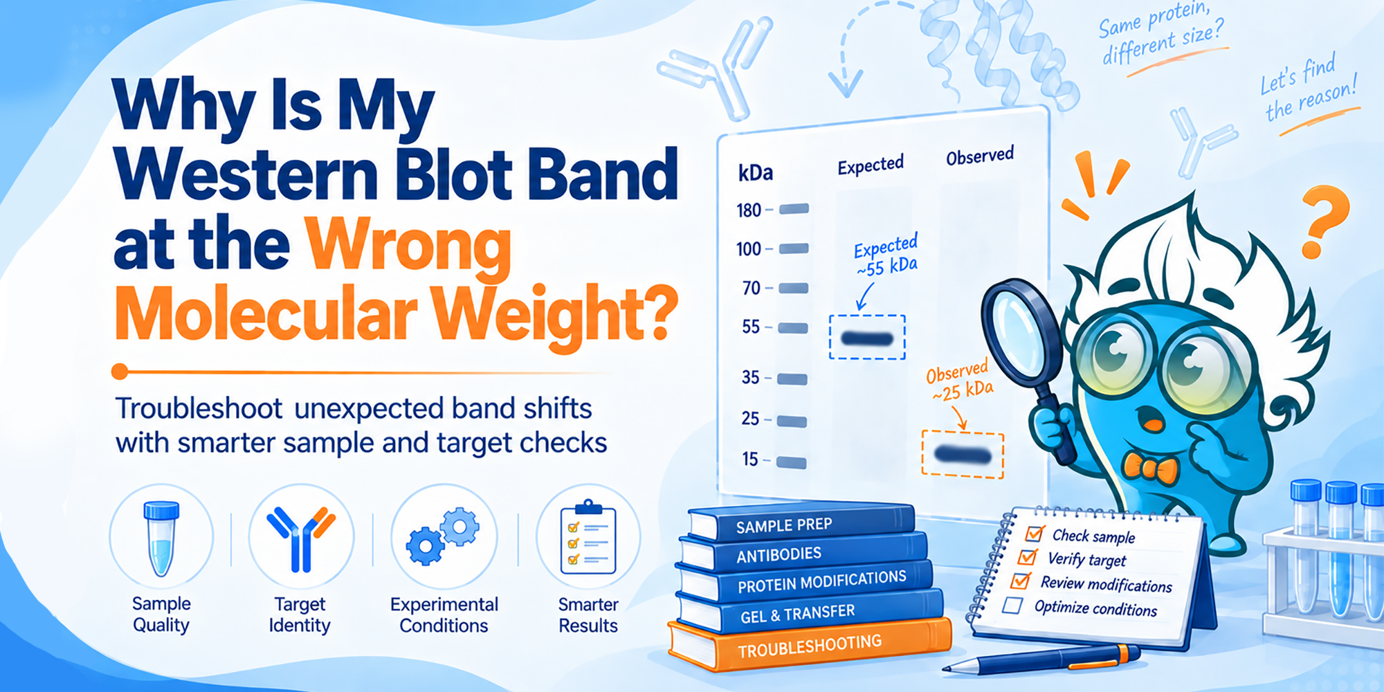
An unexpected molecular weight does not automatically mean the antibody failed. A cleaner troubleshooting sequence is to establish band identity first, then use the shift pattern to choose a mechanism-specific control.
A Western blot band appearing above or below the predicted molecular weight is easy to treat as a technical failure. Sometimes it is. But the number calculated from a protein sequence is not always the number that appears on an SDS-PAGE gel.
Glycosylation can increase or broaden apparent molecular weight. Proteolytic processing can convert a precursor into a smaller mature protein or generate stable fragments. Poor sample handling can produce lower-molecular-weight bands through degradation. And some proteins migrate anomalously even when none of those mechanisms is involved.
The useful question is therefore not simply, “Why is this band at the wrong size?” It is, “What control would distinguish one explanation from another?” Boster’s Western Blotting Troubleshooting Guide lists wrong band size among common WB problems; this article focuses on the next step: choosing controls that make the shift interpretable.
Core workflow: Identity → Pattern → Mechanism → Interpretation. First establish that the unexpected band is target-derived; then use the band pattern to decide which biological or technical mechanism to test.
Glycosylation and cleavage are attractive explanations because they are biologically interesting. But a shifted band is still only an observation until there is evidence that it comes from the intended target.
A KO/KD sample or a biologically true negative is particularly useful when band identity is uncertain. If the unexpected band disappears when the target is absent, it is much more likely to be target-derived. If it remains unchanged, nonspecific recognition moves higher on the list.
A second primary antibody against a non-overlapping epitope can provide another layer of evidence, especially when cleavage is suspected. Boster’s Western Blotting Principle and Control Design guide summarizes positive and negative controls that help separate antibody specificity from sample biology.
This does not mean every experiment must begin by repeating a full specificity-validation study. If the antibody has already been well validated in the same sample type and the shifted band changes predictably with a mechanism-specific treatment, that evidence may be sufficient. But when band identity itself is uncertain, confirm that before building a biological explanation around the shift.
The molecular weight calculated from the full amino-acid sequence often becomes the default “expected” Western blot size. That can be misleading when the database sequence represents a precursor rather than the mature protein.
Signal peptides, propeptides, transit peptides, and other segments may be removed during maturation, and some precursors are cleaved into multiple chains. UniProt separates these annotations from the canonical sequence, so checking Sequence processing and PTM/processing information is more informative than relying on sequence length alone.
Also check the antibody epitope. If maturation removes an N-terminal propeptide, an antibody against that removed region and an antibody against the retained C-terminal region are not expected to show the same pattern. Epitope location becomes part of the interpretation as soon as cleavage or processing is plausible.
Before troubleshooting the shift, write down three things: predicted precursor MW, expected mature/processed MW if known, and the antibody epitope region.
Once there is reasonable evidence that the band is target-derived, look at the direction, shape, and reproducibility of the shift. These features do not prove a mechanism, but they tell you which matched control is most informative next.
| What the blot looks like | First hypotheses to consider | Best next mechanism-specific control |
|---|---|---|
| Band runs higher than predicted; sometimes broad or heterogeneous | Glycosylation or another PTM; incomplete reduction/aggregation if the shift is large | Untreated vs. deglycosylated aliquots under otherwise identical conditions |
| One reproducible lower band or a defined doublet | Mature processed form, biological cleavage, or isoform | Check precursor/mature annotation; compare antibodies recognizing different protein regions |
| Several lower bands, smear, or fragments that vary between preparations | Sample degradation | Protected rapid cold lysis vs. the preparation associated with the fragment pattern |
| Shift persists but none of the above controls changes it | Other PTMs, splice variants, aggregation, anomalous migration, or residual nonspecific recognition | Broaden troubleshooting rather than forcing the original hypothesis |
There is intentional overlap here. A lower band can be a real cleavage product or a degradation fragment; a high band can reflect glycosylation or incomplete denaturation. The pattern narrows the field. The matched control makes the interpretation.
Glycosylated proteins often migrate more slowly than the unmodified polypeptide. The effect is not always a neat one-step increase: heterogeneous glycan occupancy can produce a broad band, a cluster of bands, or a smear around the expected region. Secreted and membrane proteins are common examples where the apparent Western blot size can exceed the mass calculated from the amino-acid sequence.
For suspected N-linked glycosylation, a strong WB control is a side-by-side comparison of the same starting sample with and without PNGase F treatment. If N-linked glycans account for the shift, the treated band should move downward and may collapse into a sharper species.
| Matched sample | Expected result if N-glycosylation explains the shift | Interpretation |
|---|---|---|
| Untreated aliquot | Higher, broader, or heterogeneous band | Native glycosylated form remains |
| PNGase F-treated aliquot | Band shifts lower and/or becomes sharper | N-linked glycans contributed to apparent molecular weight |
| PNGase F-treated aliquot with no change | No meaningful mobility shift | Look beyond N-linked glycosylation; O-linked glycans, other PTMs, processing, or anomalous migration may still be relevant |
Use aliquots from the same starting sample, keep protein load and electrophoresis conditions identical, and change only the treatment. The PNGase F protocol from New England Biolabs likewise uses a mobility change on SDS-PAGE as a straightforward readout of deglycosylation.
A negative PNGase F result does not mean the protein is universally “not glycosylated.” PNGase F addresses N-linked glycans. If O-linked glycosylation is the hypothesis, the control needs to match that chemistry.
This is usually the harder distinction because both mechanisms remove sequence from the full-length protein. The useful question is whether the smaller species follows a reproducible biological rule or follows how the sample was handled.
A true processing product is often discrete and reproducible. It may appear only in a tissue, activation state, differentiation stage, or treatment condition in which the relevant protease or maturation pathway is active. If the cleavage site is known, the fragment size should also be compatible with the portion of the protein that remains.
Region-specific antibodies can turn that idea into a direct test. Suppose maturation removes an N-terminal propeptide. A C-terminal antibody may detect both precursor and mature protein, whereas an antibody raised against the removed N-terminal region should detect the precursor but not the mature species. That pattern is much more informative than simply observing that “a smaller band exists.”
When the biology is well defined, a cleavage-site mutant, a relevant protease inhibitor, or a condition known to activate or suppress processing can provide even stronger evidence. Predict the expected band change before looking at the blot: which species should appear, disappear, or shift if cleavage is real?
Degradation tends to be less orderly. You may see several lower-molecular-weight bands, a smear beneath the full-length protein, loss of the intact band, or a fragment pattern that changes between sample preparations.
The cleanest first test is a matched sample-handling comparison. Prepare one aliquot with rapid cold processing, fresh protease inhibitors, and minimal time before denaturation, then compare it with the preparation that produced the questionable fragments. If the intact band is preserved and the lower bands are reduced under the protected condition, ex vivo degradation becomes much more likely.
Boster’s Western Blot Sample Preparation Guide recommends ice-cold lysis conditions with protease inhibitors to protect proteins during extraction. That matters because proteolysis can continue after cells or tissues are disrupted, even if the degradation never occurred in the living sample.
Keep freeze-thaw history consistent as well. A “fresh versus old” comparison is only interpretable if you know what differs between the aliquots. Start from the same biological material, change one preparation variable, and run the samples side by side.
“Cleavage follows the biology; degradation follows the handling” is a useful troubleshooting pattern, not a rule. Reproducibility alone does not prove cleavage, and a real cleavage pathway can generate more than one fragment.
Keep the baseline electrophoresis, transfer, blocking, antibody incubation, protein load, and exposure conditions fixed across paired samples. Changing several variables at once makes the result difficult to interpret. The Boster Western Blot Protocol can serve as a consistent reference workflow while the biological variable is tested.
| Control outcome | Most supported explanation | What to do next |
|---|---|---|
| Shifted band moves lower after PNGase F treatment | N-linked glycosylation contributes to the high apparent MW | Compare the deglycosylated size with the predicted mature polypeptide; investigate glycoform heterogeneity if needed |
| Lower band follows the predicted N- vs. C-terminal antibody pattern | Proteolytic processing/cleavage is plausible | Confirm with processing annotation, cleavage perturbation, or an appropriate biological condition |
| Lower fragments decrease with rapid cold lysis and fresh inhibitors | Ex vivo sample degradation is likely | Standardize collection, lysis, inhibitor use, aliquoting, and storage |
| Unexpected band persists in KO/KD or a true target-negative sample | Nonspecific recognition is likely | Optimize antibody conditions or validate with another antibody |
| None of the matched controls changes the band | The original hypotheses are incomplete | Consider isoforms, other PTMs, aggregation/incomplete reduction, and anomalous SDS-PAGE migration |
Not every molecular-weight discrepancy belongs to these three categories. Alternative splicing can change polypeptide length. Other post-translational modifications can alter mobility. Incomplete reduction or denaturation can leave oligomers or compact species that migrate unexpectedly. Some proteins simply bind SDS atypically and do not track their calculated mass closely.
Once a matched control has ruled out the leading mechanism, broaden the search rather than forcing the original explanation. Boster’s Western Blotting Troubleshooting Guide covers additional causes of high-, low-, and multiple-band patterns, including aggregation, incomplete denaturation, splice variants, and nonspecific binding.
Also remember that SDS-PAGE reports apparent molecular weight. A modest mismatch between theoretical and observed size is weaker evidence than a reproducible change produced by a mechanism-specific control.
Troubleshoot an unexpected Western blot band in order. First ask whether the band is target-derived. Then check whether the expected size refers to the precursor or mature protein. Use the band pattern to choose a matched control: deglycosylation for glycosylation, region- or cleavage-specific controls for processing, and protected sample preparation for degradation. Let the control outcome—not the kDa position alone—determine the interpretation.