This website uses cookies to ensure you get the best experience on our website.
- Table of Contents
and ELISA kits, proteins related to cardiovascular disease, vascular biology, cardiac injury, inflammation, and remodeling.
Cardiovascular research covers a broad set of biological questions across vascular inflammation, atherosclerosis, endothelial dysfunction, thrombosis, hypertension, myocardial injury, fibrosis, and heart failure. Antibodies are essential tools in this field because they help detect disease biomarkers, inflammatory mediators, vascular markers, remodeling proteins, and stress-response pathways across tissue sections, cultured cells, and circulating biomarker studies. In tissue-based workflows, IHC and IF are commonly used to localize endothelial markers, fibrosis markers, cardiomyocyte injury markers, and immune-cell infiltration in heart and vessel samples. Western blot supports pathway validation in remodeling and stress-response studies, while ELISA enables quantification of soluble biomarkers such as BNP, CRP, IL-6, and galectin-3. Flow cytometry is useful for immune, platelet, and endothelial-related phenotyping. This cardiovascular antibodies hub is designed to help researchers move from biomarkers to methods, disease contexts, cell systems, pathways, and related research areas more efficiently.
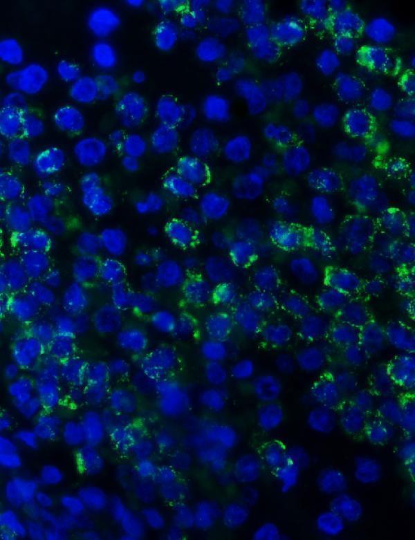
Anti-Myeloperoxidase/MPO Antibody Picoband®, Figure 5. IF analysis of MPO using anti-MPO antibody (PA1054).
MPO was detected in a paraffin-embedded section of human spleen tissue. Heat med...

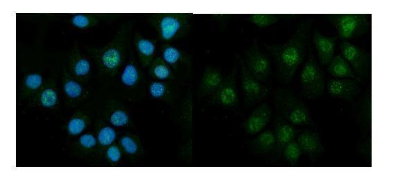
Anti-Galectin 3/LGALS3 Antibody Picoband®, Figure 6. IF analysis of Galectin-3 using anti-Galectin-3 antibody (PB9081).
Galectin-3 was detected in an immunocytochemic...
Anti-CBS Antibody Picoband®, Figure 2. IF analysis of CBS using anti-CBS antibody (A00130-3).
CBS was detected in an immunocytochemical section of MCF-7 cells. En...
| Protein Name | Gene Name | Function |
|---|---|---|
| Troponin I | TNNI3 | Marker of cardiomyocyte injury and a core readout in myocardial damage research. |
| B-type Natriuretic Peptide (BNP) | NPPB | Indicator of cardiac stress and heart failure. |
| C-reactive Protein (CRP) | CRP | Inflammation marker associated with vascular injury and atherosclerosis. |
| Low-Density Lipoprotein (LDL) Cholesterol | LDLR | Central to cholesterol handling and atherosclerosis-related lipid biology. |
| High-Density Lipoprotein (HDL) Cholesterol | APOA1 | Associated with reverse cholesterol transport and vascular protection studies. |
| Homocysteine | CBS | Elevated homocysteine metabolism is linked to endothelial dysfunction and cardiovascular risk. |
| Fibrinogen | FGA | Coagulation-related marker connected to thrombosis and vascular inflammation. |
| Myeloperoxidase (MPO) | MPO | Inflammation and oxidative stress marker often studied in plaque instability and vascular injury. |
| Lipoprotein(a) [Lp(a)] | LPA | Independent risk factor in atherosclerosis and thrombosis research. |
| Interleukin-6 (IL-6) | IL6 | Pro-inflammatory cytokine involved in cardiovascular inflammation and remodeling. |
| PCSK9 | PCSK9 | Key regulator of LDL receptor turnover and lipid-lowering target biology. |
| Galectin-3 | LGALS3 | Fibrosis and heart failure marker associated with remodeling and adverse prognosis studies. |
| ST2 | IL1RL1 | Biomarker for cardiac stress and remodeling in heart failure studies. |
| Apolipoprotein B (ApoB) | APOB | Represents atherogenic lipoprotein burden in cardiovascular risk assessment workflows. |
| Factor VII | F7 | Initiates coagulation cascade and supports thrombosis-related cardiovascular studies. |
| Lipoprotein-associated Phospholipase A2 (Lp-PLA2) | PLA2G7 | Vascular inflammation marker linked to plaque biology. |
| TIMP-1 | TIMP1 | ECM remodeling regulator relevant to fibrosis and post-injury remodeling. |
Use IHC and IF to visualize endothelial markers, cardiomyocyte injury markers, inflammatory-cell infiltration, and fibrosis-related remodeling in heart and vessel tissues.
Explore IHC/IF assay guideQuantify BNP, CRP, IL-6, galectin-3, and other soluble cardiovascular biomarkers in serum, plasma, and conditioned media.
Explore ELISA assay guideBuild panels for inflammatory-cell profiling, platelet-related studies, and vascular-cell phenotyping in cardiovascular and vascular injury models.
Explore flow cytometry guideValidate pathway activation such as Akt, AMPK, inflammatory signaling, hypoxia adaptation, and remodeling-associated proteins in heart, vessel, and cell culture lysates.
Explore Western blot guideThis area focuses on plaque initiation and progression, vascular-wall injury, endothelial activation, lipid-related risk pathways, and immune-cell recruitment. It is a core entry point for studies on vascular inflammation, plaque instability, and vessel remodeling.
Related pages: Cardiovascular Disease Antibodies · Hypertension Antibodies
Heart failure and cardiac-injury studies commonly connect cardiomyocyte damage, inflammatory activation, extracellular matrix remodeling, fibrosis, and stress biomarkers such as troponin, BNP, ST2, and galectin-3.
Related pages: Heart Disease Antibody · Cardiovascular Disease Antibodies
Hypertension research often examines endothelial dysfunction, vascular stiffness, oxidative stress, and chronic remodeling in vessel and heart models. It provides an important bridge between vascular biology, inflammation, and long-term organ damage.
Related pages: Hypertension Antibodies · Cardiovascular Disease Antibodies
This context includes coagulation-related signaling, platelet activation, vessel-wall stress, and injury-associated inflammation. It is especially relevant in studies of thrombotic risk, vascular injury, and post-damage response.
Related pages: Cardiovascular Disease Antibodies · Heart Disease Antibody
Cardiomyocytes and related muscle-cell systems are central to myocardial injury, ischemic stress, hypertrophy, and heart failure studies. These models are commonly used for injury biomarkers, survival signaling, and remodeling readouts.
Endothelial cells and fibroblasts are major entry points for studies on vessel integrity, inflammation, angiogenesis, fibrosis, and chronic tissue remodeling. This context is especially useful in atherosclerosis, hypertension, and post-injury repair workflows.
Macrophages and thrombocytes are key players in plaque progression, clot-associated injury, and vascular inflammation. These entry points are particularly useful for studies of atherosclerosis, thrombosis, and post-injury response.
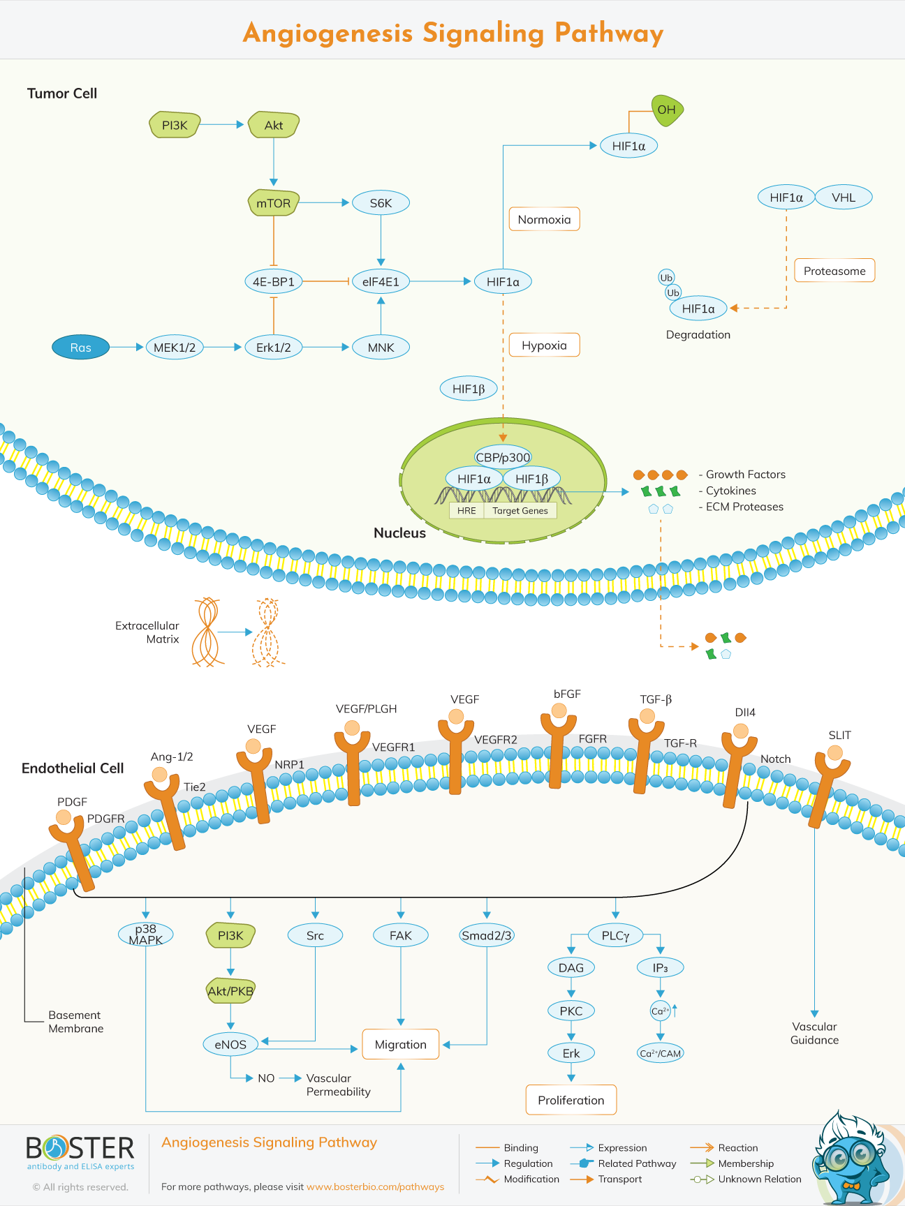
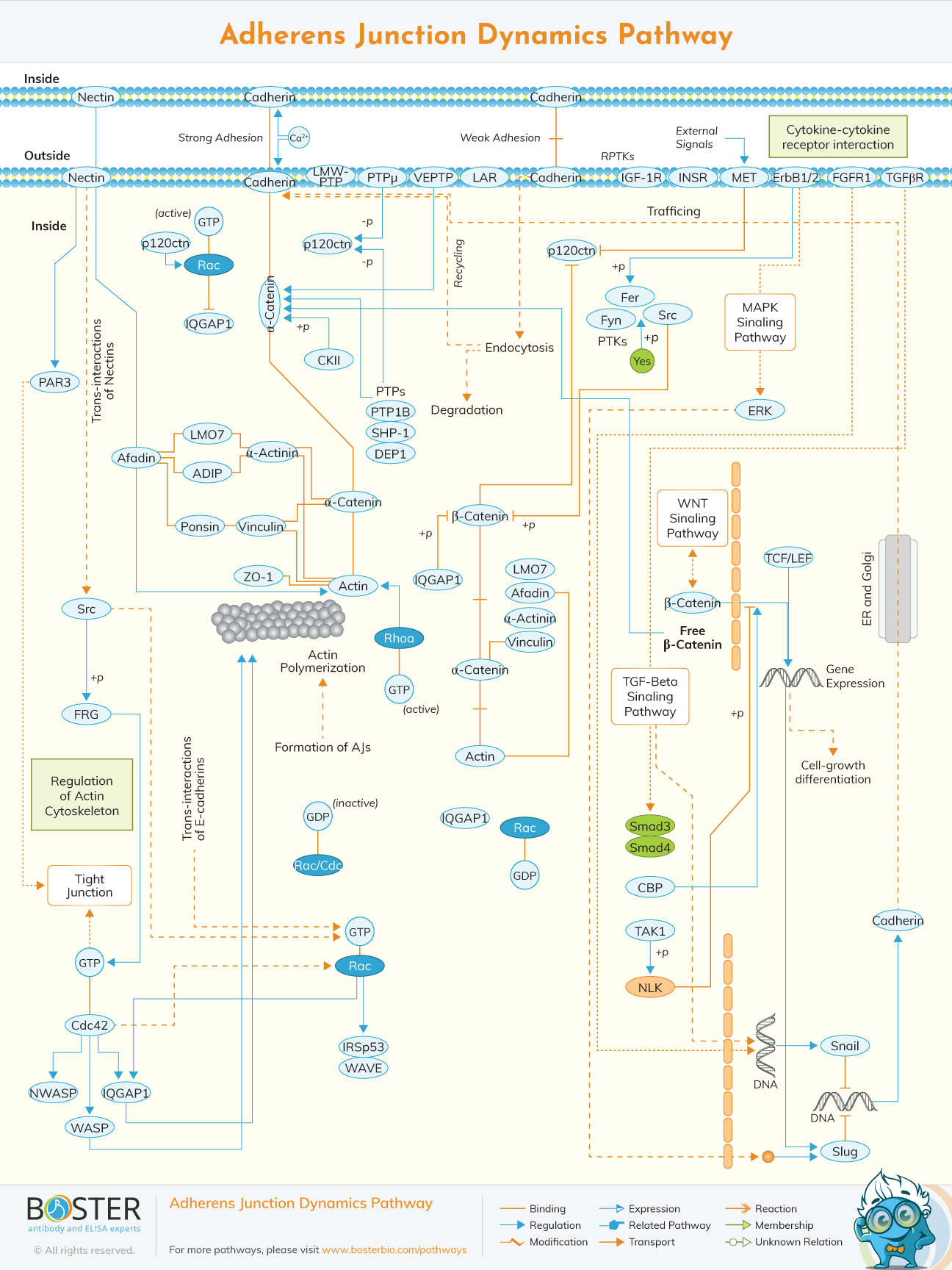
Core pathways used to study vessel growth, endothelial activation, barrier regulation, and structural remodeling.
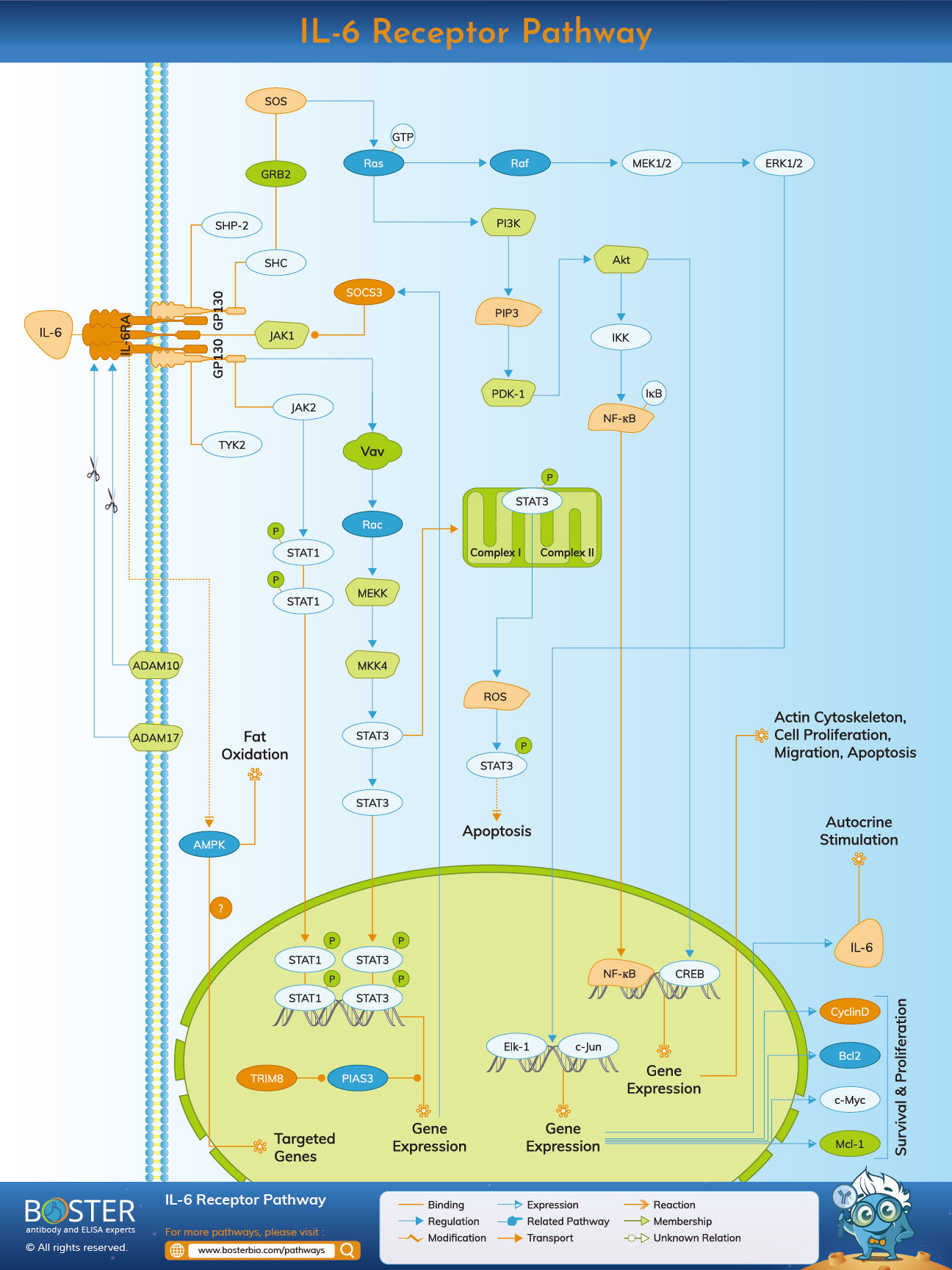
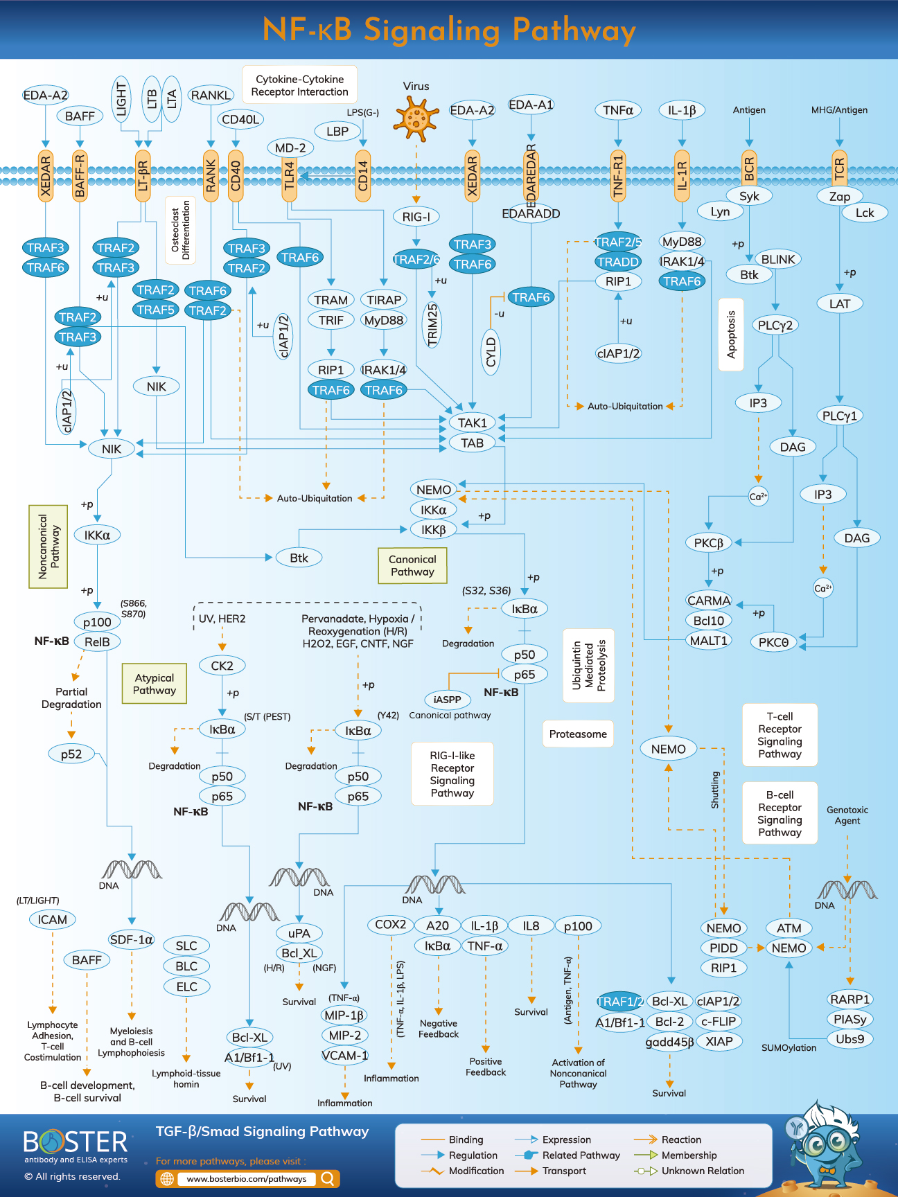
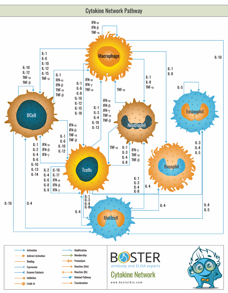
Common inflammation and stress-related pathways used in plaque biology, vascular injury, and heart failure studies.
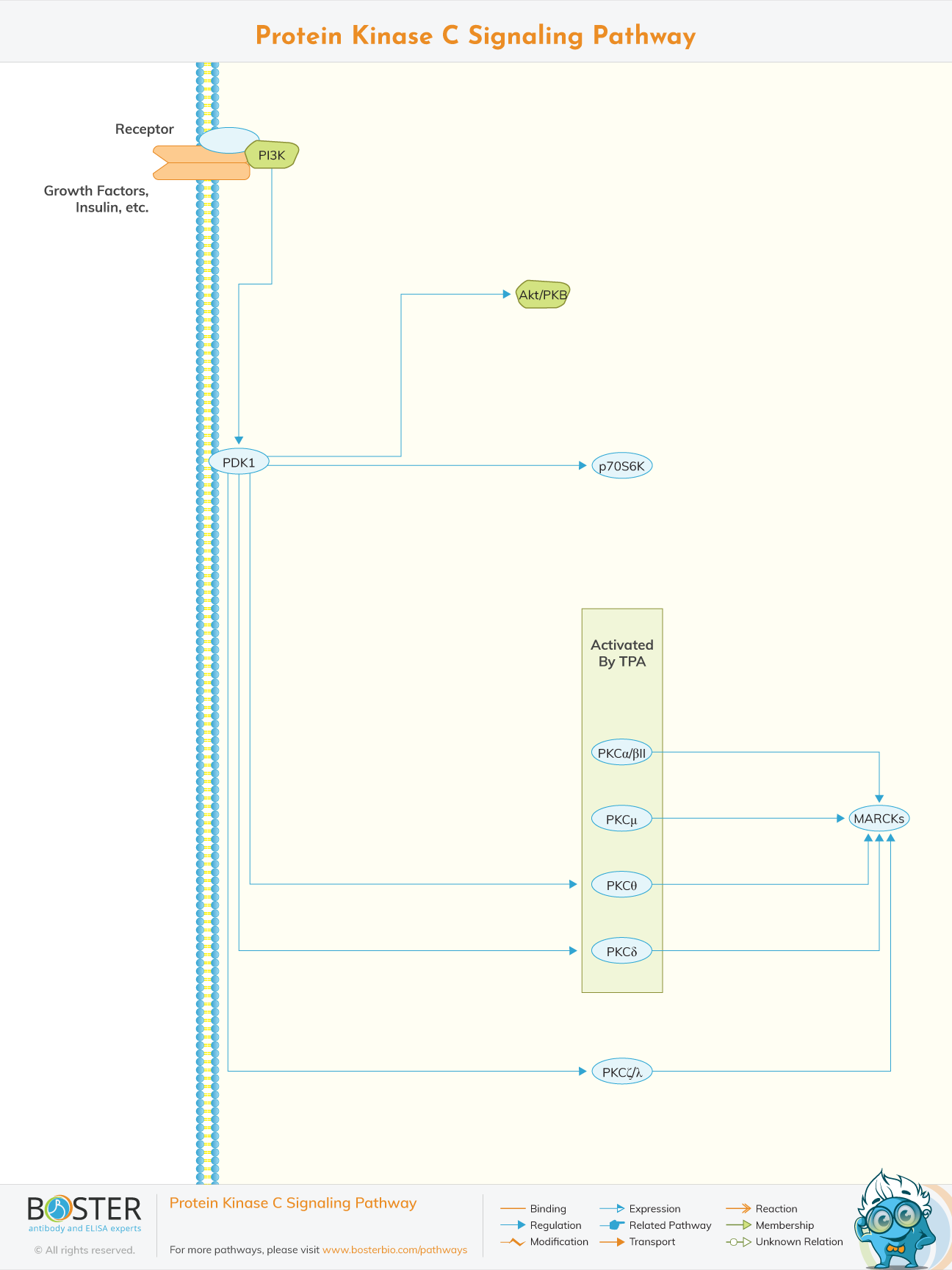
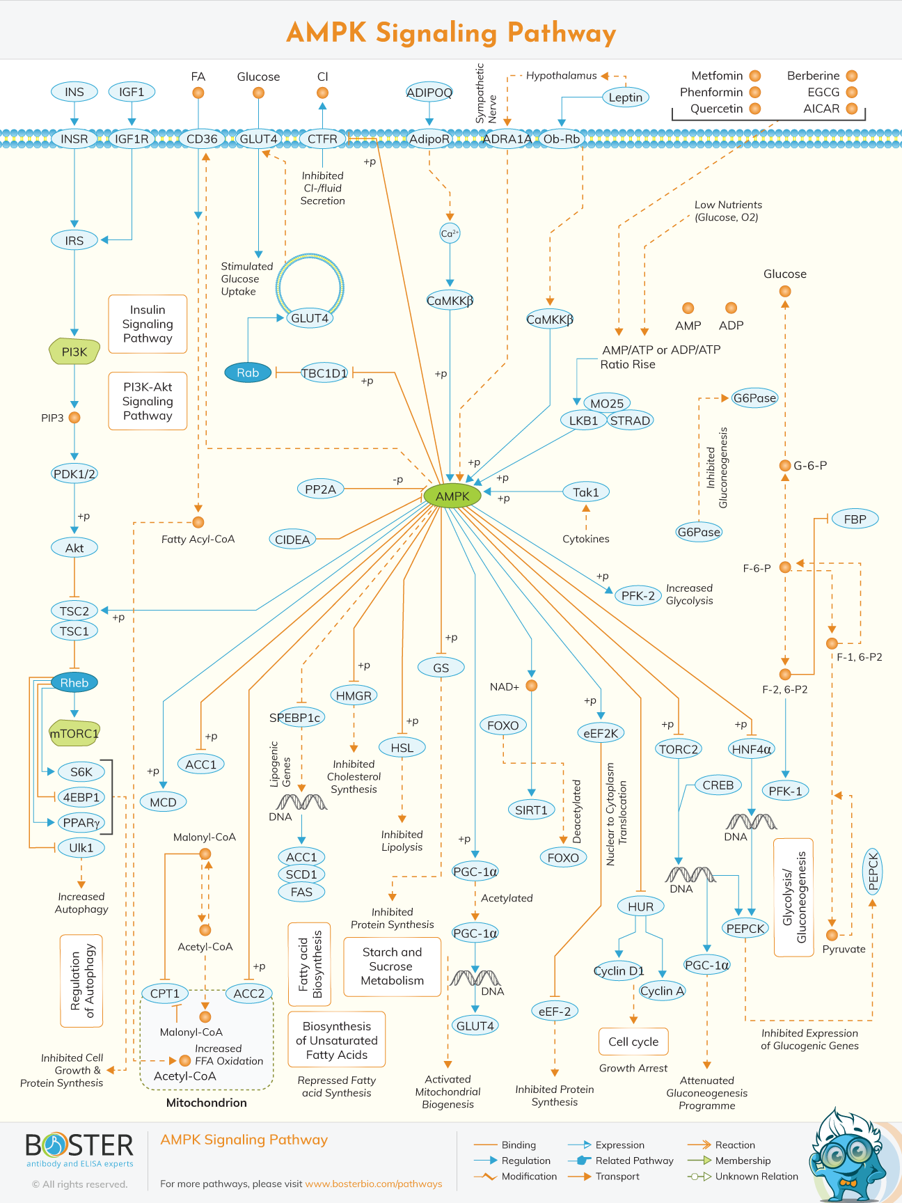
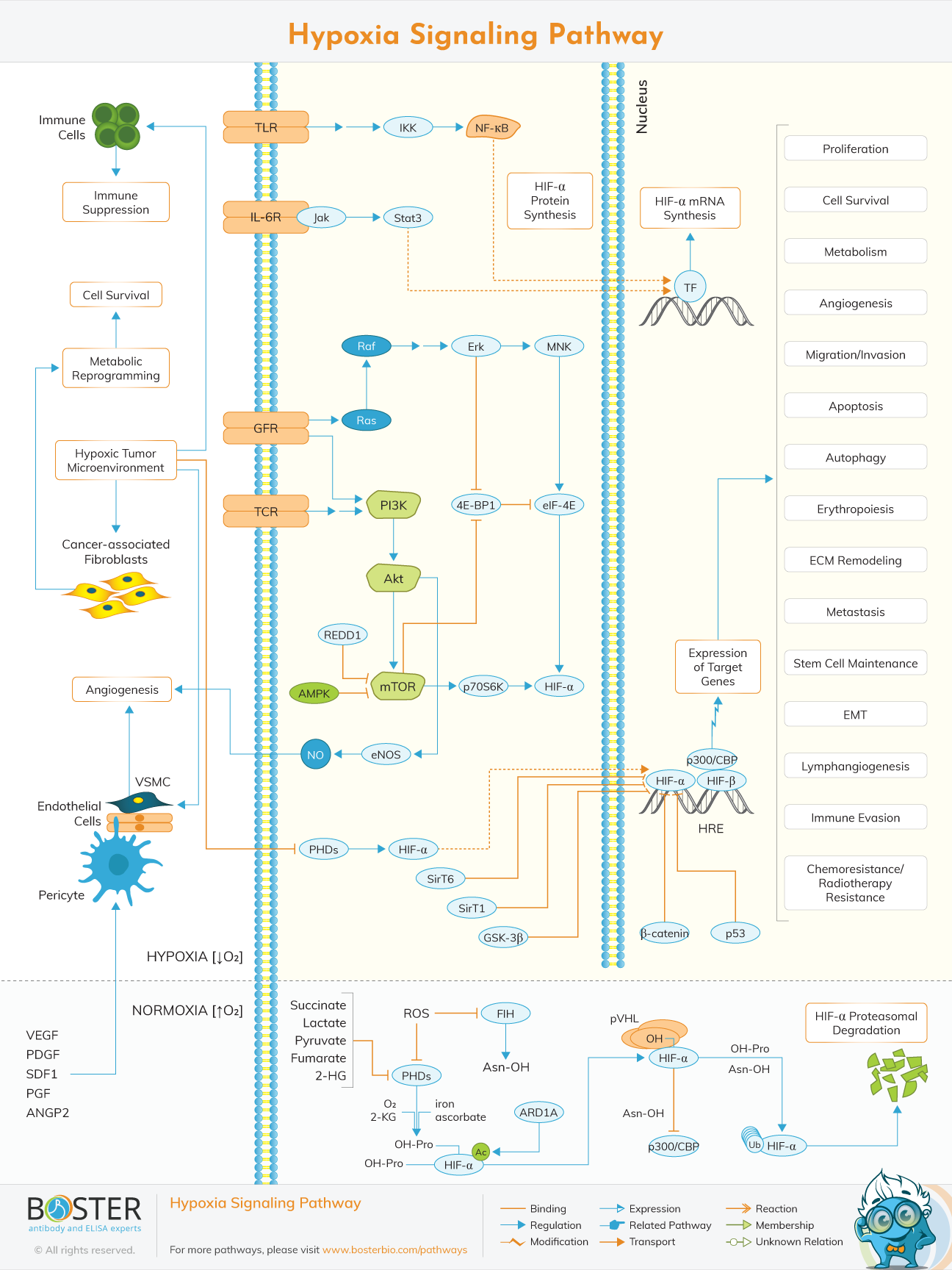
Metabolic and survival pathways relevant to ischemia, remodeling, vascular stress, and myocardial adaptation.
Atherosclerosis research focuses on how lipid accumulation, endothelial activation, oxidative stress, and immune-cell recruitment drive plaque initiation and progression. Endothelial dysfunction is an early and recurring theme that links vascular signaling imbalance to inflammation and barrier disruption.
Heart failure is closely tied to cardiomyocyte injury, fibrosis, extracellular matrix turnover, inflammatory signaling, and maladaptive remodeling. Biomarkers such as BNP, troponin, ST2, and galectin-3 help connect circulating readouts to tissue-level remodeling.
Many cardiovascular conditions converge on fibrosis, coagulation-related injury, and structural remodeling. Fibroblast activation, matrix deposition, platelet-related responses, and vessel-wall changes are common mechanisms across chronic cardiovascular disease models.