This website uses cookies to ensure you get the best experience on our website.
- Table of Contents
and ELISA kits for metabolic dysfunction, fatty liver disease, insulin resistance, and related metabolic research.
Metabolism research focuses on how cells and tissues regulate energy production, nutrient sensing, glucose homeostasis, lipid handling, and biosynthetic balance under physiological and disease conditions. In translational research, this field includes metabolic disorders such as fatty liver disease, diabetes mellitus, insulin resistance, obesity-associated inflammation, and broader metabolic syndrome. Because metabolic dysfunction is rarely confined to a single organ, researchers often study coordinated changes across hepatocytes, adipocytes, endocrine signaling pathways, inflammatory mediators, and tissue remodeling programs.
Antibodies are essential tools for metabolism research because they enable precise detection of metabolic enzymes, transporters, cytokines, adipokines, fibrosis markers, and pathway nodes across tissues, cell models, and biofluids. IHC and IF help localize metabolic and inflammatory changes in liver and adipose tissue. Western blot is widely used to monitor pathway activation such as AMPK, AKT, insulin receptor signaling, autophagy, and lipogenesis. ELISA supports quantification of soluble markers including adiponectin, leptin, IL-6, TNF-α, CRP, and other circulating readouts linked to disease severity. This metabolism antibodies hub is designed to help researchers navigate metabolic disease biomarkers, common model systems, pathway maps, and related disease areas while reaching validated antibodies and assays faster.
Metabolism biomarkers span hepatocyte injury, lipid accumulation, insulin resistance, inflammatory signaling, and tissue remodeling. Fatty liver disease is used here as the lead metabolic-disease entry point because it connects liver metabolism, adipose dysfunction, systemic inflammation, and progression toward fibrosis. Use the biomarker panel below to move from broad disease context into target-level exploration.

PA1079
PA1352
M00564-3
| Protein Name | Gene Name | Function |
|---|---|---|
| Alanine Aminotransferase (ALT) | GPT | Enzyme involved in amino acid metabolism; elevated levels indicate liver injury. |
| Gamma-Glutamyl Transferase (GGT) | GGT1 | Enzyme involved in glutathione metabolism; elevated levels indicate liver disease or bile duct issues. |
| Cytokeratin-18 (CK-18) | KRT18 | Intermediate filament protein in hepatocytes; elevated levels indicate hepatocyte apoptosis. |
| Fibronectin | FN1 | Extracellular matrix protein; increased levels associated with liver fibrosis. |
| Adiponectin | ADIPOQ | Hormone regulating glucose and lipid metabolism; lower levels linked to fatty liver disease and insulin resistance. |
| Interleukin-6 (IL-6) | IL6 | Pro-inflammatory cytokine; elevated levels associated with metabolic inflammation and liver injury. |
| Tumor Necrosis Factor-alpha (TNF-α) | TNF | Pro-inflammatory cytokine linked to insulin resistance, inflammation, and hepatocellular damage. |
| Hepcidin | HAMP | Regulates iron homeostasis; altered levels are associated with liver disease progression. |
| Retinol Binding Protein 4 (RBP4) | RBP4 | Transport protein for vitamin A; elevated levels linked to insulin resistance and metabolic dysfunction. |
| Leptin | LEP | Hormone involved in appetite regulation and energy balance; elevated levels are associated with obesity and fatty liver. |
| Ferritin | FTL | Protein that stores iron; elevated levels can reflect iron overload, inflammation, and liver damage. |
| Monocyte Chemoattractant Protein-1 (MCP-1) | CCL2 | Chemokine involved in immune-cell recruitment; elevated levels indicate inflammatory activation. |
| C-reactive Protein (CRP) | CRP | Inflammatory marker associated with systemic metabolic inflammation. |
| Fetuin-A | AHSG | Protein linked to insulin resistance and fatty liver disease. |
| Hyaluronic Acid | HAS2 | Extracellular matrix-associated marker; elevated levels indicate fibrotic remodeling. |
| Vascular Endothelial Growth Factor (VEGF) | VEGFA | Growth factor involved in angiogenesis; elevated levels are associated with liver disease progression. |
| Peroxisome Proliferator-Activated Receptor-alpha (PPAR-α) | PPARA | Nuclear receptor regulating fatty acid oxidation and lipid metabolism. |
| Sterol Regulatory Element-Binding Protein 1c (SREBP-1c) | SREBF1 | Transcription factor regulating lipid biosynthesis and de novo lipogenesis. |
| Glycogen Phosphorylase | PYGL | Enzyme involved in glycogen metabolism; altered levels may indicate changes in hepatic metabolic status. |
Use IHC and IF to localize hepatocyte injury markers, fibrosis-associated proteins, inflammatory mediators, and lipid-metabolism targets in liver and adipose tissues. These methods are especially useful for comparing steatosis, inflammation, and remodeling across disease stages.
Explore IHC / IF guideELISA is widely used in metabolism research to quantify adipokines, cytokines, liver injury markers, and circulating proteins linked to insulin resistance or metabolic syndrome. It is especially helpful for serum, plasma, and conditioned-media studies.
Explore ELISA guideWestern blot helps confirm activation of AMPK, AKT, insulin receptor signaling, autophagy, lipogenesis, and stress-response pathways. It is a core method for mechanistic studies in hepatocyte and adipocyte models.
Explore Western blot guidePCR is commonly used to profile lipogenesis, β-oxidation, inflammatory transcriptional responses, and glucose-metabolism genes across liver, adipose, and endocrine-related models.
Explore PCR guideLiver-centered metabolic disorders are one of the strongest entry points into metabolism research because the liver integrates lipid handling, glucose production, detoxification, and systemic metabolic adaptation. Fatty liver disease is especially valuable as a research model because it captures early steatosis, hepatocyte stress, inflammatory transition, and progression toward fibrotic remodeling within a single disease continuum.
This area is well suited for researchers studying lipid accumulation, hepatocellular injury, oxidative stress, cytokine signaling, extracellular matrix remodeling, and disease-stage progression in liver tissue or hepatocyte-based models.
Glucose homeostasis is a core branch of metabolism research because it connects insulin signaling, nutrient sensing, energy storage, and inter-organ communication. Diabetes mellitus and insulin resistance models are especially useful for studying how endocrine imbalance alters glucose uptake, hepatic glucose output, adipose-liver signaling, inflammatory burden, and long-term tissue stress.
This disease area complements fatty liver research by extending metabolism beyond steatosis into broader questions of insulin sensitivity, compensatory hormone signaling, adipokine biology, and the systemic consequences of disrupted glucose control.
Metabolic dysfunction does not remain confined to the liver or endocrine axis. Chronic alterations in lipid metabolism, inflammatory tone, glucose control, and adipose signaling frequently converge with vascular injury, endothelial activation, and elevated cardiovascular risk. This makes cardiometabolic biology an important expansion area for metabolism-focused research pages.
Researchers entering metabolism through obesity, diabetes, or fatty liver often need downstream access to cardiovascular and vascular-biology contexts in order to connect metabolic biomarkers with systemic disease burden and translational relevance.
Hepatocytes are central to metabolism research because the liver coordinates lipid handling, glucose output, glycogen storage, amino acid metabolism, and detoxification. In metabolism LP structure, hepatocytes should function as the primary model-context entry point because they connect directly to fatty liver disease, insulin resistance, nutrient sensing, and progression toward inflammatory liver injury.
Common sample and model contexts include liver tissue sections, primary hepatocytes, hepatocyte cell lines, steatosis-induction systems, free-fatty-acid loading models, and experiments focused on insulin response, oxidative stress, autophagy, or lipid-biosynthesis pathways.
Adipocytes provide a second major model context for metabolism research because they regulate lipid storage, endocrine signaling, leptin and adiponectin balance, inflammatory tone, and insulin responsiveness at the systemic level. Including adipocyte context prevents the page from feeling liver-only and helps frame metabolism as an inter-organ field.
Common sample and model contexts include adipose tissue sections, differentiated adipocyte cultures, obesity-associated inflammatory models, glucose-uptake experiments, lipolysis studies, and liver–adipose crosstalk systems designed to study systemic metabolic dysfunction.
Advanced metabolic disease is not driven by parenchymal cells alone. As metabolic overload persists, inflammatory mediators, recruited immune cells, endothelial dysfunction, and extracellular matrix remodeling all become part of the disease context. This broader tissue-model layer is important for explaining why metabolism research often overlaps with inflammation, vascular biology, and fibrosis-focused validation work.
Common sample and model contexts include inflammatory liver microenvironments, fibrosis-associated remodeling systems, endothelial activation studies, macrophage-linked cytokine signaling, and tissue-level progression models where metabolic stress becomes chronic injury and remodeling.
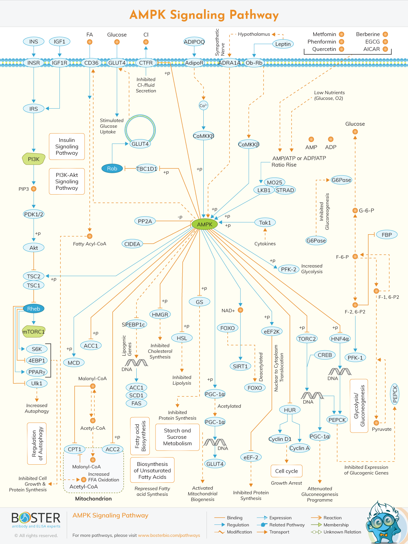
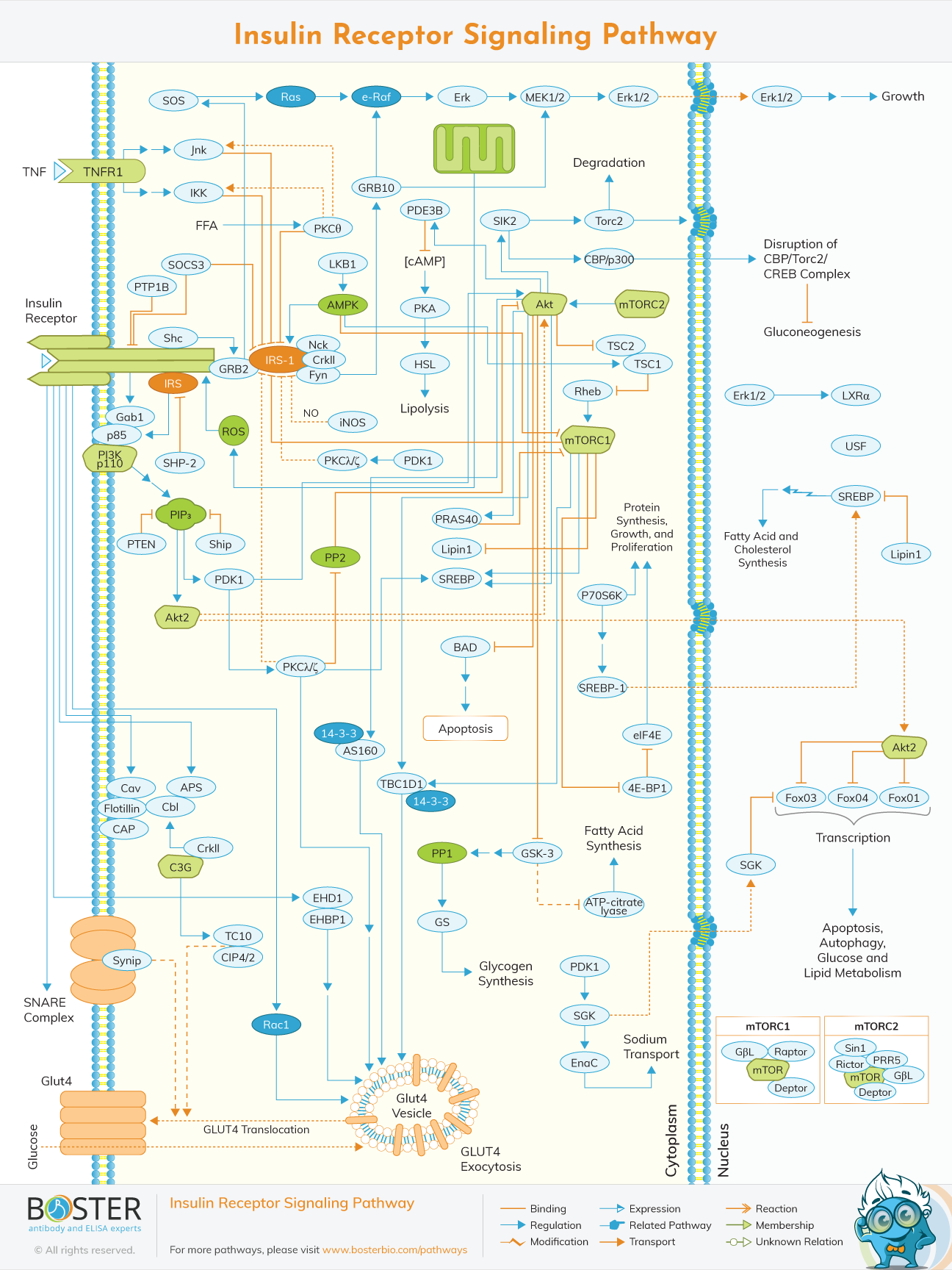
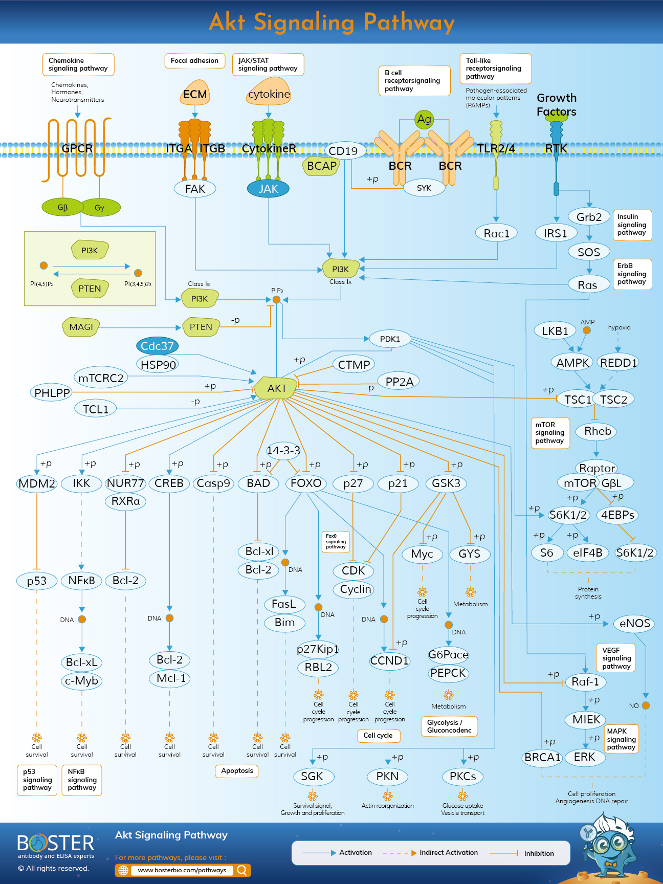
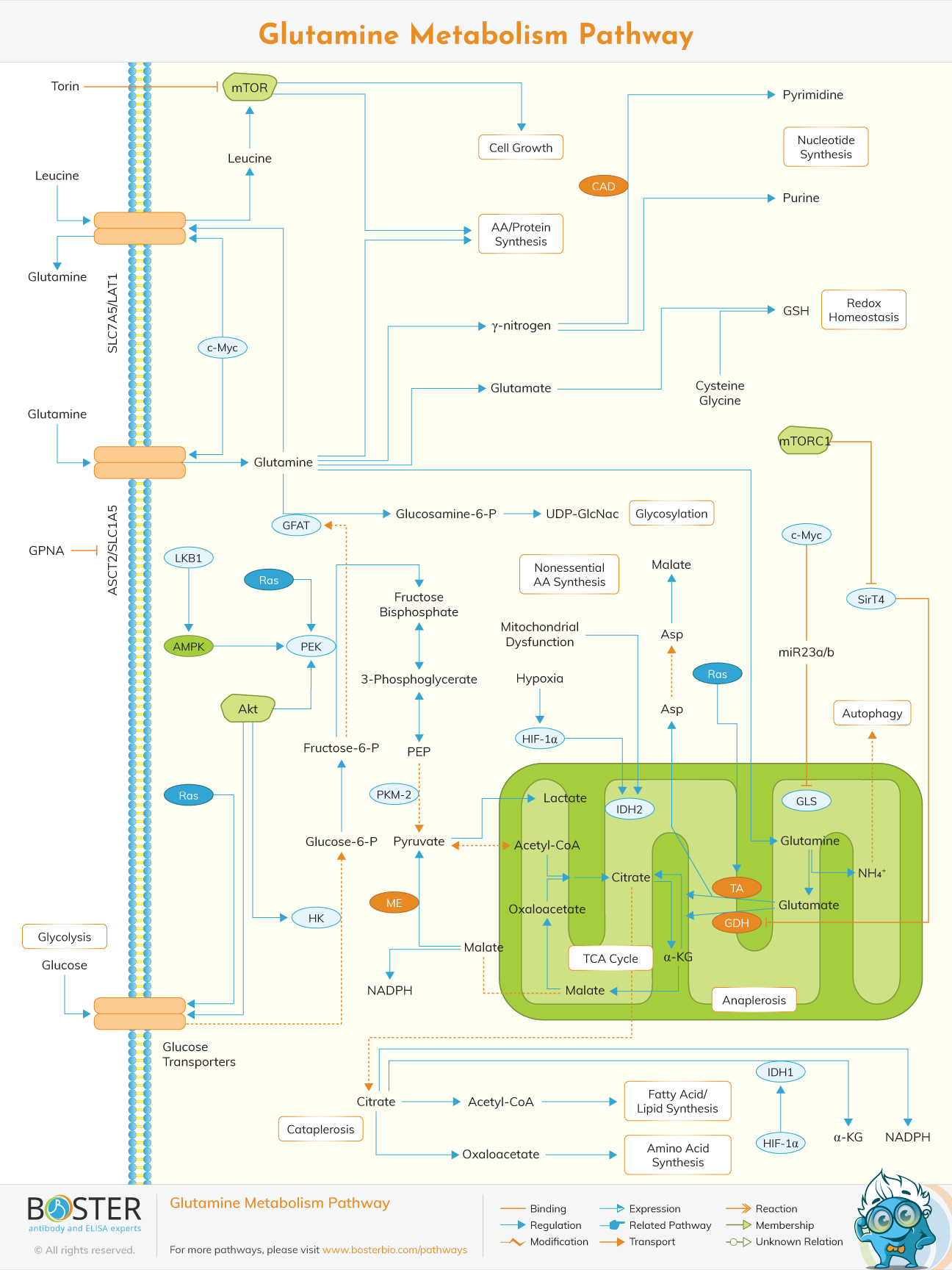
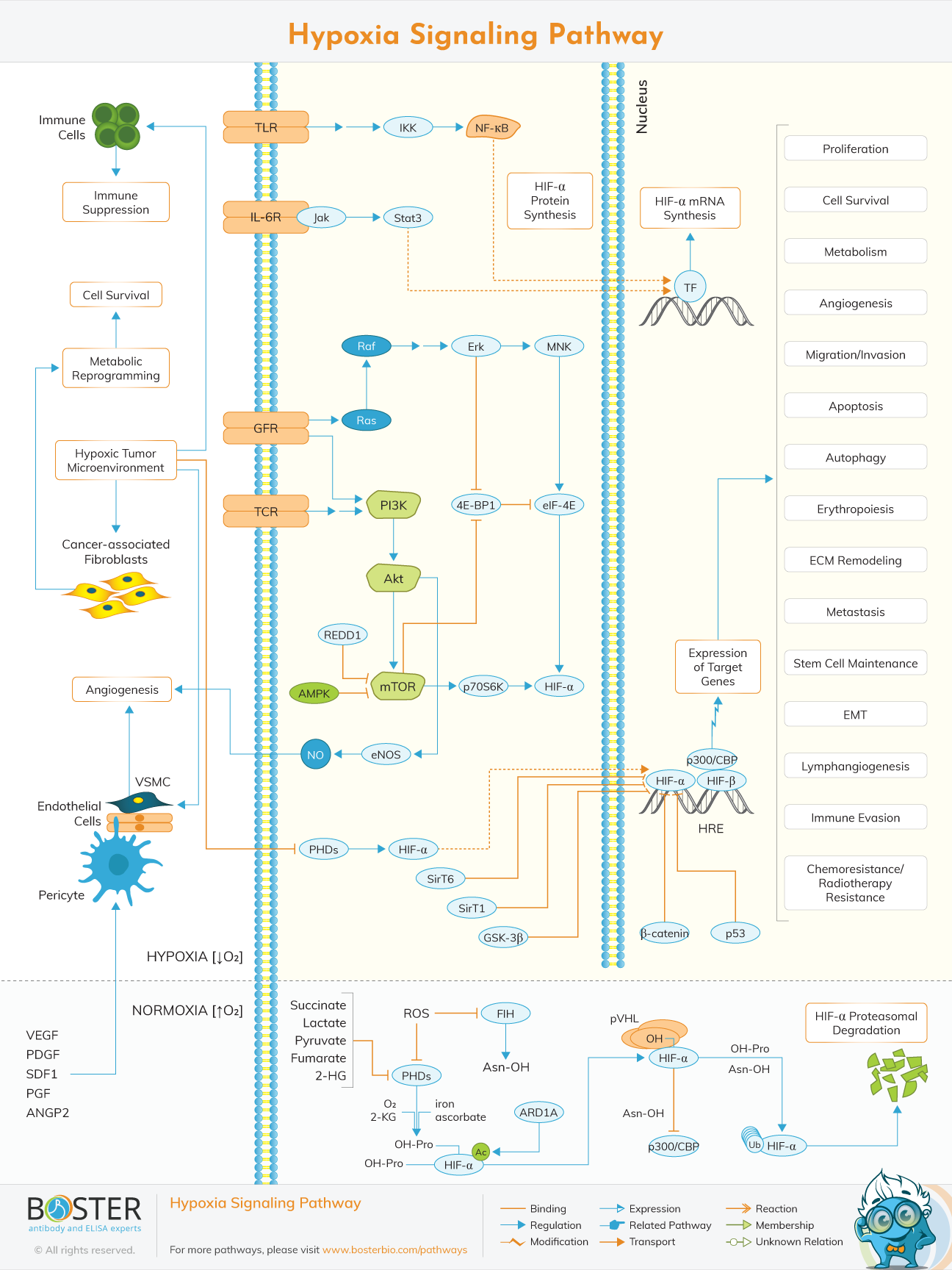
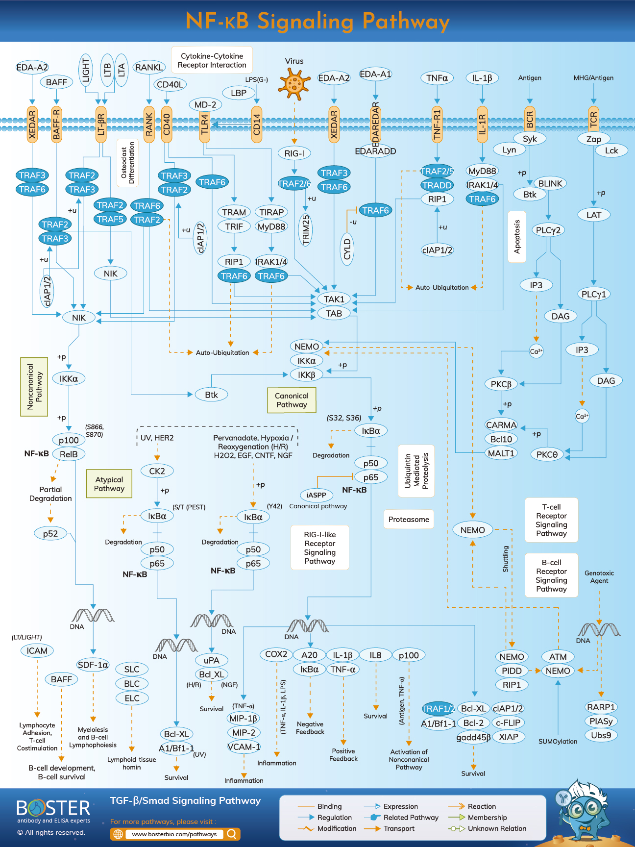
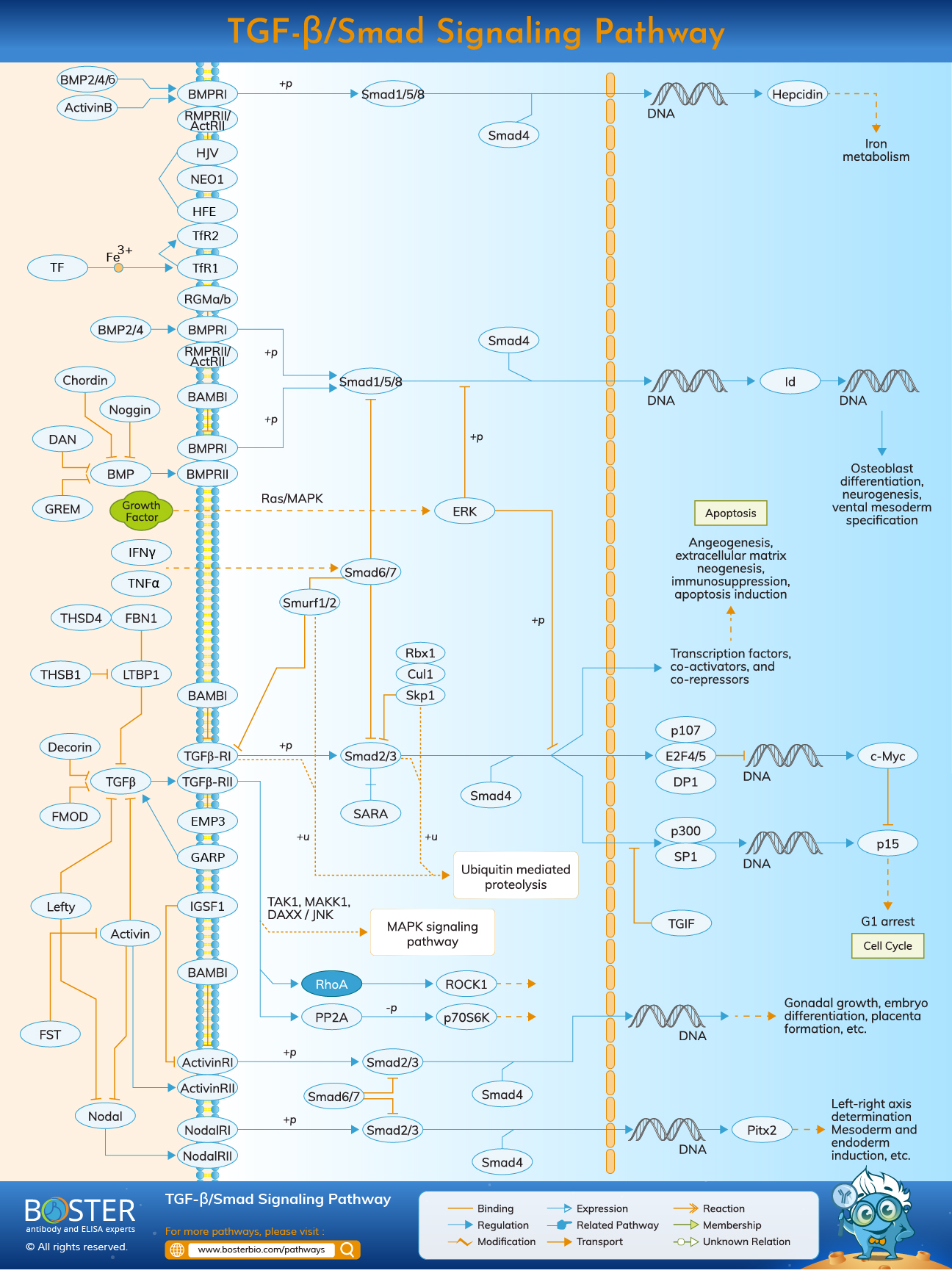
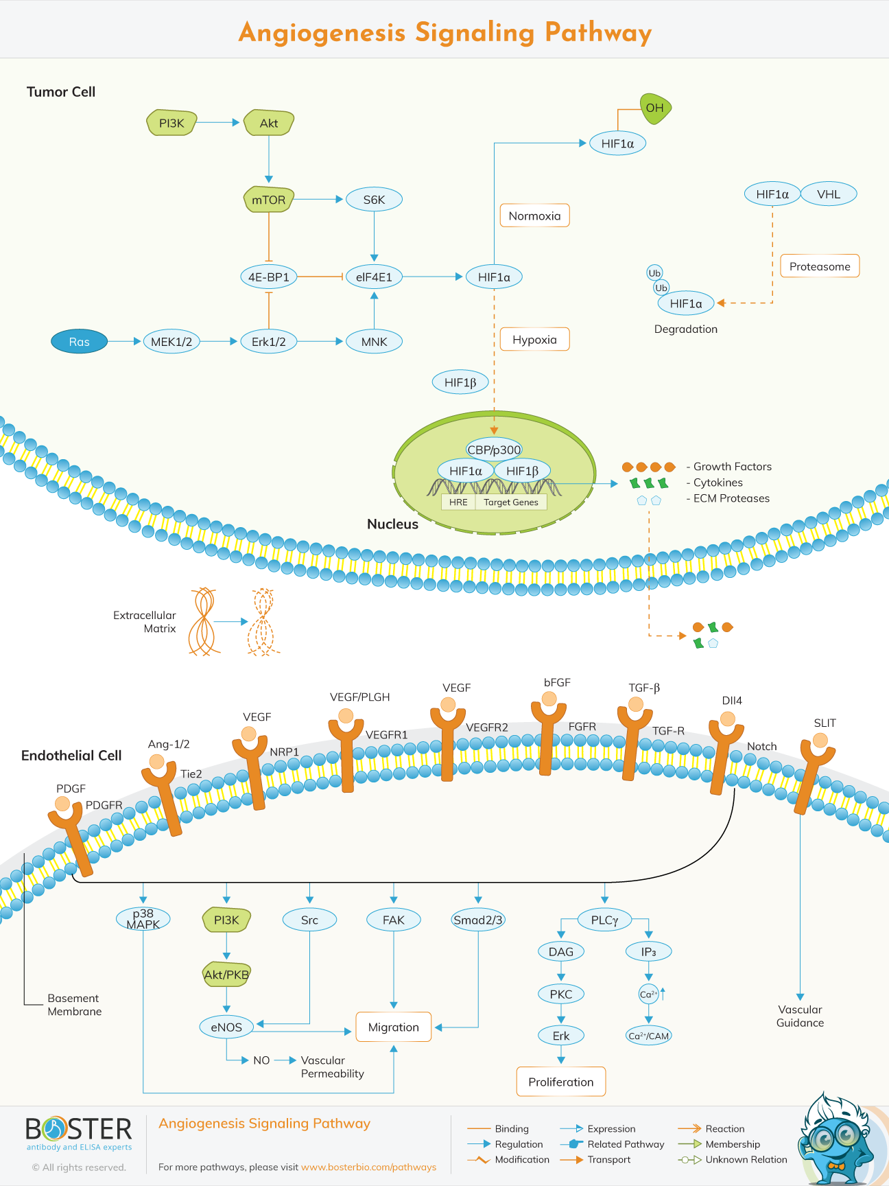
Use these pathway maps to connect biomarker changes with core metabolic control mechanisms.
These pathways help explain the shift from metabolic overload to inflammation, autophagy, and fibrosis-associated remodeling.
Insulin resistance is one of the core mechanisms linking diabetes, obesity-associated metabolic dysfunction, and fatty liver disease. When insulin signaling becomes less effective, adipose tissue lipolysis increases, hepatocytes take up excess fatty acids, and de novo lipogenesis is often enhanced. At the same time, glucose handling becomes less efficient and broader metabolic homeostasis begins to fail. This makes insulin signaling, adipokines, transporters, and pathway mediators central targets in metabolism research.
Excess lipid accumulation in hepatocytes is not only a storage problem but also a trigger for oxidative stress, organelle dysfunction, and inflammatory signaling. As hepatocyte stress increases, cytokines and chemokines recruit and activate immune and stromal cells, creating a transition from simple metabolic overload to tissue-level injury. This is why biomarkers such as KRT18, IL-6, TNF-α, CCL2, CRP, and extracellular matrix-associated proteins are frequently used to track disease progression.
Metabolic disease progression is shaped by inter-organ communication. Adipose dysfunction changes circulating leptin, adiponectin, inflammatory mediators, and lipid flux, all of which influence liver phenotype. In more advanced disease settings, persistent inflammation and tissue injury activate extracellular matrix remodeling and fibrosis-related mechanisms. This makes liver-adipose crosstalk an important framework for interpreting both biomarker panels and pathway-level readouts in metabolism studies.